|
|
 |
Fiche d'espèce de Copépode |
|
|
Calanoida ( Ordre ) |
|
|
|
Epacteriscoidea ( Superfamille ) |
|
|
|
Ridgewayiidae ( Famille ) |
|
|
|
Ridgewayia ( Genre ) |
|
|
| |
Ridgewayia marki minorcaensis Razouls & Carola, 1996 (F,M) | |
| | | | | | | Ref.: | | | Razouls & Carola, 1996 (p.48, figs.F,M); Vives & Shmeleva, 2007 (p.991, figs.F,M, Rem.); Figueroa & Hoefel, 2008 (p.145, Table 2, 3, Rem.) | 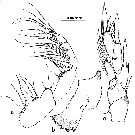 issued from : C. Razouls & M. Carola in Crustaceana, 1996, 69 (1). [p.49, Fig. a-c]. Female (from SE Minorca, Balearic Islands): a, Mxp; b, Md (mandibular blade); c, P5. Nota: Head with rounded front, showing a small dot (naupliar eye). Rostrum prominent, bent ventrally. A1 26-segmented, reaching the distal end of the caudal rami. Cephalosome and 1st thoracic segment separate, suture well marked., 4th and 5th thoracic segments separate. 5th thoracic segment with postero-lateral angles rounded, covering 1/3 of the genital segment. P5 symmetrical, biramous, exopodal segment 3 inserted in the middle of segment 2; endopod 2-segmented, the distal bearing 7 setae. mxp is more similar to the description given by Yeatman (1969) than to the one given by Esterly (1911, pl.4, fig.38), because of the rudimentary endopod not represented by Esterly, which bears 2 setae but is weakly separated from the basis.
|
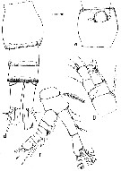 issued from : C. Razouls & M. Carola in Crustaceana, 1996, 69 (1). [p.50, Fig. d-g]. Female: d, urosome (dorsal); e, genital segment (ventral); f, A2; g, P1. Nota: Urosome 4-segmented; anal segment very short and not well visible; length ratio of urosomites and caudal rami 38.1:19.5:14.9: 5:22.5 = 100. Genital segment with a slight ventral protuberance, spermatheca located in the posterior region of the segment. Caudal rami with length 2.2 times width.The setation of A2 (Fig.1 f) differs from that given by Yeatman (1969, pl.1, fig.7); the distal segment of the endopod has 5 setae on the inner lobe, and 7 on the outer lobe instead of 5 according to Yeatman. The inner laciniae 1 and 2 of Mx1 have, respectively, 5 and 4 setae, which is in agreement with Esterly (1911, pl.3, fig.25) and Yeatman (1969, pl.1, fig.9). The proportions of the somites of the urosome, according to the figure in Esterly (1911, pl.2, fig.13), and including the anal segment and the caudal rami not observed by him, are, respectively, 35.5:17.8:14.9:5.6:26.2 = 100, giving the impression of being slightly shorter than in our specimens, except the caudal rami. The ratio length/width of the caudal rami is 2.2; Yeatman (1969, p.7) found this ratio to be 3 and Esterly (1911, p.220) 3.5; Yeatman (1969) observed fine setules at the inner margin of the caudal rami, which have not been observed in our specimens.
|
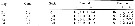 issued from : C. Razouls & M. Carola in Crustaceana, 1996, 69 (1). [p.51, Table II]. Female & Male: Setation formula of swimming legs P1 to P4. Roman numeral = spine; arabic numeral = seta. Nota: R. marki was redescribed by Yeatman (1969) who found some discrepancies in the setation of legs 1-4 with respect to the original description. He attribued these to a printing error in Table I (cf. Esterly, 1911): endopodal segment 3 of P3 with 8 setae instead of 5, and endopodal segment 3 of P4 with 7 setae instead of 6, because of one terminal spine missing. There are no difference between our setation formula for P1-P4 (in table II) and that given by Yeatman (1969).
|
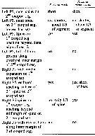
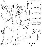 issued from : C. Razouls & M. Carola in Crustaceana, 1996, 69 (1). [p.53, Fig.2]. Male: a, right P5; b, left P5; c, urosome (dorsal). ?Nota: Prosome/urosome = 2.81; prosome length/width = 2.23. Rostrum prominent, bent ventrally. Left A1 26-segmented; right A1 24-segmented and geniculate (in agreement with Yeatman, 1969, p.8). Head and 1st thoracic segment separated (suture well marked), 4th and 5th thoracic segments separate. Urosome 5-segmented. Ratio of urosomites and caudal rami lengths: 21:16:19:12:9:24. Genital segment slightly wider than long (width/length = 1.38). Caudal rami with length/width = 2.23. The right P5 has the same general structure as reported by Esterly (1911, pl.3, fig.30) and Yeatman (1969, pl.2, fig.19) but at the basis it presents a group of 5 strong spines at the inner margin, and a weak seta at the subdistal external margin; the external spine of exopodal segment 1 is longer than the preceding spine (tis observation agrees well with that of Yeatman (1969, pl.2, fig.19). The left P5 presents one external spine on exopodal segment 1, which is longer than that depicted by Esterly (1911, pl.3, fig.34) and Yeatman (1969, pl.2, fig.20); exopodal segment 2 is similar to that described by Esterly and Yeatman, and exopodal segment 3 is too complicated to be compared in detail with descriptions by others.
|
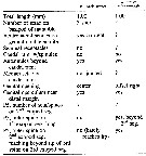 issued from : D.F. Figueroa & K.L. Hoefel in J. Crustacean Biol., 2008, 28 (1). [p.145, Table 2]. Characters female of the marki species-group.
|
 issued from : D.F. Figueroa & K.L. Hoefel in J. Crustacean Biol., 2008, 28 (1). [p.145, Table 3]. Characters male of the marki species-group.
| | | | | Ref. compl.: | | | Belmonte, 2018 (p.273, Table I: Italian zones) | | | | NZ: | 1 | | |
|
Carte de distribution de Ridgewayia marki minorcaensis par zones géographiques
|
| | | | | | | Loc: | | | Baleares: Minorca (Calan Porter submarine cave), Sardinia, Messina Strait | | | | N: | 2 | | | | Lg.: | | | (520) F: 1; M: 0,8; {F: 1,00; M: 0,80} | | | | Rem.: | Dans une excavation sous-marine. Pour Boxshall (1997, comm. pers.) cette sous-espèce est une espèce distincte de R. marki.
Voir aussi les remarques en anglais | | | Dernière mise à jour : 06/02/2020 | |
|
|
 Toute utilisation de ce site pour une publication sera mentionnée avec la référence suivante : Toute utilisation de ce site pour une publication sera mentionnée avec la référence suivante :
Razouls C., Desreumaux N., Kouwenberg J. et de Bovée F., 2005-2026. - Biodiversité des Copépodes planctoniques marins (morphologie, répartition géographique et données biologiques). Sorbonne Université, CNRS. Disponible sur http://copepodes.obs-banyuls.fr [Accédé le 27 mars 2026] © copyright 2005-2026 Sorbonne Université, CNRS
|
|
|
|


;)
;)
;)
;)
;)

