|
|
 |
Fiche d'espèce de Copépode |
|
|
Calanoida ( Ordre ) |
|
|
|
Ryocalanoidea ( Superfamille ) |
|
|
|
Ryocalanidae ( Famille ) |
|
|
|
Ryocalanus ( Genre ) |
|
|
| |
Ryocalanus infelix Tanaka, 1956 (F, M) | |
| | | | | | | Ref.: | | | Tanaka, 1956 b (p.3, Descr.M, figs.M); 1956 c (p.404, Rem.M); Chihara & Murano, 1997 (p.898, Pl.167: M); Boxshall & Halsey, 2004 (p.184); Renz & al., 2013 (p.257); Andronov, 2004 (p.33, fig.M); Renz & al., 2018 (p.13, figs.F,M, Rem.) | 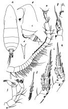 issued from : O. Tanaka in Breviora, 1956, 64. [p.2, Fig.1]. Male: a, habitus (dorsal); b, Forehead (lateral); c, last thoracic segment and urosome (lateral left side); d, rostrum; e, right A1; f, A2; g, P1 and poximal outer margin of endopodite; h, P2; i, P3; j, 3rd segment of exopodite of P4. Nota: Head and 1st thoracic segment separate; 4th and 5th separated. The urosome segments and furca are in the proportional lengths as 32:18:11:7:14:18 = 100; Right A1 forming a grasping organ, extends to the distal margin of the 2nd thoracic segment.
|
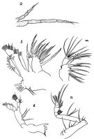 issued from : O. Tanaka in Breviora, 1956, 64. [p.4, Fig.2]. Male: k, Md; l, Mx1; m, Mx2; n, Mxp; o, P5.
|
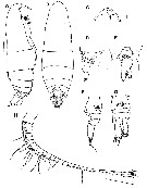 Issued from : J. Renz, E.L. Markhaseva & S. Laakmann in Zool. J. Linnean Soc., 2018, XX. [p.14, Fig.7]. Female (from 46.2333 °N, 155.5333 °E): A-B, habitus (lateral and dorsal, respectively); C, rostrum (ventral); D-E, prosome posterior corners and urosome (lateral and ventral, respectively); F-G, urosome (lateral and ventral, respectively); H, A1. Scale bars; A-B: 0.5 mm; C-G: 0.1 mm. Nota: - Prosome 5.1 times as long as urosome. - Rostrum one-pointed, slender. Cephalosome and pediger 1 separate, pedigers 4-5 separate, - Posterolateral corners of prosome (in dorsal view) asymmetrical, extended posteriorly into points, extending to distal margin of genital double-somite on right side and to distal margin of 2nd urosomal segment in left side . - Ventral inner surface of pediger 5 with short spinules. - Urosome: genital double-somite plus 3 articulated or partly articulated somites. - Genital double-somite asymmetrical , with lateral swelling on right side or left side and faint line of incomplete fusion on dorsal and ventral surface; in lateral view swollen ventromedially. Seminal receptacles in lateral view oval, turned upward. - Urosomites 2, 3 and 4 asymmetrical; 3 and 4 partly fused. - Urosome covered by viscous mass. - Caudal rami asymmetrical with right ramus longer and wider than left; both rami with row of spinules on inner margin and with 2 lateral setae (II and III), 3 terminal setae (IV-VI) and 1 dorsal seta (VII). - A1 with 24 segments (I: 1s+1ae; II:-IV: 6s+4ae; V: 2s+2ae; XXVII-XXVIII: 4s+1ae). Remarks from Renz & al. (2018, p.48): A1 of one female did show a quadrithek arrangement of appendages with segments V, VII, VIII, IX and XIII bearing 2 small, slender aesthetascs in addition to 2 setae. While the doubling of aesthetascs is unusual in female calanoid copepods, it has previopusly been observed in a few genera within the Calanidae by Fleminger (1985). He postulated that quadrithek females derive from genotypic males in which the gonad develops as an ovary and suggested that environmental factors or internal factors affect the final phenotypic sex.
|
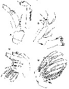 Issued from : J. Renz, E.L. Markhaseva & S. Laakmann in Zool. J. Linnean Soc., 2018, XX. [p.15, Fig.8]. Female: A, A2; B, Md (palp and gnathobase separately); C, Mx1; D, Mx2. Scale bars: A-D: 0.1 mm. Nota: A2: coxa with 1, basis with 2 setae; endopod segment 1 with 2 setae and row of spinules, segment 2 with 16 setae; exopod 8-segmented (setal formula: 1, 3, 1, 1, 1, 1, 1, 3 setae)/ - Md: gnathobase cutting edge with 8 unequal teeth plus ventral seta; basis with 3 setae; endopodal segments incompletely fused, with 6 setae; 1st endopodal segment with 4 setae (3 setae plus 1 scar), 2nd with 11 setae. - Mx1: praecoxal arthrite with 9 terminal spines, 3 posterior and 1 anterior setae; posterior surface with small spinules; coxal endite with 6 setae, coxal epipodite setae broken; proximal basal endite with 4 setae, distal basal endite with 5 setae and small surface spinules; endopod with 12 setae and patch of small surface spinules; exopod with 8 setae. - Mx2: proximal praecoxal endite bearing 3 setae plus attenuation on left limb, 5 setae in right limb, distal praecoxal endite with 3 setae and surface spinules; coxa with 1 outer seta, coxal endites with 3 setae each and surface spinules; proximal basal endite 4 setae; remaining endopod with 9 setae.
|
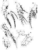 Issued from : J. Renz, E.L. Markhaseva & S. Laakmann in Zool. J. Linnean Soc., 2018, XX. [p.16, Fig.9]. Female: A, Mxp; B, P1 'with endopod figured separately); C, P2; D, P3; E, P4 (with exopod figured separately); F, P4 coxa and basis (different view). Scale bars: A-F: 0,1 mm. Nota; - Mxp: syncoxa with 1 seta on proximal praecoxal endite, 2 setae on middle endite, and 3 setae on distal praecoxal endite; coxal endite with 3 setae; basis with 3 medial setae; endopod 6-segmented (setal formula: 2, 4, 4, 4, 3+1, and 4 setae)
|
 Issued from : J. Renz, E.L. Markhaseva & S. Laakmann in Zool. J. Linnean Soc., 2018, XX. [p.18, Table 4]. Female: Seta and spine formula from P1 to P4.
|
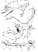 Issued from : J. Renz, E.L. Markhaseva & S. Laakmann in Zool. J. Linnean Soc., 2018, XX. [p.17, Fig.10]. Male (from 43.5666 °N, 153.9666 °E): A, habitus (lateral); B, left A1; C, right A1 (segments XX-XXVIII figured separately); D, right A1 (segments XX-XXVI); E, A1 (segments XXV-XXVIII). Scale bars: A-C: 0.5 mm; D-E: 0.1 mm. Nota: - Oral limbs and swimming legs as in original desvription by Tanaka (1956), deviating only in details. - Left A1 unmodified, 24-segmented, extending th 1st urosomal segment; armature (I: 1s+1ae; II:-IV: 5s+3ae?+ 2 short sensillae; V: 2s+2ae+ 1 short sensilla; VI: 2s+2ae; VII: 2s+2ae+1 short s in particular position; XXVII-XXVIII: 4s+1ae). Right A1 strongly modified for grasping, 22-segmentes; segments XVIII-XXII/XXIII with surgace spinules; segments XIX-XXVI strongly enlarged; segments XXI-XXII partly fused; segments XXII-XXIII fused; segment XXIV with lateral broad denticulated lamella; segment XXV with small lateral chitinized lamella; hinges occurring between segments XVIII and XIX and XXIX and XX, and XX and XXI, XXIII and XXIV, XXIV and XXV. Armature (I: 1s+1ae; II-IV: 6s+4ae; V: 2s+2ae; XXVII-XXVIII: 5s+1ae).
| | | | | NZ: | 2 | | |
|
Carte de distribution de Ryocalanus infelix par zones géographiques
|
| | |  Carte de 1996 Carte de 1996 | |
| | | | Loc: | | | Japan (Izu region: Suruga Bay), Kurile-Kamchatka Trench | | | | N: | 2 | | | | Lg.: | | | (55) M: 2,18; (1223) F: 2,60-2,65; M: 2,11; {F: 2,60-2,65; M: 1,95-2,18} | | | | Rem.: | From a vertical haul (1410-0 m).
After Renz & al. (2018, p.15-8) specimens male and females were present in the samples COI sequence analysis verified the assignment of females and males of the same species.
For Renz & al. (2018, p.18) the right A1 male differs from other Ryocalanus species in the shape of the denticulated and chitinized lamella on segments XXIV and XXVI, which is of a diggerent structure in R. brasilianus (Renz & al.,2013) and absent in R. squamatus (Renz & al., 2018) and R. bowmani (Markhaseva & Ferrari, 1996).
The morphology of the male specimens is mostly as in the original description of Tanaka (1956), except for any setae missing in P1, Mx1, Mx2, and in the left A1 male | | | Dernière mise à jour : 26/03/2019 | |
|
|
 Toute utilisation de ce site pour une publication sera mentionnée avec la référence suivante : Toute utilisation de ce site pour une publication sera mentionnée avec la référence suivante :
Razouls C., Desreumaux N., Kouwenberg J. et de Bovée F., 2005-2025. - Biodiversité des Copépodes planctoniques marins (morphologie, répartition géographique et données biologiques). Sorbonne Université, CNRS. Disponible sur http://copepodes.obs-banyuls.fr [Accédé le 02 janvier 2026] © copyright 2005-2025 Sorbonne Université, CNRS
|
|
 |
 |


