|
|
 |
Fiche d'espèce de Copépode |
|
|
Calanoida ( Ordre ) |
|
|
|
Clausocalanoidea ( Superfamille ) |
|
|
|
Scolecitrichidae ( Famille ) |
|
|
|
Macandrewella ( Genre ) |
|
|
| |
Macandrewella cochinensis Gopalakrishnan, 1973 (F,M) | |
| | | | | | | Ref.: | | | Gopalakrishnan, 1973 (p.180, figs.F,M); Bradford & al., 1983 (p.91); Ohtsuka & al., 2002 (p.544, Rem.); El-Sherbiny & Al-Aidaroos, 2013 (p.4, figs.F,M, Redescr., Rem.: p.12-following) | 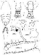 Issued from : M.M. El-Sherbiny & A.M. Al-Aidaroos in ZooKeys, 2013, 344. [p.5, Fig.2]. Female (from 27°51.234'N, 34°17.605'E): A, habitus (dorsal); B, rostrum (lateral); C, rostrum (ventral; D, posterior prosome and urosome (dorsal); E, urosome (ventral); F, genital double-somite with spermatophore (lateral, right side); G, genital double-somite (lateral, right side); H-I, A1. Scale bars in mm. Nota: Cephalosome and 1st pedigerous somite completely fused, pedigers 4 and 5 partially fused, with incomplete suture visible dorsally and ventrolaterally Posterior margin of last thoracic segment asymmetrical, left one longer; each with 1 pairs of processes on each side, postero-dorsolateral projecting on each side lamellar with serrated margin, asymmetrical ventrolateral processes curved ventromedially at tip, slightly exceeding the posterior end of genital double-somite on left side and slightly exceeding half length of genital double-somite on right. A1 symmetrical, 23-segmented, extending nearly posterior border of 2nd somite. 4th urosomite (anal somite) very short, telescoped into preceding somite. Caudal rami symmetrical with 5 caudal setae, left middle seta (V) 1.5 times as long as righjt one.
|
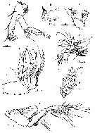 Issued from : M.M. El-Sherbiny & A.M. Al-Aidaroos in ZooKeys, 2013, 344. [p.7, Fig.4]. Female: A, A2; B, Md (gnathobase with cutting edge); C, Md (mandibular palp); D, Mx1; E, Mx2; F, endopod of Mx2; G, Mxp. Scale bars in mm. A2: Exopod 7 segmented, endopod 2-segmented; Md: gnathobase heavily sclerotized with cutting edge bearing 8 teeth (5 of them flattened with broad edge) and spinulose seta. Mx1 with praecoxal arthrite bearing 13 setae, 9 setae along terminal border, 4 setae on posterior surface and 1 seta on anterior surface. Coxal endite with 2 setae; coxal epipodite with 9 setae; basis completely fused with endopod; 1st and 2nd basal endites with 3 and 5 setae respectively; basoendopod with 7 setae terminally; exopod lobate, bearing 8 setae. Mx2: praecoxal endite 1 with 4 setae; 2nd praecoxal to 2nd coxal endites each with 3 setae; basis with 2 setae and 2 worm-like sensory setae and patch of fine spinules; Endopd indistinctly 3-segmented, bearing 3 brush-like, 2 brush-like and 3 worm-like sensory setae, respectively.
|
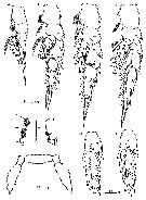 Issued from : M.M. El-Sherbiny & A.M. Al-Aidaroos in ZooKeys, 2013, 344. [p.9, Fig.5]. Female: A, P1 (anterior surface); B, medial margin of 1st and 2nd exopodal segments of P1; C, lateral distal margin of P1 endopod; D, P2 (posterior surface); E, P3 (posterior surface); F, P4 (posterior surface); G-H, 2nd and 3rd exopodal segments of P4 (anterior surface); I, P5 (anterior surface). Scale bars in mm. Nota: P5 rudimentary, 2-segmented separated at base; each terminal segment cylindrical with medial papilla-like protrusion and constriction at 1/3 distal part.
|
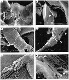 Issued from : M.M. El-Sherbiny & A.M. Al-Aidaroos in ZooKeys, 2013, 344. [p.6, Fig.3]. Female (SEM micrographs): A, rostrum and cuticular lens indicated by arrow, ventral view); B, serration of postero-dorsolateral process of prosomal end indicated by arrow, lateral view; C, urosome, anterodorsal protrusions and posterodorsal swelling on left side indicated by arrows, dorsal view; D, urosome, posterodorsal swelling on left side indicated by arrow, lateral view (left); E, maxillary endopod; F, P5 indicated by arrow.
|
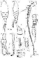 Issued from : M.M. El-Sherbiny & A.M. Al-Aidaroos in ZooKeys, 2013, 344. [p.10, Fig.6]. Male: A-B, habitus (dorsal and lateral, respectively); C, urosome (dorsal); D, 1st and 2nd urosomal segment (lateral, right side); E, left A1; F, Mxp (terminal endopod segments); G, exopod segment 3 of P2; H, left P5; I, terminal portion of left exopod of P5; J, terminal portion of left endopod of P5; K, right P5. Scale bars in mm. Nota: Rostrum bifurcated with pair of filaments; cuticular median lens present at base of rostrum. Cephalosome completely fused with 1st pediger, 4th and 5th pedigers fused with suture visible laterally; Urosome 5-segmented. Genital somite asymmetrical, with anterior dorsal knobs on right side. 2nd to 4th urosomites with thin spinules along posterior margin; 2nd urosomite slightly asymmetrical in dorsal view. Anal somite very small. Caudal rami symmetrical, each ramus bearing 4 plumose setae. A1 consisting of 18 and 19 articulated segments on right and left side, respectively. Mouth parts similar to those of female.
|
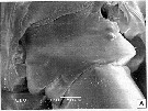 Issued from : M.M. El-Sherbiny & A.M. Al-Aidaroos in ZooKeys, 2013, 344. [p.11, Fig.7 A]. Male (SEM micrograph); A, genital somite (dorsal).
|
 Issued from : M.M. El-Sherbiny & A.M. Al-Aidaroos in ZooKeys, 2013, 344. [p.8, Table 1]. Female and Male: Armature of the swimming legs P1 to P4. Nota: 3rd exopodal segment of P2 male with different number and distribution of posterior surface setules (Fig. 5G).
|
 Issued from : T.C. Gopalakrishnan in Handbook Int. Zoopl. Coll., Vol. V, 1973 (IIOE). [p.181, Fig.1]. Female (from 10°10'N, 75°46'E): a, habitus (dorsal); b, lateral view of posterior part from left side; c, same, from right side.
|
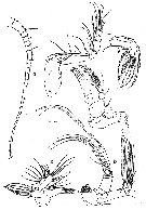 Issued from : T.C. Gopalakrishnan in Handbook Int. Zoopl. Coll., Vol. V, 1973 (IIOE). [p.182, Fig.2]. Female: a, A1 (magnification double that of the other appendages); b, A2; c, Md; d, Mx1; e, Mx2; f, Mxp.
|
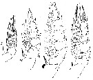 Issued from : T.C. Gopalakrishnan in Handbook Int. Zoopl. Coll., Vol. V, 1973 (IIOE). [p.183, Fig.3]. Female: a-d, P1 to P4, respectively.
|
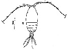 Issued from : T.C. Gopalakrishnan in Handbook Int. Zoopl. Coll., Vol. V, 1973 (IIOE). [p.184, Fig.4, a]. Male: a, habitus (dorsal).
|
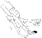 Issued from : T.C. Gopalakrishnan in Handbook Int. Zoopl. Coll., Vol. V, 1973 (IIOE). [p.184, Fig.4, b]. Male : b, P5.
|
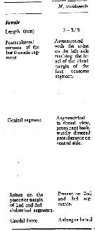 Issued from : T.C. Gopalakrishnan in Handbook Int. Zoopl. Coll., Vol. V, 1973 (IIOE). [p.185: Table]. Morphological characteristics of Macandrewella cochinensis female. Compare with M. scotti, M. chelipes, M. sewelli, M. asymmetrica, M. mera, M. agassizi, M. joanae.
|
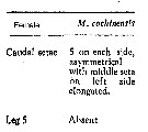 Issued from : T.C. Gopalakrishnan in Handbook Int. Zoopl. Coll., Vol. V, 1973 (IIOE). [p.186: Table]. Morphological characteristics of Macandrewella cochinensis female. Compare with M. scotti, M. chelipes, M. sewelli, M. asymmetrica, M. mera, M. agassizi, M. joanae.
|
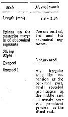 Issued from : T.C. Gopalakrishnan in Handbook Int. Zoopl. Coll., Vol. V, 1973 (IIOE). [p.186: Table]. Morphological characteristics of Macandrewella cochinensis male. Compare with M. joanae, M. chelipes, M. sewelli, M. asymmetrica, M. mera, M. agassizi, M. scotti.
|
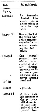 Issued from : T.C. Gopalakrishnan in Handbook Int. Zoopl. Coll., Vol. V, 1973 (IIOE). [p.186: Table]. Morphological characteristics of Macandrewella cochinensis male. Compare with M. joanae, M. chelipes, M. sewelli, M. agassizi, M. scotti. (not recorded: M. asymmetrica, M. mera).
|
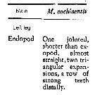 Issued from : T.C. Gopalakrishnan in Handbook Int. Zoopl. Coll., Vol. V, 1973 (IIOE). [p.188: Table]. Morphological characteristics of Macandrewella cochinensis male. Compare with M. scotti, M. chelipes, M. sewelli, M. agassizi, M. joanae. (not recorded: M. asymmetrica, M. mera),
| | | | | Ref. compl.: | | | Madhupratap & Haridas, 1986 (p.105, tab.1); Gopalakrishnan & Balachandran, 1992 (p.167, figs.1, 7, Table 1, 2); | | | | NZ: | 2 | | |
|
Carte de distribution de Macandrewella cochinensis par zones géographiques
|
| | | | | |  issued from : S. Ohtsuka, S. Nishida & K. Nakaguchi inJ. Nat. Hist., 2002, 36 [p.558, Fig.21]. issued from : S. Ohtsuka, S. Nishida & K. Nakaguchi inJ. Nat. Hist., 2002, 36 [p.558, Fig.21].
Distribution of several species of Macandrewella: 1, 2, 4, M. chelipes; 3, M. cochinensis; 4, M. scotti; 5, 8, 10, M. sewelli; 6, M. stygiana; 7, M. joanae; 8, M. asymmetrica; 8, M. mera; 9, M. agassizi; 11, M. serratipes. |
| | | | Loc: | | | India (Cochin), N Red Sea (Sharm El-Maya Bay) | | | | N: | 3 | | | | Lg.: | | | (526) F: 3,15-3; M: 2,95-2,9; (1153) F: 2,88-3,15; M: 2,83-3,21; {F: 2,88-3,15; M: 2,83-3,21} | | | | Rem.: | The species closely resembles M. stygiana. After El-Sherbiny & Al-Aidaroos (2013, p.14) this species is the first record in the family Scolecitrichidae to form a monospecific aggregation (swarm) near surface, suggesting an antipredation against visual predators or facilitating and enhancing mating opportunity. Among adults males show a slightly higher percentage in the population than females (38.1 and 34.4 % respectively).
In general, morphological characters of Macandrewella specimens collected from the northern Red Sea correspond to M. cochinensis and they are currently attributed to this species. However, their taxonomic status is expected to be proved when additional specimens from the M. cochinensis type locality will be obtained. The studied specimens from the Red Sea differ from M. cochinensis sensu Gopalakrishnan (1973) in the following characters of females (features of Gopalakrishnan given in brackets): 1- pedigers 4 and 5 fused (separate); 2- pediger 5 with 2 pairs of processes, both dorsolateral projecting lamellar with serrated margin (apparently overlooked); 3- genital double-somite with asymmetrical anterodorsal protrusion on each side and posterodorsal swelling on left side (not described, only mentioned it is asymmetrical); 4- A2 1st endopodal segment with 2 setae subterminally (1 seta); 5- endopodal middle lobe of distal segment with 8 setae (7 setae); 6- endopodal terminal lobe of distal segment bearing 7 setae (6 setae); 7- A2 2nd exopodal segment with long setae (seta shorter); 8- Md palp with basis carrying 2 setae (1 seta); 9- endopodal segment with 9 setae (10 setae); 10- Mx1 praecoxal artrite with 13 setae (9 setae); 11- Mx1 coxal epipodite bearing 9 setae (8 setae); 12- Mx1 2nd basal endite with 5 setae (4 setae); 13- Mx1 basiendopod with 7 terminal setae (6 setae); 14- Mxp endopod with setal formula 2, 4, 4, 3, 3+1, 4 (vs 2, 4, 4, 3, 2+1, 4); 15- P3 basis with lateral prominence (absent); 16- surface spinulation of swimming legs is more dense than in the original description; 17- P5 rudimentary, composed of 2 segments (apparently overlooked).
Male from the Red Sea and described by Gopalakrishnan (1973): differ in 2nd exopodal segment of left P5 with 1 seta and 1 element terminally (1 element). | | | Dernière mise à jour : 30/10/2015 | |
|
|
 Toute utilisation de ce site pour une publication sera mentionnée avec la référence suivante : Toute utilisation de ce site pour une publication sera mentionnée avec la référence suivante :
Razouls C., Desreumaux N., Kouwenberg J. et de Bovée F., 2005-2026. - Biodiversité des Copépodes planctoniques marins (morphologie, répartition géographique et données biologiques). Sorbonne Université, CNRS. Disponible sur http://copepodes.obs-banyuls.fr [Accédé le 08 janvier 2026] © copyright 2005-2026 Sorbonne Université, CNRS
|
|
 |
 |


