|
|
 |
Fiche d'espèce de Copépode |
|
|
Calanoida ( Ordre ) |
|
|
|
Clausocalanoidea ( Superfamille ) |
|
|
|
Scolecitrichidae ( Famille ) |
|
|
|
Scolecithricella ( Genre ) |
|
|
| |
Scolecithricella vittata (Giesbrecht, 1892) (F,M) | |
| | | | | | | Syn.: | Scolecithrix vittata Giesbrecht, 1892 (p.266, 283, 775, figs.F,M); Giesbrecht & Schmeil, 1898 (p.43, Rem. F,M fig.F); Fernandes, 2008 (p.465, Tabl.2);
Scolecithricella sub-vittata Rose, 1942 (p.142, figs.F, Rem.); Hure & Scotto di Carlo, 1968 (p.26); Boucher, 1970 (p.15); Kovalev & Shmeleva, 1982 (p.84); Hure & Krsinic, 1998 (p.49, 101);
Amallothrix tenuiserrata (M) : Rose, 1942 (p.163);
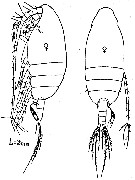
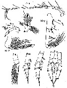
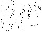
no Scolecithricella vittata (M) : Rose, 1942 (p.159, figs.M, Rem. M: n°4) | | | | Ref.: | | | Sars, 1925 (p.190, figs.F); Farran, 1926 (p.259); 1929 (p.209, 247); Rose, 1933 a (p.158, figs.F); Farran, 1936 a (p.97); Rose, 1942 (p.140, Redescr.F, figs.F, non p.159: M n°4); C.B. Wilson, 1950 (p.335, figs.F); Chiba & al., 1957 (p.308); Tanaka, 1962 (p.41, figs.F,M); Grice, 1962 (p.208, figs.F, Rem.); Chen & Zhang, 1965 (p.62, figs.F); Vervoort, 1965 (p.65, Rem.); Owre & Foyo, 1967 (p.61, figs.F,M); Mazza, 1967 (p.167, 172, figs.juv., F,M); Park, 1968 (p.555); Bradford, 1970 a (p.356, figs.F); Razouls, 1972 (p.94, Annexe: p.55, figs.F); Bradford, 1973 (p.142); Park, 1980 (p.37, figs.F,M, Rem.); Björnberg & al., 1981 (p.639, figs.F); Bradford & al., 1983 (p.103, 112, figs.F,M, Rem.); Campaner, 1984 a (p.175, Rem., figs.F); Bradford-Grieve & al., 1999 (p.881, 933, figs.F,M); Vyshkvartzeva, 1999 (2000) (p.234); Vives & Shmeleva, 2007 (p.790, figs.F,M, Rem.) | 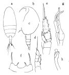 issued from : O. Tanaka in Publ. Seto Mar. Biol. Lab., 1962, X (1). [p.42, Fig.129]. Female: a, habitus (dorsal); b, head (left lateral side); c, last thoracic segment and urosome (left lateral side); d, P1; e, P2; f, 5th pair of legs; Male: g, P5; h, distal segment of right P5. Nota Female: - Abdomen contained 4 times in the length of the cephalothorax; - Posterolateral corners of the last thoracic segment broadly rounded. - Rostral filaments slender and long. - Urosome 4-segmented; proportional lengths of abdominal segments and caudal rami 42 : 19 : 17 : 3 : 19 = 100. - Genital segment as wide as long, not swollen ventrally. - Caudal rami 1.7 times as long as wide. - A1 21-segmented, extends to the distal end of the anal segment. - Mx1 with 7 setae on the outer lobe; 11 setae on the exopod; 15 (7+4+4) setae on the endopod; 4 setae on the 2nd basal; 3 setae on the 3rd inner lobe; 2 setae on the 2nd inner lobe; 13 setae on the 1st inner lobe (= praecoxal arthrite). - Mx2 with 5 bud-like setae and 3 long vermiform filaments on the endopod. - P1 without outer edge spine on the 1st exopodal segment. - P2 with the outer edge spine on the 1st exopodal segment, long and curved. P5 1-segmented. Apical spine long; there is a small spine at the base of the apical spine (this spine was not figured by Giesbrecht or by Sars). Nota Male: - A1 extends to the distal margin of the 3rd abdominal segment. ) P5 exceeds the distal end of the abdomen. Endopod of the right leg very short; 2nd exopodal segment with 2 protuberances, 1 on the middle, another on the distal margin of the segment; the 3rd segment terminates in bud-like process. Endopod of the left leg short with 1 spinule at apex; distal segment of exopod obliquely truncate, and furnished with a sort of lamella on the distal outer margin.
|
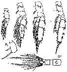
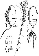
 issued from : J.M. Bradford, L. Haakonssen & J.B. Jillett in Mem. N.Z. Oceanogr. Inst., 1983, 90. [p.112, Fig.69]. Female (from Bradford, 1970 b): B, endopod of P2; C, P5. Nota Female: - A1 21-segmented , extends to end of caudal rami. - Basis of Md with 1 seta. - Mx2 endopod with 3 of 5 brush-like filaments long and thin, while other two short and thick. - P2 endopodal segment 2 elongated, with 4 strong spines on posterior surface. - P5 pallet-shaped, terminal spine bordered by hairs, the spine longer than segment itself and between one and one-half and two times as long as denticulate inner edge spine.
|
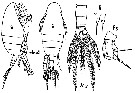
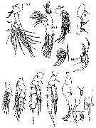
 issued from : T. Park in Antarct. Res. Ser. Washington, 1980, 31 (2). [p.39, Fig.5]. Female: a, b, forehead (lateral); c, last thoracic segment and urosome (lateral left side); d, genital segment (lateral left side); e, rostrum (anterior); f, A2; g, Md; h, Mx1; i, distal part of Mx2; j, Mxp; k, P1 (anterior); l, P2 (postrior); m, P3 (posterior); n, P4 (posterior); o, P5 (posterior). Nota: Urosome about 1/5 length of prosome. A1 reaching only as far as posterior end of caudal ramus.
|
 issued from : T. Park in Antarct. Res. Ser. Washington, 1980, 31 (2). [p.40, Fig.6]. Male: a, forehead (lateral); b, last thoracic segment with P5 and urosome (lateral left side); c, A2; d, Md; e, Mx1; f, distal part of Mx2; g, Mxp; h, P5 (anterior); i, P5 (posterior). Nota: Urosome about 1/3 length of prosome. A1 reaches close to distal end of urosome.
|
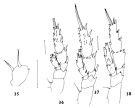 issued from : J.M. Bradford in N.Z. Jl Mar. Freshw. Res., 1970, 4 (4). [p.356, Figs 35-38]. Female (off Kaikoura, New Zealand): 35, P5; 36, P2; 37, P3; 38, P4. Scale bars represent 0.1 mm.
|
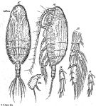
 issued from: Q.-c Chen & S.-z. Zhang in Studia Marina Sinica, 1965, 7. [Pl.19, 11-12]. Female (from E China Sea): 11, habitus (dorsal); 12, P5 (posterior).
|
 issued from : A.F. Campaner in Bolm. Zool., Univ. S. Paulo, 1984, 6 . [p.177, Fig.6, a-e]. Female (from off Rio de Janeiro): a, habitus (lateral left side); b, genital somite (with attached spermatophore); c, exopodite 3 of P2 (posterior); d, exopodite 3 of P3 (posterior); e, P5.
|
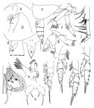
 Issued from : G.O. Sars in Résult. Camp. Scient. Prince Albert I, 69, pls.1-127 (1924). [Pl.LII, figs.15-20]. As Scolecithrix vittata. Female (from Canary Islands): 15, habitus (dorsal); 16, idem (lateral left side); 17, P2; 18, endopodal segments of P3; 19, endopodal segments of P4; 20, P5. Nota Female: - Rostrum less prominent than Scolecithricella abyssalis but of the same structure. - Posterolateral corners of the last thoracic segment regularly rounded. - Urosome about equal one third and half the prosome length. - Genital segment slightly swollen ventrally, . about as long as the three following segments together. - A1 extends back to the 2nd abdominal segments.w - Swimming legs resembles to S. abyssalis. - P5 of same shape than S. abyssalis, but shorter and the strong development of the apical spine., 2 times the length of the inner edge spine.; As in S. abyssalis there is a little tooth on the external margin opposite to the inner spine.
|
 issued from : M. Rose in Annls Inst. océanogr., Monaco, 1942, XXI (3). [p.140, Fig.31]. Female (from Alger Bay, Algeria): habitus (lateral and dorsal, respectively).
|
 issued from : M. Rose in Annls Inst. océanogr., Monaco, 1942, XXI (3). [p.141, Figs.32, 33]. Female: A1; R, rostrum; A2; Md; Mx1 (as Mx); Mx2 (as Mxp1); Mxp (as Mxp2). Nota: A1 21-segmented with sketching of subdivision on 1st segment. A2 with exopodite more longer than endopodite.
|
 issued from : M. Rose in Annls Inst. océanogr., Monaco, 1942, XXI (3). [p.142, Fig.34]. Female: P1 to P5; Ur, urosome).
|
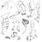
 issued from : M. Rose in Annls Inst. océanogr., Monaco, 1942, XXI (3). [p.142, Fig.35]. As Scolecithricella sub-vittata. Female (from Alger Bay, Algeria): habitus (lateral and dorsal, respectively); Ur, urosome; R, rostrum; P5.
|
 issued from : M. Rose in Annls Inst. océanogr., Monaco, 1942, XXI (3). [p.143, Figs.36, 37]. As Scolecithricella sub-vittata. Female: A1; A2; Mx1 (as Mx); Mx2 (as Mxp1); Mxp (as Mxp2); P1 to P4. Nota: A1 22-segmented (without sketching of subdivision on 1st segment).
|
 issued from : M. Rose in Annls Inst. océanogr., Monaco, 1942, XXI (3). [p.164, Fig.56]. As Amallothrix tenuiserrata. Male (from Alger Bay, Algeria): habitus (lateral and dorsal, respectively); Ur, urosome (lateral and dorsal, respectively); A1. Nota: A1 19-segmented (the 8th segment indistictly separated.
|
 issued from : M. Rose in Annls Inst. océanogr., Monaco, 1942, XXI (3). [p.165, Figs.57, 58]. As Amallothrix tenuiserrata. Male: A2; Md; Mx1 (as Mx); Mx2 (as Mxp1); detail of sensorial setae on Mx2; Mxp (as mxp2); P1 to P5. Nota: Mx1 with 3 distal setae, strong and curved; endopodite with 9 sensorial setae: 3 vermiforms falcate-like, 1 club-like, 5 button-like (3 longer, slender and curved, and 2 thick, shorter)
|
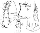
 issued from : C. Razouls in Th. Doc. Etat Fac. Sc. Paris VI, 1972, Annexe. [Fig.45, C, D]. Female (from Banyuls, G. of Lion): C, urosome (dorsal); D, P5.
|
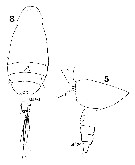 Issued from : W. Giesbrecht in Systematik und Faunistik der Pelagischen Copepoden des Golfes von Neapel und der angrenzenden Meeres-Abschnitte. Fauna Flora Golf. Neapel, 1892. Atlas von 54 Tafeln. [Taf. 37, Figs.5, 8]. As Scolecithrix vittata. Female: 5, last thoracic segment and abdomen (lateral, left side); 8, habitus (dorsal).
|
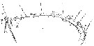
 Issued from : W. Giesbrecht in Systematik und Faunistik der Pelagischen Copepoden des Golfes von Neapel und der angrenzenden Meeres-Abschnitte. Fauna Flora Golf. Neapel, 1892. Atlas von 54 Tafeln. [Taf. 13, Figs. 23, 32]. As Scolecithrix vittata. Female: 23, exopod of P2 (part., posterior view); 32, P5.
|
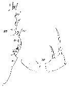
 Issued from : W. Giesbrecht in Systematik und Faunistik der Pelagischen Copepoden des Golfes von Neapel und der angrenzenden Meeres-Abschnitte. Fauna Flora Golf. Neapel, 1892. Atlas von 54 Tafeln. [Taf. 13, Fig.2]. As Scolecithrix vittata. Female: 2, A1 (dorsal view). Ae : aesthetasc; Spr = proximal spine; St = terminal spine;
|
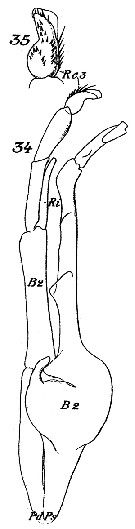 Issued from : W. Giesbrecht in Systematik und Faunistik der Pelagischen Copepoden des Golfes von Neapel und der angrenzenden Meeres-Abschnitte. - Fauna Flora Golf. Neapel, 1892. Atlas von 54 Tafeln. [Taf. 13, Figs. 34-35]. As Scolecithrix vittata. Male: 34, P5 ( anterior view); 35, Re3 of right P5. Pd = right leg; Ps = left leg; B = basipod; Ri = endopod; Re = exopod.
|
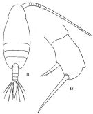
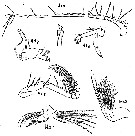
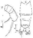
 issued from : G.D. Grice in Fish. Bull. Fish and Wildl. Ser., 1962, 61. [p.209, Pl.17, Figs.1-8]. Female (from 00°11'S, 119°58'W): 1-2, habitus (dorsal and lateral, respectively); 3, rostrum; 4, P5; 5, P2; 6, P3; 7, P4 (one endopod and one exopod missing).; 8, P5. Nota: Variation was noted in P5. The terminal seta in ione specimen was approximately twice the length of the proximal one, while in the other specimen it was somewhat shorter.
| | | | | Ref. compl.: | | | Sewell, 1948 (p.349, 502, 508, 546, 550, 551); Grice & Hart, 1962 (p.287, 293: Rem.); V.N. Greze, 1963 a (tabl.2); Unterüberbacher, 1964 (p.26); De Decker & Mombeck, 1964 (p.14); Grice & Hulsemann, 1965 (p.224); Mazza, 1966 (p.70); Pavlova, 1966 (p.43); Furuhashi, 1966 a (p.295, vertical distribution in Kuroshio region, Table 9); Fleminger, 1967 a (tabl.1); Morris, 1970 (p.2300); Park, 1970 (p.476); Deevey, 1971 (p.224); Roe, 1972 (p.277, tabl.1, tabl.2); 1972 b (p.539, Rem.); Apostolopoulou, 1972 (p.328, 352); Björnberg, 1973 (p.333, 389); Vives & al., 1975 (p.43, tab.II, III); Deevey & Brooks, 1977 (p.256, tab.2, Station "S"); Carter, 1977 (1978) (p.35); Vaissière & Séguin, 1980 (p.23, tab.1); Vives, 1982 (p.292); Kovalev & Shmeleva, 1982 (p.84); Brenning, 1984 (p.6, Rem.); Scotto di Carlo & al., 1984 (1043); Guangshan & Honglin, 1984 (p.118, tab.); Brenning, 1985 a (p.28, Table 2); Lozano Soldevilla & al., 1988 (p.59); Gopalakrishnan & Balachandran, 1992 (p.167, figs.1, 6, Table 1, 2); Shih & Young, 1995 (p.73); Errhif & al., 1997 (p.423); Hure & Krsinic, 1998 (p.49, 101); Suarez-Morales & Gasca, 1998 a (p.111); Siokou-Frangou, 1999 (p.476); Lapernat, 2000 (tabl.3, 4); Razouls & al., 2000 (p.343, Appendix); Seridji & Hafferssas, 2000 (tab.1); Lopez-Salgado & al., 2000 (tab.1); Vukanic, 2003 (139, tab.1); Hsiao & al., 2004 (p.326, tab.1); Lo & al., 2004 (p.89, tab.1); Isari & al., 2006 (p.241, tab.II: as vitata); Kuriyama & Nishida, 2006 (p.293, 300: Tab.II, p.309: Tab.III, fig.7,10, vertical distribution); Dur & al., 2007 (p.197, Table IV); Morales-Ramirez & Suarez-Morales, 2008 (p.522); Lan Y.-C. & al., 2008 (p.61, Table 1, % vs stations); Licandro & Icardi, 2009 (p.17, Table 4); Park & Ferrari, 2009 (p.143, Table 5, Appendix 1, biogeography); Hafferssas & Seridji, 2010 (p.353, Table 2, 3); Williamson & McGowan, 2010 (p.273, Table III, Pacific central gyres: N & S); Schnack-Schiel & al., 2010 (p.2064, Table 2: E Atlantic subtropical/tropical); Mazzocchi & Di Capua, 2010 (p.427); Medellin-Mora & Navas S., 2010 (p.265, Tab. 2); Hafferssas & al., 2010 (p.1281, Table III, abundance vs spatial distribution); Hsiao S.H. & al., 2011 (p.475, Appendix I); in CalCOFI regional list (MDO, Nov. 2013; M. Ohman, pers. comm.); Lidvanov & al., 2013 (p.290, Table 2, % composition); Mazzocchi & al., 2014 (p.64, Table 5, abundance); Benedetti & al., 2016 (p.159, Table I, fig.1, functional characters); Benedetti & al., 2018 (p.1, Fig.2: ecological functional group); Belmonte, 2018 (p.273, Table I: Italian zones) | | | | NZ: | 16 | | |
|
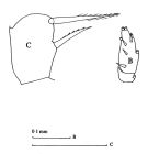
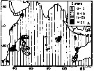
Carte de distribution de Scolecithricella vittata par zones géographiques
|
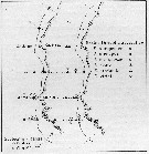
| | | | | | | | | | | | | | |  issued from : T.C. Gopalakrishnan & T. Balachandran in Oceanogr. Indian Ocean, 1992. [p.171, fig.6, A]. issued from : T.C. Gopalakrishnan & T. Balachandran in Oceanogr. Indian Ocean, 1992. [p.171, fig.6, A].
Distribution of Scolecithricella vittata in the Indian Ocean.
Nota: Main occurnces between 10° N - 30° S. |
 issued from : U. Brenning in Wiss. Z. Wilhelm-Pieck-Univ. Rostock - 33. Jahrgang 1984. Mat.-nat. wiss. Reihe, 6. [p.6, Fig.2]. issued from : U. Brenning in Wiss. Z. Wilhelm-Pieck-Univ. Rostock - 33. Jahrgang 1984. Mat.-nat. wiss. Reihe, 6. [p.6, Fig.2].
Spatial distribution for Scolecithricella vittata and other scolecithrids from 8° S - 26° N; 16°- 20° W, for different expeditions (V1: Dec. 1972- Jan. 1973; V2: Feb/Mar. 1973; VI: May 1974; IV: Jun./Jul. 1972). |
| | | | Loc: | | | sub-Antarct. (Indian, SW Pacif.), South Africa (E), Namibia, off Congo, off Morocco-Mauritania, Canary Is., off Madeira, S Brazil, off Amazon, Barbados Is., Caribbean Sea, Caribbean Colombia, G. of Mexico, Florida, Sargasso Sea, off Bermuda: Station S (32°10N, 64°30W), off Portugal, Bay of Biscay, Ibero-moroccan Bay, Medit. (Alboran Sea, Banyuls, Marseille, Ligurian Sea, Tyrrhenian Sea, Strait of Messina, Adriatic Sea, Ionian Sea, Aegean Sea, Lebanon Basin), Natal, Indian (SW & equatorial), Bay of Bengal, S Indian (subtropical convergence), China Seas (East China Sea, South China Sea), 25°30'N-122°30'E, Taiwan (SW, E, N: Mienhua Canyon), Japan, Pacif. (W equatorial), Pacific (central gyres: N & S), Australia (Great Barrier), New Zealand, California, W Costa Rica, Hawaii, Pacif. (central subtropical N), Fidji Is., Galapagos, N Chile | | | | N: | 90 | | | | Lg.: | | | (1) F: 1,65; (9) F: 1,75; (34) F: 1,74-1,66; (35) F: 1,64; (38) F: 1,7-1,5; (47) F: 1,65; M: 1,6; (72) F: 1,8-1,74; (101) F: 1,66; (117) F: 1,75; M: 1,62; (147) F: 2; (199) F: 1,75-1,52; M: 1,6; (207) F: 2; (290) F: 1,55-1,6; (313) F: 1,7; (523) F: 1,82-1,6; M: 1,72-1,62; (1000) F: 1,7 ± 0,1; M: 1,7 ± 0,3; (1111) F: 1,63; 1,82; {F: 1,50-2,00; M: 1,60-2,0} | | | | Rem.: | épi-mésopélagique.
Sampling depth (sub-Antarct.) : 0-1200 m.
Voir aussi les remarques en anglais | | | Dernière mise à jour : 19/01/2021 | |
|
|
 Toute utilisation de ce site pour une publication sera mentionnée avec la référence suivante : Toute utilisation de ce site pour une publication sera mentionnée avec la référence suivante :
Razouls C., Desreumaux N., Kouwenberg J. et de Bovée F., 2005-2026. - Biodiversité des Copépodes planctoniques marins (morphologie, répartition géographique et données biologiques). Sorbonne Université, CNRS. Disponible sur http://copepodes.obs-banyuls.fr [Accédé le 09 mai 2026] © copyright 2005-2026 Sorbonne Université, CNRS
|
|
|
|


;)
;)
;)
;)
;)
;)
;)
;)
;)
;)
;)
;)
;)
;)
;)
;)
;)
;)
;)
;)

;)
;)
