|
|
 |
Fiche d'espèce de Copépode |
|
|
Calanoida ( Ordre ) |
|
|
|
Spinocalanoidea ( Superfamille ) |
|
|
|
Spinocalanidae ( Famille ) |
|
|
|
Mimocalanus ( Genre ) |
|
|
| |
Mimocalanus cultrifer Farran, 1908 (F,M) | |
| | | | | | | Syn.: | no Mimocalanus cultrifer : Tanaka, 1956 c (p.387);
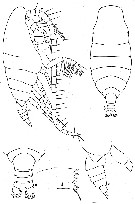
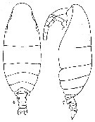
Microcalanus cultrifer : Björnberg & al., 1981 (p.628, [lapsus calami]) | | | | Ref.: | | | Farran, 1908 b (p.23, Descr.F, figs.F); 1926 (p.244); Rose, 1933 a (p.86, figs.F); Vervoort, 1946 (p.156, Rem.); Farran & Vervoort, 1951 h (n°40, p.3, figs.F); Vervoort, 1957 (p.42, figs.F, Rem.); Minoda, 1971 (p.22); Grice, 1971 (p.279, ? figs.F); Damkaer, 1975 (p.68, figs.F); Brodsky & al., 1983 (p.305, Rem.F,M, figs.F); Roe, 1984 (p.356); Bradford-Grieve, 1994 (p.99, figs.F, Rem.); Bradford-Grieve & al., 1999 (p.878, 913, figs.F); Bode & al., 2017 (p.600, Table I, III, fig. 2, 3, 4, morphology vs genetic). | 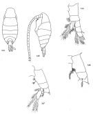 issued from : D.M. Damkaer in NOAA Technical Report NMFS CIRC-391, Seattle, 1975. [p.68, Fig.164-168]. Female: 164, habitus (dorsal); 165, idem (left lateral side); 166, P1; 167, P2; 168, P4. P2-4: incomplete legs.
|
 issued from : Brodsky K.A., Vyshkvartseva N.V., Kos M.S. & Markhaseva E.L. in Opred. Faune SSSR, 1983, 135. [p.306, Fig.148]. Female. P. md = mandibular palp.
|
 issued from : G.D. Grice in Cah. Biol. Mar., 1971, XII. [p.278, Fig.3 C-D]. With doubt. Female (from Medit.): C, habitus (lateral left side); D, Mxp (coxa and basis). Nota: Rostrum absent. 2nd endopod segment of Mxp less than twice the length of 1st segment. Posterior surfaces of P2-P4 without spines.
|
 issued from : W. Vervoort in B.A.N.Z. Antarct. Res. Exped., Rep.-Ser. B, III, 1957. [Fig.12]. Female (from 61°44'S, 77°59'E): a-b, habitus (lateral and dorsal, respectively); c-e, posterior part cephalothorax and urosome, respectively); d, right Md (cutting edge). Nota: Proportional lengths of prosome and urosome as 9:2. Cephalon and 1st thoracic somite almost completely fused (line of separation visible on the dorsal surface only), 4th and 5th completely separated. Frontal organ scarcely distinguishable in lateral aspect but can be recognized by a pair of fine hairs. Postero-lateral thoracic corners covering about 3/4 of the genital somite. Urosome 4-segmented; proportional lengths of urosomal segments and caudal rami 30:20:16:13:21 = 100. Anal somite with distinct, rounded anal flap. A1 24-segmented (8th-9th are partly and 24th-25th segments completely fused) reaching beyond the caudal rami by the last 4 segments.
|
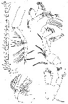 issued from : W. Vervoort in B.A.N.Z. Antarct. Res. Exped., Rep.-Ser. B, III, 1957. [Fig.13]. Female: a, right A1; b, right Mxp; c, left Mx1; d, left A2; e, right Mx2; f, right Md (mandibular palp). Nota: 1st endopodal segment 4 times as long as wide; terminal segment of exopod with 3 apical setae (1 seta at the middle of the segment has not been observed, such a seta has been figured by Farran (1908, Pl.I, fig.2).
|
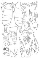
 issued from : W. Vervoort in B.A.N.Z. Antarct. Res. Exped., Rep.-Ser. B, III, 1957. [Fig.14]. Female: a, posterior part cephalothorax and urosome (ventral view); b, right P1 (anterior aspect).
|
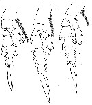 issued from : W. Vervoort in B.A.N.Z. Antarct. Res. Exped., Rep.-Ser. B, III, 1957. [Fig.15]. Female: a-c, P2 to P4 (right legs).
|
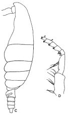
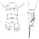
 issued from : G.P. Farran in Fish. Ire. Sci. Invest., 1906, II [1908]. [Pl. I, Figs.6-7]. Female: 6-7, habitus (dorsal and lateral, respectively).
|
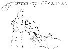 issued from : G.P. Farran in Fish. Ire. Sci. Invest., 1906, II [1908]. [Pl. I, Figs.5, 8, 9]. Female: 5, A1; 8, P1; 9, Mx2.
|
 Issued from : C. Razouls in Ann. Inst. océanogr., Paris, 1994, 70 (1). [p.60]. Caractéristiques morphologiques de Mimocalanus cultrifer femelles adultes. Terminologie et abbréviations: voir à Calanus propinquus.
|
 Issued from : M. Bode, S. Laakmann, P. Kaiser, W. Hagen, H. Auel & A. Cornils in J. Plankton Res., 2017, 39 (4). [p.603, Table I]. Species identified via morphological and molecular analyses, including the number of specimens used for each identification method (MI: morphological identification, COI: cytochrome c oxydase subunit I gene fragment, MS: MALDI-TOF analysis, 18 S: ribosomal 18 S gene fragment). Cryptic lineages were revealed only after molecular analysis. Sampling depth, latitude, total length (TL) and Prosome/Urosome ratio (Pr:Ur) of taxon were recorded. morphological characters are only listed here, if the identification was ambiguous due to missing body parts or if cryptic or pseudocryptic lineages were revealed during molecular analyses. For diagnostic character of species refer to the references: Park, 1970; Grice, 1971; Damkaer, 1975; Schulz, 1989, 1996. C = coxa; End = endopodite; P = swimming legs; End = endopodite; Exp = exopodite.
| | | | | Ref. compl.: | | | Sewell, 1948 (p.483, 499, 545, 550); Grice, 1963 a (p.495); Furuhashi, 1966 a (p.295, vertical distribution in Oyashio/Kuroshio transitional area, Table 7); Grice & Hulsemann, 1965 (p.223, 227); 1967 (p.14); Fleminger, 1967 a (tabl.1); Park, 1970 (p.475); Deevey, 1971 (p.224); Roe, 1972 (p.277, tabl.1, tabl.2); Björnberg, 1973 (p.322, 388); Kovalev & Schmeleva, 1982 (p.83); Vives, 1982 (p.290); Guangshan & Honglin, 1984 (p.118, tab., Rem.: p.165); Greze & al., 1985 (p.7, as cultifer); Hopkins, 1985 (p.197, Table 1, gut contents); Rudyakov, 1986 (tab.1); Lozano Soldevilla & al., 1988 (p.58); Shih & Young, 1995 (p.74); Razouls & al., 2000 (p.343, tab. 5, Appendix); Holmes, 2001 (p.41); Schnack-Schiel & al., 2008 (p.1046: Tab.2); Raybaud & al., 2008 (p.1765, Table A1); Park & Ferrari, 2009 (p.143, Table 4, Appendix 1); Schnack-Schiel & al., 2010 (p.2064, Table 2: E Atlantic subtropical/tropical); Mazzocchi & Di Capua, 2010 (p.427); Medellin-Mora & Navas S., 2010 (p.265, Tab. 2); Michels & al., 2012 (p.369, Table 1, occurrence frequency); in CalCOFI regional list (MDO, Nov. 2013; M. Ohman, comm. pers.); Belmonte, 2018 (p.273, Table I: Italian zones) | | | | NZ: | 17 | | |
|
Carte de distribution de Mimocalanus cultrifer par zones géographiques
|
| | | | | | | | | | | | | | | 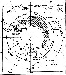 issued from : W. Vervoort in B.A.N.Z. Antarctic Reseach Expedition, Reports - Ser. B, Vol. III, 1957 [Fig.11] issued from : W. Vervoort in B.A.N.Z. Antarctic Reseach Expedition, Reports - Ser. B, Vol. III, 1957 [Fig.11]
Chart showing the geographical distribution (white triangle) in the seas surrounding the Antarctic continent.
Nota: In this chart the area frequented by whaling vessels has been hatched. The Antarctic circle (66°.5 S) has been drawn as a broken line. The numbers I to VI refer to the sectors into which the Antarctic seas are divided according to Mackintosh (1942) (after Vervoort, 1951). |
| | | | Loc: | | | Antarct. (Croker Passage, Peninsula, W Weddell Sea, Indian), sub-Antarct. (Indian), Atlant. (SE, NE, NW), Brazil, off Amazon, Canary Is., off W Cabo Finisterre, Bay of Biscay, Caribbean Sea, Caribbean Colombia, G. of Mexico, off Bermuda, off S Cape Cod, off W Ireland, Medit. (Alboran Sea, Thyrrenian Sea, Ligurian Sea, Ionian Sea, S Adriatic), Indian, Indonesia-Malaysia, China Seas (East China Sea, South China Sea), Japan, Pacif. (N & SW), Bering Sea, off S Aleutian Is., off San Francisco, California, Pacif. (W equatorial), S Tasmania, Chile | | | | N: | 36 | | | | Lg.: | | | (13) F: 1,7-1; (24) F: 1,44; (25) F: 1,7-1,6; (28) F: 1,91-1,17; (38) F: 1,6-1,3; (199) F: 1,6-1,22; M: 1,14; (202) F: 1,6-1,7; (208) F: 1,95-1,55; (1252) F: 1,36-1,62; 1,05-1,40; {F: 1,00-1,95; M: 1,14}
(1252) F: Pr/Ur = 3,3-4,7; 2,8-4,4. | | | | Rem.: | méso à abyssopélagique.
Sampling depth (Antarct., sub-Antarct.) : 0-1000 m.
Le mâle a été signalé mais non encore décrit.
Voir aussi les remarques en anglais | | | Dernière mise à jour : 25/03/2020 | |
|
|
 Toute utilisation de ce site pour une publication sera mentionnée avec la référence suivante : Toute utilisation de ce site pour une publication sera mentionnée avec la référence suivante :
Razouls C., Desreumaux N., Kouwenberg J. et de Bovée F., 2005-2026. - Biodiversité des Copépodes planctoniques marins (morphologie, répartition géographique et données biologiques). Sorbonne Université, CNRS. Disponible sur http://copepodes.obs-banyuls.fr [Accédé le 08 janvier 2026] © copyright 2005-2026 Sorbonne Université, CNRS
|
|
|
|


;)
;)
;)
;)
;)
;)
;)
;)
;)
;)

;)
