|
|
 |
Fiche d'espèce de Copépode |
|
|
Calanoida ( Ordre ) |
|
|
|
Clausocalanoidea ( Superfamille ) |
|
|
|
Stephidae ( Famille ) |
|
|
|
Stephos ( Genre ) |
|
|
| |
Stephos canariensis Boxshall, Stock, Sanchez, 1990 (F,M) | |
| | | | | | | Ref.: | | | Boxshall & al., 1990 (p.33, figs.F,M); Huys & Boxshall, 1991 (p.49, 86, 359, figs.F); Bradford-Grieve, 1999 a (p.25, Table1: Rem.); Vives & Shmeleva, 2007 (p.857, figs.F,M, Rem.) | 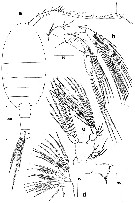 issued from : G.A. Boxshall, J.H. Stock & E. Sanchez in Stygologia, 1990, 5 (1). [p.34, Fig.1]. Female (from lanzarote: Canary Islands): a, habitus (dorsal); b, A2; c, Md; d, Mx2. Nota: Head and 1st pedigerous somite separate, 4th and 5th fused. Rostrum absent. Urosome 4-segmented. Genital double somite comprising completely fused genital and 1st abdominal somites, slightly asymmetrical, expanded anteriorly on right side; single genital opening located ventrally on right side; genital aperture oriented obliquely, closed off by a single unarmed operculum derived from the fused P6.; remains of 2 discharged spermatophores emerging from beneath operculum (fig.5b). A2 with coxa and basis incompletely fused; endopod 2-segmented; exopod 6-segmented. Mx2 small, comprising praecoxa, coxa, and basis each armed with 2 endites, and a 3-segmented endopod
|
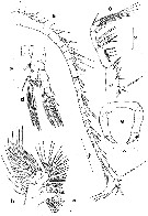 issued from : G.A. Boxshall, J.H. Stock & E. Sanchez in Stygologia, 1990, 5 (1). [p.35, Fig.2]. Female: a, A1; b, Mx1; c, Mxp; d, P1; e, P5. Nota: A1 24-segmented; segments I and II incompletely fused, segment II representing a triple segment; segment VIII representing a triple segment. Mx1 with well defined praecoxa produced into powerful endite bearing 8 stout spines and 4 slendersetae; Coxa with 3 setae on endite; outer lobe (epipodite) represented by 9 setae on surface segment; basis fused to endopod, lacking outer seta; proximal endite discrete, bearing 4 setae;distal endite incorporated into segment , represented by 5 setae; fused endopod armed with proximal and distal groups of 4 setae on inner margin; free apical segment with 7 setae; exopod 1-segmented, with 11 setae along distal and lateral margins. mxp 7-segmented; 1st segment (syncoxa) comprising fused praecoxa and coxa, armed with a hirsute lobe and 3 groups of setae along medial margin; basis ornamented with a row of stout spinules and some fine denticles; free endopod 5-segmented. P5 reduced, uniramous; basal part representing fused coxae and intercoxal sclerite, ornamented with minute denticles
|
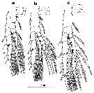 issued from : G.A. Boxshall, J.H. Stock & E. Sanchez in Stygologia, 1990, 5 (1). [p.36, Fig.3]. Female: a-c, P2 to P4.
|
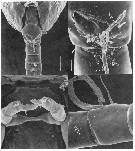 issued from : G.A. Boxshall, J.H. Stock & E. Sanchez in Stygologia, 1990, 5 (1). [p.38, Fig.5]. Female (Scanning Electron Micrographs): a, urosome (ventral); b, genital aperture, showing spent spermatophores (arrowed); c, P5; d, A1 segments XI and XII, showing comb of spinules (arrowed) on ventral surface of segment XII. Scale bars: a = 0.050 mm, b-d = 0.010 mm.
|
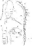 issued from : G.A. Boxshall, J.H. Stock & E. Sanchez in Stygologia, 1990, 5 (1). [p.37, Fig.4]. Male: a, habitus (lateral); b, urosome (ventral); c, A1; d, P5 (anterior view). head and 1st pedigerous somite marked by complete suture, 4th and 5th fused. Urosome 5-segmented. Genital segment slightly asymmetrical, single functional genital opening on left side; 4th and urosome somite ornamented with row of stout spinules along posterior border. A1 24-segmented; Other mouthparts and swimming legs 1 too 4 as in female in segmentation and setation. P5 as grasping organ, uniramous, markedly asymmetrical; right leg slender 4-segmented; 1st and 2nd segments short; 3rd segment very long; 4th segment comprising 2 processes oriented at right angles, both uramed; left leg 5-segmented, 1st to 3rd segments short, unarmed; 4th segment robust ornamented with row of short spinules distally along inner margin; 5th segment complex, trifid, innermost branch tapering and unarmed; middle and outer branches separate distally, outer branch spoon-like and bearing small denticles around rim of concave tip.
|
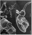 issued from : G.A. Boxshall, J.H. Stock & E. Sanchez in Stygologia, 1990, 5 (1). [p.40, Fig.6]. Male (Scanning Electron Micrographs): a, P5 (anterior view); b, tip of right leg; c, distal segments of left leg; d, apex of outer process of left leg. Scale bars: a = 0.040 mm, b = 0.010 mm, c = 0.020 mm, d = 0.004 mm.
| | | | | NZ: | 1 | | |
|
Carte de distribution de Stephos canariensis par zones géographiques
|
| | | | Loc: | | | Canary Islands (Lanzarote Is.: anchialine lava pool) | | | | N: | 1 | | | | Lg.: | | | (552) F: 0,695-0,641; M: 0,641-0,595; {F: 0,64-0,70; M: 0,60-0,64} | | | | Rem.: | salinité de 27 p.1000.
Voir aussi les remarques en anglais | | | Dernière mise à jour : 14/08/2015 | |
|
|
 Toute utilisation de ce site pour une publication sera mentionnée avec la référence suivante : Toute utilisation de ce site pour une publication sera mentionnée avec la référence suivante :
Razouls C., Desreumaux N., Kouwenberg J. et de Bovée F., 2005-2025. - Biodiversité des Copépodes planctoniques marins (morphologie, répartition géographique et données biologiques). Sorbonne Université, CNRS. Disponible sur http://copepodes.obs-banyuls.fr [Accédé le 11 juillet 2025] © copyright 2005-2025 Sorbonne Université, CNRS
|
|
 |
 |


