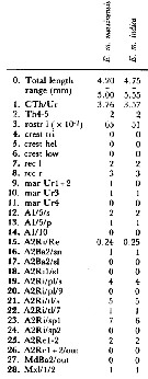
|
|
 |
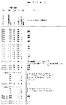
Fiche d'espèce de Copépode |
|
|
Calanoida ( Ordre ) |
|
|
|
Clausocalanoidea ( Superfamille ) |
|
|
|
Aetideidae ( Famille ) |
|
|
|
Euchirella ( Genre ) |
|
|
| |
Euchirella messinensis (Claus, 1863) (F,M) | |
| | | | | | | Syn.: | Undina messinensis Claus, 1863 (p.187, figs.F,M);
? E. messinensis : T. Scott, 1894 b (p.45);
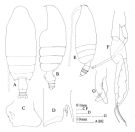
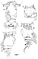
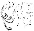
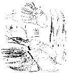
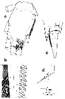
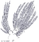
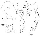
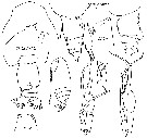
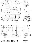
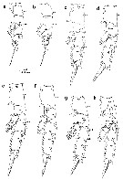
Euchirella indica : Vervoort, 1949 (p.23, figsF, Rem.); Bradford & Jillett, 1980, figs.F,M, Rem.); Dur & al., 2007 (p.197, Table IV); Hsiao S.H. & al., 2011 (p.475, Appendix I) | | | | Ref.: | | | Giesbrecht, 1892 (p.232, 244, 772, figs.F,M); Giesbrecht & Schmeil, 1898 (p.35, Rem. F,M); Thompson & Scott, 1903 (p.233, 244); Esterly, 1905 (p.151, figs.F,M); Farran, 1908 b (p.37); A. Scott, 1909 (p.56, Rem.); Wolfenden, 1911 (p.237); With, 1915 (p.122, figs.F,M); Pesta, 1920 (p.509); Lysholm & Nordgaard, 1921 (p.16); Sars, 1925 (p.65, figs.F,M); Farran, 1926 (p.252, Rem.); Rose, 1929 (p.20); Sewell, 1929 (p.107, 115, fig.M); Wilson, 1932 a (p.56, figs.F,M); Rose, 1933 a (p.103, figs.F,M); Jespersen, 1940 (p.22); Lysholm & al., 1945 (p.17); Sewell, 1947 (p.69, 70, 77, fig.M); Vervoort, 1949 (p.28, figs.M, Rem: p.17); Brodsky, 1950 (1967) (p.173, figs.F,M); Vervoort, 1952 f (n°47, p.3, figs.F); Tanaka, 1957 b (p.180, figs.F,M); Vervoort, 1963 b (p.136, Rem.); Mazza, 1965 a (p.296, figs.juv., F,M); 1967 (p.132, 139, figs. juv., F,M); Owre & Foyo, 1967 (p.47, figs.F,M); Mazza, 1968 (p.533, figs.); Tanaka & Omori, 1969 (p.51, figs.F,M, Rem.); Vaupel Klein, 1972 (p.501, 504, fig.M); Razouls, 1972 (p.94, Annexe: p.44, figs.F); Park, 1976 a (p.113, figs.F,M); Bradford & Jillett, 1980 (p.41, figs.F,M, Rem.: 2 species); Björnberg & al., 1981 (p.605, 632, fig.F,M); Chahsavar-Archad & Razouls, 1982 (p.28, fig.M); Vaupel Klein, 1982 b (p.4, 5, figs.F,M, Rem.); 1984 (p.110, fig.F, Rem.: anomalie); Vaupel Klein, 1984 a (p.34, fig.F, Rem., Table II: characters); Roe, 1984 (p.357); Koomen, 1991 (p.437: organes tégumentaires buccaux); 1992 (p.113, figs.F, Rem.); Markhaseva, 1996 (p.156: E. messinensis indica, figs.F,M, p.160: E. messinensis messinensis, figs.F,M, Rem.); Bradford-Grieve & al., 1999 (p.879, cf. E. messinensis messinensis); Boxshall & Halsey, 2004 (p.55: figs.F); Mulyadi, 2004 (p.48, figs.F, Rem.: E. messinensis indica); Yamaguchi & al., 2005 (p.835, Tabl. VIII); Ferrari & Dahms, 2007 (p.95, 96, Rem., fig.32: P1, Table XXIV, XXVII); Vives & Shmeleva, 2007 (p.567, figs.F,M, Rem.) | 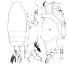 issued from : Mulyadi in Crustaceana, 2001, 74 (9). [Fig.5, p.819]. As Euchirella messinensis indica. Female: a, habitus (dorsal view); b, Cephalon (lateral view); c, last thoracic segment and Urosome (lateral left side); d, A2; e, right P4 (incomplete); f, left P4 (incomplete).
|
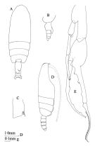
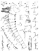
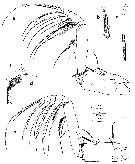
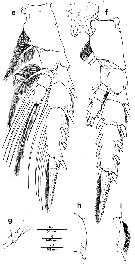
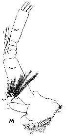
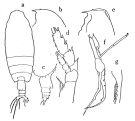 issued from : Tanaka O. in Publ. Seto Mar. Biol. Lab., 1957, 6 (2). [Fig.46, p.181]. As E. messinensis, but probably as E. messinensis indica. Female: habitus (dorsal aspect); b, head (lateral aspect); c, last thoracic segment and urosome (lateral aspect); d, P4. Male: e, head (lateral aspect); f, P5; g, a part of distal joint of exopodite of right P5. Nota Female: - Cephalothorax about 3.42 times the abdomen length (4.07 : 1.19). - Rostrum rather slender and acute, directs downwards. - Abdomen 4-segmented; segments and caudal rami in proportional lengths 49 : 18 : 12: 6 : 15 = 100. - Dorsal protuberance of genital segment differs from the figure given by Giesbrecht or Sars: it is much smaller and exceeds slightly the distal margin of genital segment, and situated rather on the middle of the dorsal surface. - A1 extends to the middle of 2nd abdominal segment. - Mx1 endopod with 4 setae. - P4 with 2 spines on the inner margin of coxa; terminal spine of the exopod with 20 teeth. Nota Male: - Cephalothorax about 2.7 times the abdomen length. - Forehead with low crest. - Rostrum moderately long. - Lateral corners of last thoracic segment rounded. - 2nd and 3rd abdominal segments furnished with coarse teeth on the distal margin. - A1 22-segmented, extends to the middle of 2nd abdominal segment. - Mouthparts and P1 to P3 as those of E. pulchra.P4 with basis with 2 minute spines near the inner distal margin. - P5 resemble those of E. pulchra, but endopod of right leg longer than the combined lengths of two basal segments.
.
|
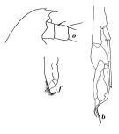
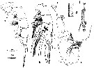
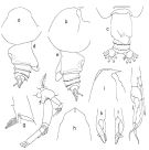 issued from : T. Park in Contr. Mar. Sci., 1976, 20. [p.114, Fig.5] Female: a, b, forehead (dorsal, lateral, respectively); c, d, e, posterior part of metasome and urosome (dorsal, left side, right side, respectively); f, A2; g, coxa of fourth leg (posterior). Male: h, i, forehead (dorsal, lateral); j, fifth pair of legs (anterior); k, l, distal part of left fifth leg (anterior, posterior, respectively).
|
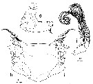
 issued from : J.C. von Vaupel Klein in Zool. Verh. Leiden, 1982, 198. [p.7, Fig.1]. As Euchirella messinensis f. typica. Female (from Tyrrhenian Sea): habitus (dorsal view); b, idem (left side); c, frontal region of head with rostrum (left lateral view); d, rostrum (anterior view). Nota : Proportional lengths cephalothorax urosome as 79 : 21 = 100 ; length of urosome contained 3.76 times in that of cephalothorax. Cephalon and 1st pedigerous somite partially fused (the line of fusion being faintly visible in dorsal and pratically invisible to obsolescent in lateral view), 4th pedigerous somite and the strongly reduced of the 5th pedigerous somite entirely coalescent (a faint line of fusion being discernable just anteriad to the attachment of the urosome).
|
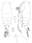
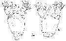
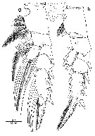
 issued from : J.M. Bradford & J.B. Jillett in Mem. N.Z. Oceanogr. Inst., 86, 1980. [p.38, Fig.23]. As Euchirella indica. Female: A, habitus (dorsal); B, idem (lateral left side); C, genital segment (lateral left side); D, basipod 1 of P4. Male: E, habitus (lateral left side); F, P5; G, terminal part of left exopod of P5.
|
 issued from : J.M. Bradford & J.B. Jillett in Mem. N.Z. Oceanogr. Inst., 86, 1980. [p.41, Fig.25]. Female: A, habitus (dorsal); B, last thoracic segment and urosome (lateral left side); C, basipod 1 of P4. Male: D, habitus (lateral right side); E, P5.
|
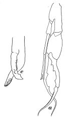
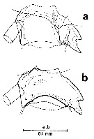
 Issued from : W. Vervoort in Zool. Verh., Leiden, 1949, 5. [p.24, Fig.11]. As Euchirella indica. Female (from Celebes Sea): a, habitus (lateral right side); b, distal part of thorax and urosome (dorsal). Nota: A1 (24 -free segments) reaches the distal margin of the 2nd urosomal segment. The abdominal segments and furca in the proportional lengths 55:14:11:18:12 = 100. This species approaches E. msessinensis, but differs by the structure of the urosome in the female and the shape of P5 in the male.
|
 Issued from : W. Vervoort in Zool. Verh., Leiden, 1949, 5. [p.25, Fig.112]. As Euchirella indica. Female (from Celebes Sea): a, urosome (lateral right side; b, idem (lateral left side); c, forehead (lateral).
|
 Issued from : W. Vervoort in Zool. Verh., Leiden, 1949, 5. [p.18, Fig.8, b; 19, Fig.9b,f]. As Euchirella indica. Male (from Sawoe Sea): o, forehead (lateral right side); b, P5; f, terminal part of the left P5. Nota: P5 resembles that of E. messinensis closely, but there are small differences in the structure of the right exopod and endopod segments, as well as in the shape of the terminal portion of the left P5.
|
 Issued from : W. Vervoort in Zool. Verh., Leiden, 1949, 5. [p.19, Fig.9, a,e]. As Euchirella messinensis. Male (from Indonesian seas): a, P5; e, terminal part of of the left P5.
|
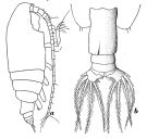
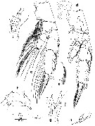
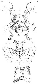
 Issued from : G.O. Sars in Résult. Camp. Scient. Prince Albert I, 69, pls.1-127 (1924). [Pl.XIX, figs.6-13]. female : 6, habitus with two ovisacs (dorsal); 7, idem witout ovisacs (lateral left side); 8, A2; 9, P1; 10, basal part of P4. Male: 11, habitus (dorsal); 12, forehead (lateral); 13, P5.
|
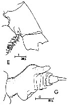
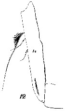
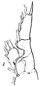
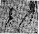
 issued from : R.B.S. Sewell in The John Murray Expedition, 1933-34, Scientific Reports, VIII (1), 1947. [p.78, Fig.15, A]. Male: A, forehead (lateral).
|
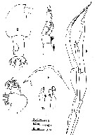
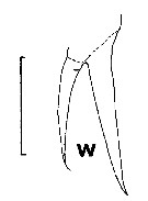
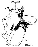
 issued from : F. Giron-Reguer in Thèse Fac. Sc. Paris, 1963. [Fig.9]. Female (from Alboran Sea): urosome (detail of genital segment and fixation of spermatophore; lateral left side).
|
 issued from : J.C. von Vaupel Klein in Crustaceana, Supplt 9, Studies on Copepoda, III, 1984. [p.76, Fig.14, c, l]. As Euchirella messinensis indica. Female: c, accessory seta of endopodite 4th segment of Mxp (left appendage from lateral) (Note the very feebly developed seta); l, larrangement of spinules on the lateral tubercle of endopod of left P1 (organ of Vaupel Klein) (anterior face). Nota: Note measurements taken to compute the ratio: length of central hairs/length of segment, shown on a edopod of P1 (left, anterior). Scale bar 0.1 mm for c and l.
|
 issued from : J.C. von Vaupel Klein in Crustaceana, Supplt 9, Studies on Copepoda, III, 1984. [p.63, Fig.5, e]. Female: e, setal armature of A1 segment 10 (note: proximal small seta totally absent, see normal type I small seta in Euchirella rostrata p.63, fig.5 f). Scale bar 0.1 mm.
|
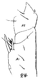
 issued from : C.O. Esterly in Univ. Calif. Publs Zool., 1905, 2 (4). [p.152, Fig.18]. Female (from San Diego Region): a, forehead (lateral); b, last thoracic segment and urosome (lateral). Nota: Head without crest. Genital segment asymmetrical, with sac-like appendage on left side of dorsal surface Male: c, forehead (lateral); d, P5. Nota: Head with a low and rather long crest. Right P5 over 7-times as long as the 2nd basal is broad.
|
 issued from : C. Razouls in Th. Doc. Etat Fac. Sc. Paris VI, 1972, Annexe. [Fig.44, E, G]. Female (from Banyuls, G. of Lion): E, basipodal segment 1 of P4; G, urosome (lateral).
|
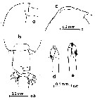 issued from : O. Tanaka & M. Omori in Publ. Seto mar. Biol. Lab., 1969, XVII, 1. [p.49, Fig.6, a-e]. Female (from Atlantic): a, forehead (lateral); b, last thoracic segment and urosome (dorsal). Male: c, forehead (lateral); d, left P5 (distal segment of exopod); e, idem (another view).
|
 issued from : O. Tanaka & M. Omori in Publ. Seto mar. Biol. Lab., 1969, XVII, 1. [p.49, Fig.6, f-m]. As Euchirella messinensis indica. Female (from Pacific): f, forehead (lateral); g, last thoracic segment and urosome (dorsal). Male: h, forehead (lateral); ; i, Mx1; j, P1; k, P5; l, left P5 (distal segment of exopod); m, idem (from another specimen).
|
 issued from : J.C. von Vaupel Klein in Crustaceana, Supplt 9, Studies on Copepoda, III, 1984. [p.82, Fig.17, a]. As Euchirella messinensis messinensis. Female: a, arrangement of hair-sensilla on the posterior face of the proximal segments of right P4. Scale bar 0.2 mm. Nota: 10 long sensilla, viz., 5 on basipodal segment 1, 3 on ba2, 1 on endopodal segment 1 and 1 on exopodal segment 1.
|
 issued from : J.C. von Vaupel Klein in Crustaceana, Supplt 9, Studies on Copepoda, III, 1984. [p.80, Fig.16, w]. As Euchirella messinensis indica. Female: w, arrangement of medial spines on the posterior face of basal segment 1 of right P4 . Scale bar 0.1 mm. Nota: 2 unequal stout spines on a common base.
|
 issued from : J.C. von Vaupel Klein in Zool. Verh., 1982, 198. [p.74, Fig.16, a-f]. As Euchirella messinensis f. typica. Female: a-c, semi-schematic drawings of genital somite , showing internal lumina of seminal receptacles in, respectively, left lateral, dorsal, and right lateral view; d-f, genital somite of aberrant female from ''Bache'' sta.10176, note structures on the left being normal, including the recurved seminal duct, while the right receptacle has apparently got stuck, resulting in a separate swelling for its accomodation. R = right seminal duct plus receptacle; L = same (left); open circle represents sperm-plug.
|
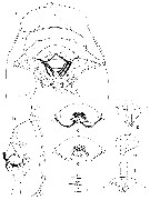 issued from : J.C. von Vaupel Klein in Zool. Verh., 1982, 198. [p.11, Fig.3]. As Euchirella messinensis f. typica. Female: a, ventral aspect of the oral field, with outline of cephalic region indicated; b, left lateral aspect of the oral field; c, central part of the upper lip in ventral view, showing integumental structures of the ventral wall of the oral cavity proper,as visible through the transparent cuticle; c', map of same presenting chaetotaxie code, positions of papillae (p) and rows of urn-shaped pores (1-3) d-f, urn-shaped pores (1, 2m, 3, respectively), as observed under the same conditions as in c.. medially on the upper lip. Nota : Oral field delimited by a large, semi-dome-shaped upper lip (Labrum) and a small, bifid lower lip (labium). The labrum arises smoothly from the sternal iontegument ; its bulbously rounded antero-medial portion attenuates postero-laterally to form a strong, transverse ridge which extends over the entire width of the body. The postero-central part is distinctly set-ott from the remainder of the lip and roughly of a semi-circular shape. The free caudal edge of this part is weakly bilobed and fringed with various rows of hairs. Close to the midline, the labrum bears a pair of short, rounded papillae (the exact nature of which could not be established as yet). The lower lip, closed-in between the wide proximal part of the Md gnathobases, consists of a largely flattened, semi-circular part of the sternites which bears a pair of blunt, turret-shaped protrusions. The labium and the oral atrium are profusely beset with rows and patches of hairs of different types. The inner face of labrum is equipped with 3 rows of urn-shaped, apparently glandular , pores (fig3 c-f). Between upper and lower lip there is room for the slender distal parts of the Md gnathobases to reach each other in the mid-sagittal plane where the masticatory edges perform their grinding function : the largest monocuspidate teeth ventralmost, the setiform teeth entering into the oral atrium. To accomodate the Md, the adjoinning faces of upper and lower lips are strongly arched in lateral aspect. The posterior limitation of the oral field is marked by the smoothy rounded sternal surface which runs from between the base of Mx1 to caudad. These sternites show various scattered patches of integumental hairs.
|
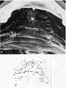 issued from : J.C. von Vaupel Klein in Zool. Verh., 1982, 198. [p.127, Pl.6]. As Euchirella messinensis f. typica. Female: a, overall view of the ventral wall of the oral cavity (the inner face of the upper lip, showing chaetotaxy and urn-shaped pores; a', map of same indicating code-nos. Scale bar 0.020 mm.
|
 issued from : J.C. von Vaupel Klein in Zool. Verh., 1982, 198. [p.127, Pl.5]. As Euchirella messinensis f. typica. Female: a, detail of oral field chaetotaxy: hairs and spinules of groups 17-25, as indicated; b, the pair of median papillae on the upper lip; c-e, urn-shaped pores on the inner face of the upper lip; c, pore 1; d, pore 2-l; e, pore 2-m. Scale bars: a = 0.040; b = 0.010; c, d, e = 0.005 mm.
|
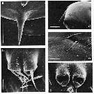 issued from : J.C. von Vaupel Klein in Zool. Verh., 1982, 198. [p.127, Pl.1]. Female: a, anterior view of rostrum and frontal organ; b, frontal organ with both hairs in situ; c, frontal organ with hairs detached, note the concave distal faces of the tubercles and the unpaired midventral pore; d, left lateral view of anterior part of cephalon, showing hair-sensillum (arroww); e, the same sensillum, enlarged. Scale bars: a, d = 0.100; b, c, e = 0.010 mm. Nota : The lateral aspect of the head shows a slightly vaulted frontal part, smoothly merging into the rostrum. This rostrum is a heavily chitinized, entire structure, comparatively large and syout, and acutely pointed. It is directed almost straight downward, only its tip showing a faint curve to caudad. The frontal organ consists of a pair of semi-tubular outgrowths, smoothly arising from the extreme anterior integument of the head. Their distal ends are shallowly concave and have a short hair (0.030 mm long) in their centre. A small, rounded elevation provided with an elongate pore is situated mid-ventrally to the tubercles.
|
 issued from : J.C. von Vaupel Klein in Zool. Verh., 1982, 198. [p.15, Fig.4, a-d]. As Euchirella messinensis f. typica. Female: a, posterior part of cephalothorax and urosome (dorsal); b, same (left lateral view); c, genital double-somite (genital complex), right lateral view with outline of rest urosome indicated; d, genital double-somite with vulva (ventral view). Nota : Urosome composed of 4 somites and the caudal rami. The genital double-somite (or fused complex urosomites 1 + 2) arrticulates freely with the cephalothorax. In all somites the dorsal, lateral, and ventral sclerites have coalesced to form a smooth ring. The urosome is short, i.e. rostro-caudally compressed. Relative lengths of urosomites + caudal rami 52 : 13 : 12 : 8 : 15 = 100. Genital double-somite strongly asymmetrical, showing a large sxemming on its left side. The swelling is produced into a robust dorso-lateral outgrowth, the basal part of which is directed dorso-caudally, followed by a more posteriorly pointing terminal portion ; a rather abrupt bend in the outgrowths dorsal outline marks the site where both parts meet. In dorsal aspect the asymmetrical structure appears as a slightly curved, caudo-medially directed hump which covers a large portion of urosomites 3 and 4, and which reaches most commonly to or even beyond the caudal edge of the latter urosomite (anal). Place abd shape of the outgrowth are highly characteristic for this species. From observations on specimens with filled seminal receptacles, it is clear that both left and right receptacles are situated in the outgrowth, i.e. the left receptacle in the anterior elevation, and the originally right receptacle in the posterior part. Both receptacles differ in shape but are approximately the same size. The anterior part of the genital double-somite, rostrad to the genital prominence, shows an almost completely annular field of tiny protuberances (warts), interrupted only mid-dorsally. The ventral outline of the genital double-somite is broadly rounded. The genital field dis located on the longer, smoothly sloping face of the genital prominence, posteriad ti its apex and bordered caudally by a small, transverse swelling. The vulva is trapezoidal, with the longer parallel side anteriorly. It is covered entirely by the genital operculum, which hinges anteriorly while its remaining sides are free. The surface of the operculum is produced into several ridges and wrinkles ; the posterior edge is indented medially to leave a small opening. The caudal slit between operculum and sternite gives access tio the underlying vulval cavity (here, the fertilization tube of the spermatophore may enter to reach the underlying pores).
|
 issued from : J.C. von Vaupel Klein in Zool. Verh., 1982, 198. [p.18, Fig.5]. As Euchirella messinensis f. typica. Female: a1-a3, left A1 in lateral view; b, segment 23 of right A1 (lateral view), showing normal arrangement of integumental organs; c, detail of hinge joint 17/18 of right A1 (medial view); d, antero-distal corner of segment 1 of left A1, with peg-sensillum; e, anterior margin of segments 3 through 8+9 of left A1, showing anteriorly placed brushes of wrinkled hairs; f, detail of segment 12 of left A1, with short, spiniform seta; g, details of proximal parts of large articulating setae of segment 7 (left) and 23 (right), respectively showing presence and absence of a breaking plane ; h, detail of distal small seta of segment 5; i, detail of normal small seta , i.e. the distal one of segment 11 (Type I); j, detail of distal small seta on segment 19 (Type II). Nota : A1 comparatively short ; when fully streched backwards they reach the posterior edge of the genital-double-simite ; 24 free segments, 8th and 9th, 25th and 26th, respectively, being wholly coalesced (though in both cases remnants of the former sutures are discernable). In complex segment 2, traces of sutures between the three original components can be detected only by SEM ; the fusion of segments 24 and 25 is intimate but not quite complete. The relative lengths of the segments are : 74 : 49 : 27 : 21 : 24 : 23 : 24 : 39 : 27 : 29 : 28 : 46 : 48 : 65 : 60 : 67 : 60 : 68 : 52 : 43 : 44 : 36 : 33 : 13 = 1000.
|
 issued from : J.C. von Vaupel Klein in Zool. Verh., 1982, 198. [p.18, Table II]. As Euchirella messinensis f. typica. Female: Setal armature of A1. Small setae are present along the frontal edges of most other segments than 1 and 2. Most of these are referable to either one of two distinct types: Type I is comparatively long and slender, rounded in cross-section, with a distinctly visible wall, and with a breaking region comparable to that of the large setae; the internal organization of type I setae, in preserved condition, shows like a coarse and irregularly transverse striation. Type II seate are shorter and broader, with thinner walls, and showing longitudinal ridges indicating a flattened cross-section; their breaking planes are narrow, annular regions only, and their internal structure looks like an evenly granulated matrix. Aberrant small setae are present on segments nos. 5, 12, and 25+26; A single aesthetasc is present on each of the segments 2, 5, 8+9, 12, 14, 19, and 25+26; Integumental organs and structures include slit-shaped glandular pores, peg- and pit-sensilla, circular pores, and wrinkled hairs. On segments 1 to 8+9, minute brushes of wrinkled hairs are present, viz., one each on segments 1 and 3-7, disto-caudally, and on segment 2, 3 to 6 brushes along its caudal margin; disperse brushes are found on the anterior margins of segments 3 through 8+9. See Table V.
|
 issued from : J.C. von Vaupel Klein in Zool. Verh., 1982, 198. [p.48, Table V]. As Euchirella messinensis f. typica. Female: Types of integumental organs. Aspect: dependent upon the way the segment is flattened, i.e., either laterally or rostro-caudally. ae = anterior edge; If = lateral face; mf = medial face. Individual numbers are assigned to the organs of each of the four aspects of a segment separately, in a proximal to distal, and lateral to medial or posterior to anterior order. On antennula, all organs designated as in the ''ae'' position are situated directly mediad to the anterior row of setae. ** = sites A 1 -3-ae-1 and A1 -7-ae-1 are placed rather medially but this is due entirely to the strong development of the large seta, at the base of which the organ is situated.
|
 issued from : J.C. von Vaupel Klein in Zool. Verh., 1982, 198. [p.22, Fig.6, a-f]. As Euchirella messinensis f. typica. Female: a, right A2 (medial view); b, left A2 (lateral view); c, medial lobe of basis 1 of right A2 of a anoyher specimen, showing second brush of hairs; d, endopodite of right A2 (medial view); d' same, detail of lobular outgrowth on terminal lobe; e-f, distal part of exopodal segment 7 with insertion of the three large setae of right (e) and left (f) A2, in medial, respectively lateral view, arrows indicate distal ring of sclerotized integument. Scale d' = scale d x 4. Nota : A2 are composed of a short basipodite, a well developed exopodite, and a markedly reduced endopodite. The two basipodal segments are largely fused, the former suture still being distinct anteriorly but faint to obsolescent posteriorly ; the 1st segment with 1 strongly plumose seta ; the lobe bears a dense brush of long, thin hairs posteriorly ; the 2 nd segment is swollen , it bears 1 small, naked seta,, inserted near the basis of the endopodite. Endopodite measures 0.24 the length of the exopodite. It consists of 2 distinct segments which exhibit any fusion ; the proximal segment bears 1 small, naked subapical seta ; the 2 nd segment is composed of the completely coalesced segments 2 and 3, it is produced into a proximal and terminal lobe ; the proximal one with an apical row of 4 slender, finely plumose setae which increase gradually in length to distad ; the terminal lobe bears 1 apical row of 5 plumose setae, finely drawn-out tips, also increasing sequentially in length to distad ; the medial edge of the terminal lobe is produced into an acutely pointed, lobular outgrowth in front of the row of setae ; a subapical row of 6-9 short spinules is present on the posterior margin. Exopodite of 6 free segments, the nos. 1 and 2 being coalesced to form a long, cylindrical structure ; the site of fusion is indicated by a blunt angle in the anterior outline of the combined segment, as well as by some faint, transverse grooves ; the anterior angle is not produced into a lobular outgrowth, the segment without setae. The following 4 segments are short, annular structures, the nos. 3 to 5 about equal in length, segment 6 slightly longer ; each segment bears 1 large, smoothly curved plumose seta rostro-medially ; the terminal, 7th segment is long and cylindrical, its bears 3 strong, gently curved plumose setae apically ; in the thin membrane surrounding these setae a small, incompletely annular sclerite is present ; Two slit-shaped glandular pores are the only integumental organs present ; 1 on endopodal segment 1 and 1 on exopodal segment 4.
|
 issued from : J.C. von Vaupel Klein in Zool. Verh., 1982, 198. [p.22, Fig.6, g-j]. As Euchirella messinensis f. typica. Female: g, corpus of left Md (venral view), with outline of proximal part of palp indicated; h-i, masticatory edge of right Md in posterior (h) and anterior (i) view; j, palp of right Md (postero-medial view), showing relative lengths of setae. Nota : Gnathobasis of Md heavily chitinized. Two fields of short spinules are present on the steep sloping medial face ; the anterior surface of the corpus is smooth. The masticatory edge is composed of 5 groups of teeth. There is a multicuspidate molariform complex proximally, in the apical part of which 1 spinulose setiform tooth and 1 smaller, serrate setiform tooth are included ; the spinulose tooth is equipped with an extensive row of long spinules anteriorly, and 2 shorter rows of smaller spinules, posteriorly ; the smaller setiform tooth has multiple rows of serrations in its middle portion. The remainder of the multicuspidate complex consists of 4 to 6 digitiform projections, some of which are smooth, others finely serrated. The complex is limited ventrally by a slender, bicuspidate ridge. The base of the complex bears 3 groups of slender spinules : a row of 10 and a group of 10-20, posteriorly, and 1 small group of about 6 spinules on the anterior face. Next, there are 2 smaller and 1 larger bicuspidate molariform complexes, each of which has a setiform protrusion in its central part (these protrusions have not been observed in all specimens). An extremely heavy, monocuspidate molariform tooth terminates the toohthed edge distally. Asymmetry in development of the left and right Md has not been observed.
|
 issued from : J.C. von Vaupel Klein in Zool. Verh. Leiden, 1982, 198. [p.26, Fig.7, a-b]. As Euchirella messinensis f. typica. Female (from Tyrrhenian Sea): a-b, left and right palpus mandibularis (anterior, posteerior, respectively), arrow indicates vestigial seta on endopodal segment 1. Nota : The Md palp is weakly developed and shows a moderately swollen basal segment fitting by a constricted proximal part to the ventral face of the gnathobasis ; this segment is devoid of setae. The two segments of the endopodite are small ; the proximal one bears an extremely small, spiniform protrusion which represents the remnants of a single seta. The distal segment is equipped with an apical row of 9 slender setae (3 delicate and finely plumose, followed by 5 long, stronger, curved, and densely plumose setae, while the 9th set ais again shorter and also more slender. The penultimate seta, i.e. n° 8, is inserted differently, its base does not articulate directly with the segment but is supported by a well-delimited pedestal-like structure. The originally 6-segmented exopodite exibits incomplete fusion of all segments, to varying degrees. In all joints, the intersegmental sutures are most clear-cut anteriorly and less distinct to obsolescent posteriorly. The exopodite bears 6 long and slender, gently curved plumose setae, one on every segment. A single slit-shaped glandular pore has been found on the basis 2 and a small granular area is present on the anterior face of rndopodal segment 2.
|
 issued from : J.C. von Vaupel Klein in Zool. Verh. Leiden, 1982, 198. [p.30, Fig.8]. As Euchirella messinensis f. typica. Female (from Tyrrhenian Sea): a, left Mx1 (posterior view); b, right Mx1 (anterior); c, detail of the terminal part of the endite of basal segment 2 of left Mx1 (showing 3 setae and the blunt tooth, with hairs and plumosity omitted; d, idem of right Mx1. Nota : 1st basal complex of Mx1 produced into 1 outer and 2 inner lobes. 1st inner lobe developed into an arthrite, capable of some motility relative to the corpus (as evidenced by a distinct anterior suture and the course of large striated muscles). 2 nd inner lobe plus the outer lobe together form a unit which is distinctly marked posteriorly but largely fused with the corpus along the anterior part of their boundary. 1st inner lobe bears 9 setae along its internal margin : 2 proximal ones comparatively long and slender, with blunt tips, and sparsely spinulose ; there are long and slender spinules which are distributed irregularly, as well as short and broad, blade-like spinules arranged serially in various longitudinal rows ; the four apical spinules in such a row are much larger than the rest. All spinules are widely spaced and inserted all around the circonference of the seta. These 2 setae have a breaking plane at 0.40 their length, approximately at the boundary between the smooth proximal and the spinulose distal part. The remaining 7 setae are stout, with broad bases, and taper to more acute tips. They alternate along the anterior and posterior sides of the edge. All the 7 setae are bipectinate distally whereas the anterior ones are also sparsely spinulose in their medial parts. Double pectinations are confined to the anterior face of the seta. The long, stout spinules of the anterior setae concentrate posteriorly in a close brush at the basis of the spinulose part, while this type of spinulosity constinues distally along the the inner an douter edges ; anteriorly such spinules are also present on a more proximal stretch. Next, a small, bipectinate set ais inserted submarginally on the anterior face of this lobe.. 3 submarginal setae are present posteriorly ; these are both bipectinate and spinulose ; the proximal set ais stout, with a broad base, the other 2 are more slender. The 1st inner lobe is also equipped with distinct patches of hairs and spinules, viz., 7 on its anterior face and 8 posteriorly. 2 nd inner lobe is slender and markedly shorter than the 1st. This lobe bears a close group of 4 apical setae which are moderately long and apparently rigid. 3 setae are bipectinate distally and sparsely spinulose along most of their length. The 4th, proximalmost, is slightly shorter and it is spinulose only. A small patch of short , slender spinules is present proximally on the anterior face of this lobe in some specimens. The outer lobe of basipodal segment 1 shows a squarish outline an dis hardly protruding, with 8 slender, delicate setae in a submarginal row posteriad to its outer lobe. The setae vary in size and in plumosity. The seta n°5 is markedly reduced in length and in thickness (this reduction is characteristic for the genus). 2 nd basal segment long and slender (length/width ratio : 3.6). It is only moderately well delimited against basipod 1 anteriorly but a distinct suture is present on the posterior side. With 3 apical setae : 1 long and slender, finely bipectinate and partly spinulose, and 2 small and delicate, plumose setae. The small and only partly free endite of basipod 2 does not reach as far as the 2 nd inner lobe of basipod 1 ; it bears 3 delicate setae apically, finely but densely plumose (1 long, the others are about 1/3 and ¼ the length of the large seta, respectively : fig.8 c, d) ; the terminal part of the endite is produced into a blunt tooth, which bears a large tubular pore on the posterior side of its apex. 2 extensive brushes of long, thin hairs are present anteriorly on the andite (one elongate, the other shorter). The minute endopodite is faintly delimited against basipod 2 (suggesting some degree of fusion. It bears 4 long and slender setae, bipectinate distally and sparsely spinulose proximally Exopodite apparently well-articulating with basipod 2, with 11 slender, smoothly curved setae, all strongly but coarsely plumose ; the distalmost set ais the shortest. On the anterior face of the segment a shallow submarginal ridge follows the distal outline. It is interrupted about halfway by a small tubular pore. A few slender spinules are present on the antero-medial edge of this ramus. Next to the tubular pores mentioned, 3 other integumental organs have been found, all slit-shaped glandular pores. Granular areas have been indicated on the corpus, the 1st outer lobe, and on the 2 nd inner lobe.
|
 issued from : J.C. von Vaupel Klein in Zool. Verh. Leiden, 1982, 198. [p.26, Fig.7, c-j]. As Euchirella messinensis f. typica. Female (from Tyrrhenian Sea): c, left Mx2 (postero-medial view); d, idem (detail of first endite showing apical hook-shaped outgrowth; e, right Mx2 (antero-lateral view, ornamentation of setae omitted); f, idem (detail of spinules at the base of the primary seta on endite 5); g-i, detail of the primary seta of the fifth endite to show arrangement of the five rows of denticles: g, this seta of the left Mx2 (postero-medial view); h: idem schematic drawing showing denomination of individual pectines; i, the same seta of the right Mx2 (antero-lateral view); j, ventral view of sternal thorn, caudal to insertion of left Mx2 (bar: 0.01 mm). Nota : Mx2 consist of a basal complex bearing 4 endites, the 2nd basal segment produced into a single endite, and a small endopodite. Possibly, basipod 1 and basipod 2 are fused to some degree. The lateral outline of basipod 1 is strongly arched proximally ; adjacent to this margin, the posterior integument is covered with small, rounded warts. All endites bear 3 apical setae. The small endopodite apparently is composed of 4 free segments, nos. 1-3 short, annular structures, the 4th being minute and globular. Segments 1-3 equipped medially with 1 bipectinate and spinulose seta each, while 2 and 3 also bear a small, smooth seta posteriorly ; the terminal segment bears 3 slender, smoothly curved setae which are finely bipectinate only. 2 slit-shaped glandular pores have been located on basipod 1 and 2 (one each) .
|
 issued from : J.C. Von Vaupel Klein in Zool. Verh. Leiden, 1982, 198. [p.34, Fig.9]. As Euchirella messinensis f. typica. Female (from Tyrrhenian Sea): a, right Mxp (medial view); b, idem, detail of comb of spinules on basipod 2; c, left Mxp (lateral view, ornamentation of setae partly omitted); d, idem, detail of antero-distal corner of basipod 1, showing field of short spinules; e, detail of the shorter distal seta of basipod 2 of the left Mxp (in outer view). Nota : Mxp strongly developed. They are curved in the way characteristic for most Aetideidae. While the 1st basal segment has kept its original position, the segments distad of hinge-joint (Basipod 1-2) have rotated to mediad whereby their originally anterior margin is now taking a postero-medial position if Mxp is completely stetched. These appedages are usually depicted in this position, hence the reversed terminology of the distal parts with regard to anterior and posterior as compared to most other calanoids. However, in situ the Mxp are held in a curved and also twisted position, with the secondary postrior margins facing antero-ventrally. 1st basal segment (L/W = 2.2) bears 7 setae, arranged in 4 groups (1, 2, 3 and 3, respectively). The 1st group consists of a single, smooth seta inserted medially ; the other setae are situated along the anterior margin. There is an extensive patch of short and sharp, slightly curved spinules on the antero-distal corner of basipod 1. 2 nd segment is long and slender (L/W = 5.5), bears 5 setae along its (secondary) posterior margin : 1 smaller, distally bipectinate seta about halfway its length ; 1 shorter and 1 longer seta at two-thirds tthe shorter bipectinate only, the longer bipectinate distally and densely spinulose in its proximal half ; and, extremely distally, 1 short, blunt and 1 long, slender seta (the shorter coarsely bipectinate, the long one multipectinate and spinulose proximally. Proximo-medially on basipod 2 a longitudinal combi s present which consists of a single row of 29-35 densely set spinules. Endopodite composed of 5 segments, nos. 1-4 short, annular structures and n° 5 minute (setal formula : 4, 3, 3, 3 and 3 setae, respectively). In addition, endopodal segments 4 and 5 are equipped with 1 small, naked seta on the anterior margin, viz., halfway on segment 4 and subterminally on 5.
|
 issued from : J.C. von Vaupel Klein in Zool. Verh. Leiden, 1982, 198. [p.36, Fig.10, a-f]. As Euchirella messinensis f. typica. Female (from Tyrrhenian Sea): a, right P1 with intercoxal plate (postrior view); b, left P1 (anterior); c, detail of the endopodite and the specialized curved seta of basipod 2 of the right leg (anterior); c', detail of tubercle (posterior view); , showing complex of five tubular pores and internal tissue strand; d, detail of terminal seta of exopod 3; e, detail of terminal spiniform outgrowth of exopod 3; f, detail of one of the terminal setae of the endopod, showing two modified sites and the serially arranged, contiguous setules. Nota : Basipodal segment 1 of P1 with 1 medial brush of long, coarse hairs. Basipodal segment 2 with 1 medial brush of very long, coarse hairs ; and the specialized, bdoubly curved, plumose seta inserted at the articulation with the endopodite, anteriorly in the segments disto-medial corner. Endopodite comprising 1 free segment ; rostro-lateral tubercle well developed, bearing c. 16-23 medium-sized, smooth, slightly curved spinules placed in a single, partly alternativing row ; 3-5 slender central hairs are present, with lengths ranging from 0.14 to 0.24 times the length of the segment ; the undivided distal hairbrush consists of moderately coarse hairs, is slender and curved, and hardly reaches the fringe of coarser hairs on the disto-lateral margin ; proximall, the medial margin bears 1 seta ; there are also 2 medial,1 subterminal and 1 terminal setae. A specialized integumental organ is found on the postero-medial face of the tubercle, where a complex of 4 to 5 tubular (glandular ?) pores is found ; internally, a tissue strand runs from these pores to proximad, to be traced as far as the proximal half of basipodal segment 2 (this structure is named Von Vaupel Kleins organ). Exopodite with segments 1 and 2 completely fused ; with 2 short and smooth lateral spines, the shorter apically ; the margin between these spines finely hairy ; two separate rows of long, coarse hairs are present, viz., one each on both the proximal and distal sections of the biconvex inner margin. Exopodal distal segment with whole lateral margin beset with short, fine hairs ; 1 outer apical spine, minutely pectinate along its inner an douter margin, and flanked by a short and acute, subtriangular outgrowth of the segment ; medial margin with 3 setae ; 1 long terminal seta, finely plumose along its inner margin and equipped with a serrate edge laterally, which is composed of about 80 serially contiguous, trapezoid denticles, gradually decreasing in size to distad.
|
 issued from : J.C. von Vaupel Klein in Zool. Verh. Leiden, 1982, 198. [p.36, Fig.10, g-h]. As Euchirella messinensis f. typica. Female (from Tyrrhenian Sea): g, left P2 (anterior view); h, right P2 with intercoxal plate (posterior aspect).
|
 issued from : J.C. von Vaupel Klein in Zool. Verh. Leiden, 1982, 198. [p.39, Fig.11, a-d]. As Euchirella messinensis f. typica. Female (from Tyrrhenian Sea); a, endopod of P2 (anterior view); b, detail of the terminal spine of exopod 3 of P2; c, detal of the short disto-lateral spine of exopod 2 of P2 (anterior view), with its adjacent large closing-flap pore; d, right basipod 2 of P2 (posterior), detail of the disto-lateral pointed outgrowth with underlying (?) pore structure (arrow);.
|
 issued from : J.C. von Vaupel Klein in Zool. Verh. Leiden, 1982, 198. [p.39, Fig.11, e-i]. As Euchirella messinensis f. typica. Female (from Tyrrhenian Sea); e, left P3 (anterior view); f, right P3 with intercoxal plate (posterior); g, right basipod 3 of P3 (posterior), detail of the disto-lateral outgrowth with (?) pore (arrow); h-i, lateral margin pf endopod 2 of P3: h, of left leg (anterior); i, of right leg (posterior).
|
 issued from : J.C. von Vaupel Klein in Zool. Verh. Leiden, 1982, 198. [p.41, Fig.12]. As Euchirella messinensis f. typica. Female (from Tyrrhenian Sea): a-b, endopod 1 of P3: a, of right leg (postrior view); b, of left leg (anterior view).
|
 issued from : J.C. von Vaupel Klein in Zool. Verh. Leiden, 1982, 198. [p.41, Fig.12, c-i]. As Euchirella messinensis f. typica. Female (from Tyrrhenian Sea): c, left P4 (anterior); d, right P4 (posterior); e, endopod 2 of left P4, detail of lateral margin in anterior view; f, endopod 1 of left P4 (anterior); g, detail of the two postero-medial spines of basipod 1 of P4; h, basipod 2 of right P4 (posterior), deatal of disto-lateral pointed outgrowth, arrow indicates presumed underlying pore-structure; i, situation on basipod 1 of P4 in an aberrant specimen, where three instead of two spines are present.
|
 Issued from : W. Giesbrecht in Systematik und Faunistik der Pelagischen Copepoden des Golfes von Neapel und der angrenzenden Meeres-Abschnitte. Fauna Flora Golf. Neapel, 1892, 19 , Atlas von 54 Tafeln. [Taf.36, Fig.24]. As Euchirella messinensis messinensis. Female : 24, head (lateral).
|
 Issued from : W. Giesbrecht in Systematik und Faunistik der Pelagischen Copepoden des Golfes von Neapel und der angrenzenden Meeres-Abschnitte. Fauna Flora Golf. Neapel, 1892, 19 , Atlas von 54 Tafeln. [Taf.15, Fig.2]. As Euchirella messinensis messinensis. Female : 2, A1.
|
 Issued from : W. Giesbrecht in Systematik und Faunistik der Pelagischen Copepoden des Golfes von Neapel und der angrenzenden Meeres-Abschnitte. Fauna Flora Golf. Neapel, 1892, 19 , Atlas von 54 Tafeln. [Taf.15, Fig.16]. As Euchirella messinensis messinensis. Female : 16, A2.
|
 Issued from : W. Giesbrecht in Systematik und Faunistik der Pelagischen Copepoden des Golfes von Neapel und der angrenzenden Meeres-Abschnitte. Fauna Flora Golf. Neapel, 1892, 19 , Atlas von 54 Tafeln. [Taf.15, Fig.15]. As Euchirella messinensis messinensis. Female : 15, Mx1.
|
 Issued from : W. Giesbrecht in Systematik und Faunistik der Pelagischen Copepoden des Golfes von Neapel und der angrenzenden Meeres-Abschnitte. Fauna Flora Golf. Neapel, 1892, 19 , Atlas von 54 Tafeln. [Taf.15, Fig.12]. As Euchirella messinensis messinensis. Female : 12, Mxp.
|
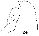
 Issued from : W. Giesbrecht in Systematik und Faunistik der Pelagischen Copepoden des Golfes von Neapel und der angrenzenden Meeres-Abschnitte. Fauna Flora Golf. Neapel, 1892, 19 , Atlas von 54 Tafeln. [Taf.15, Fig.24]. As Euchirella messinensis messinensis. Female: 24, P4. B1 = coxa; B2 = Basis.
|
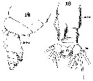 Issued from : W. Giesbrecht in Systematik und Faunistik der Pelagischen Copepoden des Golfes von Neapel und der angrenzenden Meeres-Abschnitte. Fauna Flora Golf. Neapel, 1892, 19 , Atlas von 54 Tafeln. [Taf.36, Figs.14, 18]. As Euchirella messinensis messinensis. Female: 14, last thoracic segments and urosome (lateral); 18, urosome (dorsal). Ab 1-2 = genital double-somite.
|
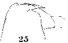 Issued from : W. Giesbrecht in Systematik und Faunistik der Pelagischen Copepoden des Golfes von Neapel und der angrenzenden Meeres-Abschnitte. Fauna Flora Golf. Neapel, 1892, 19 , Atlas von 54 Tafeln. [Taf.36, Fig.25]. As Euchirella messinensis messinensis. Male: 25, forehead (lateral).
|
 Issued from : W. Giesbrecht in Systematik und Faunistik der Pelagischen Copepoden des Golfes von Neapel und der angrenzenden Meeres-Abschnitte. Fauna Flora Golf. Neapel, 1892, 19 , Atlas von 54 Tafeln. [Taf.15, Fig.1]. As Euchirella messinensis messinensis. Male: A1.
|
 Issued from : W. Giesbrecht in Systematik und Faunistik der Pelagischen Copepoden des Golfes von Neapel und der angrenzenden Meeres-Abschnitte. Fauna Flora Golf. Neapel, 1892, 19 , Atlas von 54 Tafeln. [Taf.15, Fig.17]. As Euchirella messinensis messinensis. Male: 17, Mx1 (anterior view). Le = outer lobe; Li1 = inner lobe; Ri= endopodite; Re = exopodite.
|
 Issued from : W. Giesbrecht in Systematik und Faunistik der Pelagischen Copepoden des Golfes von Neapel und der angrenzenden Meeres-Abschnitte. Fauna Flora Golf. Neapel, 1892, 19 , Atlas von 54 Tafeln. [Taf.15, Figs.14, 21]. As Euchirella messinensis messinensis. Male: 14, portion of edge of exopodal segment 3 of right P5; 21, P5 (anterior view). Ps = left leg; Pd = right leg; B1 = basal segment 1 (= praecoxa + coxa); B2 = basipodal segment 2 (= basis); Ri = endopodite; Re = exopodite.
|
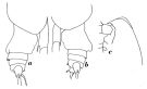
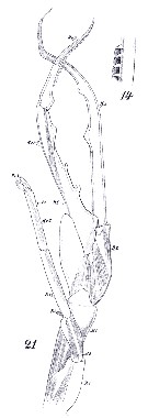
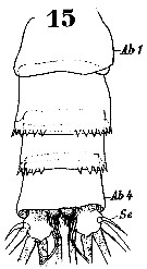 Issued from : W. Giesbrecht in Systematik und Faunistik der Pelagischen Copepoden des Golfes von Neapel und der angrenzenden Meeres-Abschnitte. Fauna Flora Golf. Neapel, 1892, 19 , Atlas von 54 Tafeln. [Taf.36, Fig.15]. As Euchirella messinensis messinensis. Male: 15, urosome. Ab1 = genital somite; Ab4 = abdominal somite 4; Se = outer seta.
|
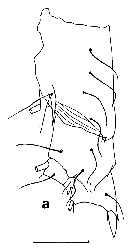
 Issued from : J.C. von Vaupel-Klein in Zool. Meded., Leiden, 19972, 47 (41). [p.509, Fig.4, c]. Male (from 1°30'N, 10°10'W): c, endopodite of right P1 (anterior view). Nota: 'organ of Vaupel Klein' (see explanation to Euchirella curticauda.)
|
 issued from : C. With in The Danish Ingolf-Expedition, Copepoda I, 1915, III, 4. [Pl. IV, Fig.2, a-c]. Female (from 61°30'N, 17°08'W):a, labrum (oral view); b, lamina labialis; c, area labialis and lobi labiales.
|
 issued from : C. With in The Danish Ingolf-Expedition, Copepoda I, 1915, III, 4. [Pl. VIII, Fig.1]. Male: 1, right P1 (anterior view).
|
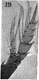 issued from : H.B. Owre & M. Foyo in Fauna Caribaea, 1967, 1, Crustacea, 1: Copepoda. [p.48, Fig.275]. Female (from Florida Current): 275, P4.
|
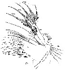
 issued from : H.B. Owre & M. Foyo in Fauna Caribaea, 1967, 1, Crustacea, 1: Copepoda. [p.48, Figs.273-274]. Male: 273, P5; 274 same (magnified).
|
 issued from : E.L. Markhaseva in Proc. Zool. Inst. RAN, St. Petersburg, 1996, 268. [p.162, Fig.124]. As Euchirella messinensis messinensis. Female (from Northern Atlantic). Nota: Left side of genital segment with large projection , covering 2 next urosomal segments (dorsal and lateral view); genital segment nearly 1.2 times longer than wide. A1 24-segmented, slightly longer than cephalothorax. Endopod of A2 about 5 times shorter than exopod. Male (from Bradford & Jillett, 1980). Nota: Differs from female in low crest. Rostrum well developed. Exopodal segment 3 of left P5 not reaching the 1st projection of the endopod of right leg.
|
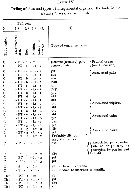
 Euchirella messinensis messinensis Euchirella messinensis messinensis female: 1 - Genital segment asymmetrical. 2 - Crest absent. 3 - Genital segment with left lateral side swelled (dorsal view). 4 - Coxopodite of P4 with 2 spines. 5 - Left and right lateral sides of genital segment (dorsal view) without swellings.Projection present on the dorsal side in the left half of genital segment. Endopodal segment 1 of Md with 1 small seta. 6 - Projection on the dorsal side of genital segment highly large, exceeding the posterior border of genital segment and covering 2 following urosomal segments.
|
 Euchirella messinensis messinensis Euchirella messinensis messinensis male: 1 - Left P5 with endopod rudimentary. Exopodal segment 2 and endopod of right P5 elongated and sharpened in their distal parts forming tongs. Endopod of right P5 significantly exceeding distal border of exopodal segment 1 of right P5. 2 - Crest present, low. 3 - Left P5 basipodite shorter than right coxopodite of P5. 4 - Exopodal segment 1 of right P5 with 4 tooth-like projections. 5 - Exopodal segment 3 of left P5 not reaching the first projection of endopod of right P5.
|
 issued from : E.L. Markhaseva in Proc. Zool. Inst. RAN, St. Petersburg, 1996, 268. [p.161, Fig.123]. As Euchirella messinensis indica. Female: C P4 (from Bradford & Jillett, 1980). Nota frm Markhaseva (1996, p.159): Body shape similar to E. messinensis messinensis; differs in the shape of genital segment, but with projection in left posterior part of the segment smaller than in E/ messinensis messinensis (dorsal view). All other features in structure of body and limbs close to that in E. m. messinensis. Male: Ce, P5 (a) (from Bradford & Jillett, 1980); other figures (from 27°48'N, 130°40'E). Nota: Cephalon with low crest. Body shape close to E. m. messinensis. Exopod of left P5 exceeding the first projection on endopod of right leg.
|
Euchirella messinensis indica female: 1 - Genital segment asymmetrical. 2 - Crest absent. 3 - Genital segment with left lateral side swelled (dorsal view). 4 - Coxopodite of P4 with 2 spines. 5 - Left and right lateral sides of genital segment (dorsal view) without swellings.Projection present on the dorsal side in the left half of genital segment. Endopodal segment 1 of Md with 1 small seta. 6 - Projection on the dorsal side of genital segment small only slightly exceeding the posterior border of genital segment.
|
Euchirella messinensis indica male: 1 - Left P5 with endopod rudimentary. Exopodal segment 2 and endopod of right P5 elongated and sharpened in their distal parts forming tongs. Endopod of right P5 significantly exceeding distal border of exopodal segment 1 of right P5. 2 - Crest present, low. 3 - Left P5 basipodite as long as right coxopodite of P5. 4 - Basipodite of left P5 nearly as long as coxopodite of right P5. 5 - Parts of exopodal segment 3 of left P5 tongs equal in length.
|
 issued from : Tanaka O. in Publ. Seto Mar. Biol. Lab., 1957, 6 (2). [p.181]. As E. messinensis but probably as E. messinensis indica. Female A1: proportional lengths of segments. - A1 22-segmented, extends to the middle of 2nd abdominal segment. Segments 24-25 are 1.06 times as long as the 23rd, segment 18 shorter than the 17th in the present specimen.
|
 Issued from : J.C. von Vaupel Klein in Crustaceana, (Supplement) 9, 1984. [p.93, Table II]. Euchirella messinensis Female: Datamatrix stating observed states of characters from Table I (p.87-90) presently examined; nos. refer to the input nos. used in Table I (see to the family Aetideidae).
|
 issued from : J.C. von Vaupel Klein in Crustaceana, Supplt 9, Studies on Copepoda, III, 1984. [p.68, Fig.8, k]. Euchirella messinensis: Structure and setal complement of endopod 1 of Md (left, anterior). A very minute setal vestige (arrow), as in Euchirella speciosa.
|
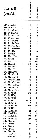 Issued from : J.C. von Vaupel Klein in Crustaceana, (Supplement) 9, 1984. [p.94, Table II (cont' d)]. Datamatrix stating observed states of characters from Table I (p.87-90) presently examined; nos. refer to the input nos. used in Table I (see to the family Aetideidae).
|
 Issued from : J.C. von Vaupel Klein in Crustaceana, (Supplement) 9, 1984. [p.95, Table II (cont' d)]. Datamatrix stating observed states of characters from Table I (p.87-90) presently examined; nos. refer to the input nos. used in Table I (see to the family Aetideidae).
|
 Issued from : J.C. von Vaupel Klein in Zool. Verhanddelingen, 1982, N°198. [p.19]. Euchirella messinensis Female: Relative lengths of the segments of A1. Nota: 24 free segments, segments 8 and 9 nad 25-26, respectively, wholly coalesced, though in both cases remnants of the former sutures are discernable. In complex segment 2, traces of sutures between the three original composants can be detected only by S.E.M. fully coalesced, fusion of segments 24 and 25 iintimate, but not quite complete.
|
Issued from : J.C. von Vaupel Klein in Zool. Verhanddelingen, 1982, N°198. [p.21, Table II]. Euchirella messinensis Female: Structure of the antennula; Legends.- f=:frontal, c = caudal, t = terminal, (p) = plumose to some extent, p = plumose, s = spiniform, d = delicate, lo = long, (no indication) = normal.
|
 Issued from : J.C. von Vaupel Klein in Zool. Verhanddelingen, 1982, N°198. [p.24]. Euchirella messinensis Female: Relative lengths of the endopodal segments of the A2 (in lateral aspect).
|
 Issued from : J.C. von Vaupel Klein in Zool. Verhanddelingen, 1982, N°198. [p.33]. Euchirella messinensis Female: Relative lengths of the segments of Mxp.
|
 Issued from : J.C. von Vaupel Klein in Zool. Verhanddelingen, 1982, N°198. [p.35]. Euchirella messinensis Female: Relative length of the legs, excluding and including (betweenj brackets) the terminal spine of the exopodite, if as follows (standard = P3 + termunal spine = 100).
|
 Issued from : J.C. von Vaupel Klein in Zool. Verhanddelingen, 1982, N°198. [p.37]. Euchirella messinensis Female: Proportional lengths of the segments of legs. The setae of the swimming legs are of two main types: A, setae founded disto-medially on Ba1 (Coxa) of P2-P4, while the proximo-medial setae of P1-P4 endodpod are of this type as well. Setae gently curved, relatively thick proximally, and narrowing abruptly at about 1/3rd their length to a slender distal part. The plumose is dense, contiguous, and long; on the thick basal part the setules are coarser whereas finer setules are found terminally;
Type B: Setae rather straight, apparently more rigid structures, denselyn but finely plumose with relatively short setules. The remaining setae of the endopod and all setae of the exopod are essentially of this type; some of these, however, may be only feebly developed.
Specialized setae, viz. on Basis of P1, on exopod 3 of P1, and the terminal spines of exopd3 of P2-P4 must be separately.
The setae and spines of the legs often show one or more modified sites along their length (cf. Fig.10 f), probably referable to articulations.
In Type A: setae such a site is either absent or present at 1/3rd their length.
Type B: setae have 1 or 2 such sites, at 1/2 and/or 2/3rds, but in reduced setae no such site was observed.
The S-curved seta on basis (Ba2) of P1 has no modified site, whereas the terminal setaz on exopod3 of P1 and the terminal spines on exopod3 of P2-P4 show such structure at 1/3rd their length.
|
 Issued from : J.C. von Vaupel Klein in Zool. Verhanddelingen, 1984 (1982), N°198. [p.42, Fig.13]. Euchirella messinensis Female: a-f, distribution and coding of sites of integumental organs of the body; a, cephalothorax (distally); a', enlarged detail of the right portion of Th4+5 (in posterior view); b, cephalothorax (left laterally); c-f, urosome (respectively ventrally, right laterally, dorsally, and left-laterally); g, explanation of symbol-code (adapted from Fleminger, 1973), and enlarged. For explanation of site-codes and abbreviations, see Table IV. Broken lines in figures a, b, c-f, indicate parts which are not visible in the other aspects shown.
|
 Issued from : J.C. von Vaupel Klein in Zool. Verhanddelingen, 1984 (1982), N°198. [p.43, Table IV]. Euchirella messinensis Female: Coding of sites of integumental organs on the body. See legends.
|
 Issued from : J.C. von Vaupel Klein in Zool. Verhanddelingen, 1984 (1982), N°198. [p.44, Table IV (continued)]. Euchirella messinensis Female: Coding of sites of integumental organs on the body. See legends.
|
 Issued from : J.C. von Vaupel Klein in Zool. Verhanddelingen, 1984 (1982), N°198. [p.48, Table V]. Euchirella messinensis Female: Coding of sites and types of integumental organs of the appendages (excluding of the swimming legs). See legends and also to Table IV.
|
 Issued from : J.C. von Vaupel Klein in Zool. Verhanddelingen, 1984 (1982), N°198. [p.43, Table IV]. Euchirella messinensis Female: Coding of sites of integumental organs on the body.
|
 Issued from : J.C. von Vaupel Klein in Zool. Verhanddelingen, 1984 (1982), N°198. [p.49, Table V (continued)]. Euchirella messinensis Female: Coding of sites and types of integumental organs of the appendages (excluding of the swimming legs) and legends (See also legends to Table IV).
|
 Issued from : J.C. von Vaupel Klein in Zool. Verhanddelingen, 1984 (1982), N°198. [p.50, Fig.15]. Euchirella messinensis Female: a-h, distribution and coding of sites of integumental organs of the swimming legs. a, b, left and right P1 (anterior respectively posterior view); c, d, left and right P2 (anterioly cq. posteriorly); e, f, left and right P3 (anterior c.q. posterior view); g,h,left and right P4 (anterior and posterior aspect) For explanation of site- and symbol-code, see fig.13 and Tables IV to VI.
|
 Issued from : J.C. von Vaupel Klein in Zool. Verhanddelingen, 1984 (1982), N°198. [p.53, Table VI]. Euchirella messinensis Female: Presence or absence of integumental organs on the swimming legs and legends.
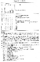
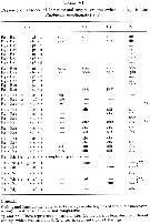
| | | | | Ref. compl.: | | | Cleve, 1904 a (p.190); Rose, 1925 (p.152); Wilson, 1942 a (p.185); Massuti Alzamora, 1942 (p.110); Sewell, 1948 (p.348, 500, 508, 516, 519, 520, 545, 556, 566); C.B. Wilson, 1950 (p.225); Fagetti, 1962 (p.20); Ganapati & Shanthakumari, 1962 (p.8, 15); Giron-Reguer, 1963 (p.29); Duran, 1963 (p.15); V.N. Greze, 1963 a (tabl.2); Grice, 1963 a (p.495); Unterüberbacher, 1964 (p.22); De Decker & Mombeck, 1964 (p.12); Grice & Hulsemann, 1965 (p.223); 1967 (p.15); Shmeleva, 1965 b (p.1350, lengths-volume -weight relation); Furuhashi, 1966 a (p.295, vertical distribution in Kuroshio region, Table 10) ; Pavlova, 1966 (p.43); Mazza, 1966 (p.70); Ehrhardt, 1967 (p.738, geographic distribution, Rem.); Mazza, 1967 (p.367); Vinogradov, 1968 (1970) (p.268); Evans, 1968 (p.13); Berdugo & Kimor, 1968 (p.448); Morris, 1970 (p.2300); Park, 1970 (p.475); Deevey, 1971 (p.224); Bainbridge, 1972 (p.61, Appendix Table I: vertical distribution vs day/night, Table III: occurrence); Roe, 1972 (p.277, tabl.1, tabl.2); 1972 a (p.339); Apostolopoulou, 1972 (p.327, 347); Vives & al., 1975 (p.42, tab.II, III); Gaudy, 1975 (p.109, fig.1, Table I, 4, respiration); Deevey & Brooks, 1977 (p.256, tab.2, Station "S"); Carter, 1977 (1978) (p.35); Vaissière & Séguin, 1980 (p.23, tab.2); Vives, 1982 (p.291); Kovalev & Schmeleva, 1982 (p.83); Dessier, 1983 (p.89, Tableau 1, Rem., %); Brenning, 1983 (p.3, 4, spatial distribution, T-S diagram, Rem.); Scotto di Carlo & Ianora, 1983 (p.150); Scotto di Carlo & al., 1984 (p.1042); Petipa & Borichenko, 1985 (tab.1); Brenning, 1985 a (p.28, Table 2); Madhupratap & Haridas, 1986 (p.105, tab.1); Lozano Soldevilla & al., 1988 (p.58); Madhupratap & Haridas, 1990 (p.305, fig.5: vertical distribution night/day; fig.7: cluster); Suarez & al., 1990 (tab.2); Hattori, 1991 (tab.1, Appendix); Suarez & Gasca, 1991 (tab.2); Suarez, 1992 (App.1); Scotto di Carlo & al., 1991 (p.271); Seguin & al., 1993 (p.23); Hays & al., 1994 (tab.1); Suarez-Morales & Gasca, 1997 (p.1525); Hure & Krsinic, 1998 (p.101); Alvarez-Cadena & al., 1998 (tab.3); Suarez-Morales & Gasca, 1998 a (p107); Lopez-Salgado & al., 2000 (tab.1); Lapernat, 2000 (tabl.3, 4); Holmes, 2001 (p.48); Beaugrand & al., 2002 (p.179, figs.5, 6); Schnetzer & Steinberg, 2002 (p.89, figs.); Vukanic, 2003 (139, tab.1); Pusch & al., 2004 (251, tab.3); Ikeda & al., 2006 (p.1791, Table 2); Hwang & al., 2007 (p.23); Kosobokova & al., 2007 (p.929: Tab.7); Gaard & al., 2008 (p.59, Table 1, N Mid-Atlantic Ridge); Raybaud & al., 2008 (p.1765, Table A1); Ayon & al., 2008 (p.238, Table 4: Peruvian samples); Licandro & Icardi, 2009 (p.17, Table 4); Hafferssas & Seridji, 2010 (p.353, Table 3); Williamson & McGowan, 2010 (p.273, Table 3, Pacific central gyres: N and S); Homma & Yamaguchi, 2010 (p.965, Table 2); Mazzocchi & Di Capua, 2010 (p.423); Medellin-Mora & Navas S., 2010 (p.265, Tab. 2); Homma & al., 2011 (p.29, Table 2, abundance, feeding pattern: suspension feeders); Miloslavic & al., 2012 (p.165, Table 2, transect distribution); in CalCOFI regional list (MDO, Nov. 2013; as E. messinensis messinensis; M. Ohman, comm. pers.); Hirai & al., 2013 (p.1, Table I, molecular marker); Sano & al., 2013 (p.11, Table 2, 7, fig.5, feeding habits); Lidvanov & al., 2013 (p.290, Table 2, % composition); Bonecker & a., 2014 (p.445, Table II: frequency, horizontal & vertical distributions); Fierro Gonzalvez, 2014 (p.1, Tab. 3, 5, occurrence, abundance); Benedetti & al., 2016 (p.159, Table I, fig.1, functional characters); El Arraj & al., 2017 (p.272, table 2); Belmonte, 2018 (p.273, Table I: Italian zones) | | | | NZ: | 20 | | |
|
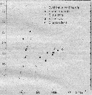
Carte de distribution de Euchirella messinensis par zones géographiques
|
| | | | | | | | | | | | | | | 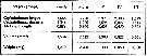 issued from : A.A. Shmeleva in Bull. Inst. Oceanogr., Monaco, 1965, 65 (n°1351). [Table 6: 18 ]. Euchirella messinensis (from South Adriatic). issued from : A.A. Shmeleva in Bull. Inst. Oceanogr., Monaco, 1965, 65 (n°1351). [Table 6: 18 ]. Euchirella messinensis (from South Adriatic).
Dimensions, volume and Weight wet. Means for 50-60 specimens. Volume and weight calculated by geometrical method. Assumed that the specific gravity of the Copepod body is equal to 1, then the volume will correspond to the weight. |
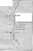 issued from : U. Brenning in Wiss. Z. Wilhelm-Pieck-Univ. Rostock - 32. Jahrgang 1983. Mat.-nat. wiss. Reihe, 5. [p.1, Fig.1]. issued from : U. Brenning in Wiss. Z. Wilhelm-Pieck-Univ. Rostock - 32. Jahrgang 1983. Mat.-nat. wiss. Reihe, 5. [p.1, Fig.1].
Spatial distribution for Euchirella curticauda, E. messinensis, E. pulchra, E. rostrata, E. splendens from 8° S - 26° N; 16°- 20° W. |
 issued from : U. Brenning in Wiss. Z. Wilhelm-Pieck-Univ. Rostock - 32. Jahrgang 1983. Mat.-nat. wiss. Reihe, 5. [p.3, Fig.3]. issued from : U. Brenning in Wiss. Z. Wilhelm-Pieck-Univ. Rostock - 32. Jahrgang 1983. Mat.-nat. wiss. Reihe, 5. [p.3, Fig.3].
T-S Diagram for Euchirella curticauda, E. messinensis, E. pulchra, E. rostrata, E. splendens from 8° S - 26° N; 16°- 20° W. |
 Issued from : J.M. Bradford & J.B. Jillett in New Zealand Ocean. Inst. Memoir, 86, 1980. [p.88-89, Figs.65-67]. Issued from : J.M. Bradford & J.B. Jillett in New Zealand Ocean. Inst. Memoir, 86, 1980. [p.88-89, Figs.65-67].
Distribution of several species of Euchirella in the Tasman Sea and around New Zealand.
Nota: Euchirella indica (= Euchirella massinensis indica). |
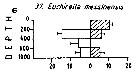 Issued from : M. Madhupratap & P. Haridas in J. Plankton Res., 12 (2). [p.312, Fig.5]. Issued from : M. Madhupratap & P. Haridas in J. Plankton Res., 12 (2). [p.312, Fig.5].
Vertical distribution of calanoid copepod (mean +1 SE), abundance No/100 m3. 37- Euchirella messinensis.
Night: shaded, day: unshaded.
Samples collected from 6 stations located off Cochin (India), SE Arabian Sea, November 1983, with a Multiple Closing Plankton Net (mesh aperture 300 µm), in vertical hauls at 4 depth intervalls (0-200, 200-400, 400-600, 600-1000 m). |
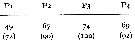
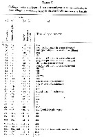
| | | | Loc: | | | South Africa (E), off E Tristan da Cunha Is., Namibia, Congo, G. of Guinea, off Lagos, off NE St. Paul Is., Cape Verde Is., off Mauritania-NW Cape Verde Is., Morocco-Mauritania, Canary Is., Azores, off Madeira, off Portugal, S Bazsil, off Amazon, Caribbean Sea, Caribbean Colombia, Yucatan, G. of Mexico, Cuba, Florida, Sargasso Sea, off Bermuda: Station "S" (32°10'N, 64°30'W), off Woods Hole, Iceland, Norwegian Sea, off W Ireland, North Sea, Bay of Biscay, off W Cape Finisterre, Ibero-moroccan Bay, Medit. (Alboran Sea, Banyuls, G. of Lion, Ligurian Sea, Thyrrhenian Sea, Strait of Messina, Malta, Adriatic Sea, Ionian Sea, Aegean Sea, Lebanon Basin), Minicoy Is., off E Sri Lanka, Indian, India (Lawson's Bay), Indonesia-Malaysia, Philippines, China Seas (East China Sea, South China Sea) , Taiwan, Kuroshio Current, Japan (Izu, Sagami Bay, off Sanriku), off Hokkaido SE, S Bering Sea, S Aleutian Basin, Alaska, Pacif. (W equatorial), New Zealand, Hawaii, Pacif. (central subtropical N), California, Pacific (central gyres: N and S), off Galapagos, off Peru, Easter Is., Chile | | | | N: | 140 (Arct.: 6; S Atlant.: 3; N Atlant.: 87; Medit.: 26; Indian: 18; Pacif.: 31) | | | | Lg.: | | | (1) F: 5,4; M: 4,4; (7) F: 5,7; M: 5,46; (14) F: 5,5-4,42; M: 4,5-2,8; (29) M: 4,88; (38) F: 6,2-5,35; M: 4,98-4,74; (45) F: 5-4,5; M: 4-3,75 (46) F: 4,75; M: 3,95; (56) F: 5,26; M: 4,62; (142) F: 4,5; M: 4; (199) F: 5,44-4,36; M: 4,96-4,26; (201) [as indica] F: 5,3-3,85; M: 4,2-4; [as messinensis] M: 4,8; (202) F: 4,36-5,25; M: 3,12-3,4; (207) F: 5-4,4; M: 4,56-4,25; (235) F: 5,25-4,36; M: 4,28-4; (242) F: 5,55-4,05; (260) F: 5-4,4; M: 4,56-4,25; (432) F: 4,7-3,8; (449) F: 4,75; M: 3,95; (1109) F: 4,56-4,7; M: 4,63; {F: 3,80-6,20; M: 2,80-5,46}
The mean female size is 4.866 mm (n = 28; SD = 0.5879), and the mean male size is 4.260 mm (n = 25; SD = 0.6016). The size ratio (male : female) is 0.87. | | | | Rem.: | épi-bathypélagique.
Pour Bradford & Jillett (1980, p.37, 39) les deux formes messinensis et indica sont considérées comme des espèces séparées.
Vaupel Klein (1984 a, p.35) souligne les distributions allopatriques des formes atlanto-méditerranéennes et indo-pacifiques : les premières correspondent à la sous-espèce E. messinensis messinensis et les secondes à E. messinensis indica , à lexception de la forme observée en Californie. Les deux formes se distinguent par l'importance de la protubérance sur le côté gauche du somite génital: plus développée chez la sous-espèce messinensis que chez indica.
Euchirella messinensis indica Vervoort,1949 (F,M)
Syn.: E. messinensis : A. Scott, 1909 (p.56); Tanaka, 1957 b (p.180, figs.F,M); E. indica Vervoort, 1949 (p.23, 28, figs.M); 1963 b (p.134); Grice & Hulsemann, 1968 (tab.2); Bradford & Jillett, 1980 (p.37, figs.F,M); Guangshan & Honglin, 1984 (p.118, tab.); Heinrich, 1990 (p.17); Shih & Young, 1995 (p.67); Hsiao & al., 2004 (p.325, tab.1)
Ref.: Tanaka & Omori, 1969 (p.48, figs.F,M, Rem.); 1969 a (p.157); Vaupel Klein, 1972 (p.501, 505, figs.F, Rem.F); 1984 a (p.35, Rem.); Markhaseva, 1996 (p.158, figs.F,M); Chihara & Murano, 1997 (p.685, Pl.37,39: F,M); Mulyadi, 2001 (p.818, figs.F, Rem.)
Loc.: Afr. S (S), Indien, Mer de Célèbes, mers de Chine, Taiwan E, Japon (Izu), Pacif. (tropical), Nouvelle-Zélande, Pacif. SE (tropic.), off Is. Juan Fernandez. Type locality: 4°21'N, 120°01'E.
N: 18
Lg.: (37) F: 5,3-3,85; M: 4,2-4; (41) F: 5,05-4,25; M: 3,4; (56) F: 5,26; M: 4,62; (110) F: 4,9-4,4; M: 4,8-3,91; (855) F: 4,5; (866) F: 4,25-5,3; M: 3,4-4,8; { F: 3,85-5,30; M: 3,40-4,80}
Euchirella messinensis messinensis (Claus,1863) (F,M)
Syn.: Undina messinensis Claus,1863 (p.187, figs.F,M); Euchirella messinensis : Giesbrecht, 1892 (p.232, 239, 244, figs.F,M); Esterly, 1905 (p.151, figs.F,M); Tanaka & Omori, 1969 (p.51, fig.F,M: a-e); Bradford & Jillett, 1980 (p.39, figs.F,M)
Ref.: Vaupel Klein, 1972 (p.501, 504, figs.F); 1982 b (p.6 & suiv.); 1984 a (p.34, 35, Rem.); Markhaseva, 1996 (p.160, figs.F,M); Lapernat, 1999 (p.8); Bradford-Grieve & al., 1999 (p.879, 921, figs.F,M)
Ref. compl.: Lapernat & Razouls, 2001 (tab.1)
Loc.: Médit., Atlant., Californie. Type locality: Mediterranean Sea.
Lg.: (37) F: 6,2-4,4; M: 5,46-2,8; (340) F: 4,9; M: 3,9
Rem.: épi-bathypélagique.
Voir aussi les remarques en anglais | | | Dernière mise à jour : 21/10/2022 | |
|
|
 Toute utilisation de ce site pour une publication sera mentionnée avec la référence suivante : Toute utilisation de ce site pour une publication sera mentionnée avec la référence suivante :
Razouls C., Desreumaux N., Kouwenberg J. et de Bovée F., 2005-2026. - Biodiversité des Copépodes planctoniques marins (morphologie, répartition géographique et données biologiques). Sorbonne Université, CNRS. Disponible sur http://copepodes.obs-banyuls.fr [Accédé le 22 juin 2026] © copyright 2005-2026 Sorbonne Université, CNRS
|
|
|
|


;)
;)
;)
;)
;)
;)
;)
;)
;)
;)
;)
;)
;)
;)
;)
;)
;)
;)
;)
;)
;)
;)
;)
;)
;)
;)
;)
;)
;)
;)
;)
;)
;)
;)
;)
;)
;)
;)
;)
;)
;)
;)
;)
;)
;)
;)
;)
;)
;)
;)
;)
;)
;)
;)
;)
;)
;)
;)
;)
;)
;)
;)
;)
;)
;)
;)
;)
;)
;)
;)
;)
;)
;)

;)
;)
;)
