|
|
 |
Fiche d'espèce de Copépode |
|
|
Misophrioida ( Ordre ) |
|
|
|
Misophriidae ( Famille ) |
|
|
|
Benthomisophria ( Genre ) |
|
|
| |
Benthomisophria cornuta Hulsemann & Grice, 1964 (F,M) | |
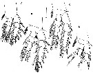
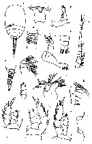
| | | | | | | Syn.: | Misophria maculata Tanaka, 1966 a (p.55, figs.F) | | | | Ref.: | | | Hulsemann & Grice, 1964 (p.259, Descr.F, figs.F); Boxshall & Roe, 1980 (p.23, figs.F,M, juv.); Boxshall, 1982 (p.126, figs.); 1983 (p.121); 1986 (p.520); Chihara & Omori, 1997 (p.932, Pl.191: F,M); Boxshall & Halsey, 2004 (p.217: figs.F,M) | 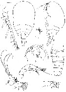 issued from : K. Hulsemann & G.D. Grice in Zool. Anz., 1964, 173 (4). [p.262, Fig. 1-9]. Female (from E Azores Islands): 1-2, habitus (dorsal and lateral, respectively); 3, anterior portion of the head (ventral); 4, last thoracic segment and abdomen (lateral); 5, A1; 6, A2; 7, Md (mandibular blade); 8, Md (mandibular palp); 9, Mx1. Nota: Forehead ending ventrally in a large pointed rostrum. The upper labrum is long and prominent. Head and 1st thoracic segment fused, and together comprise about 2/3 of the cephalothorax. 4th thoracic segment with posterior lateral corner pointed. The 5th thoracic segment forming part of the abdomen. Abdomen 4-segmented; genital segment divided into a slightly widened anterior and a posterior portion; genital lobe bears 2 short setae. Caudal rami longer than wide, of its 6 setae the 2nd and 3rd from inside are the strongest. narrow horizontal \"scales\" cover the surface of the abdominal segments. A1 17-segmented, about as long as the head; aesthetascs present on the segments 4, 6, 8, 11, 15 and 17. A2 biramous, endopod 3-segmented, exopod 5-segmented, about half as long as the endopod. Mandibular palp biramous, endopod 2-segmented, exopod 4-segmented.
|
 issued from : K. Hulsemann & G.D. Grice in Zool. Anz., 1964, 173 (4). [p.263, Fig. 10-15]. Female: 10, Mx2; 11, Mxp; 12- 15, P1 to P4. (P3 and P4 incomplete). Nota: Mx2 has 4 lobes on the 1st basal segment; lobe 5 ends directly in a strong spine; endopod 3-segmented. Mxp has dilated basipods; endopod 3-segmented.
|
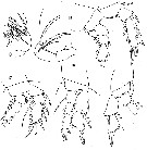
 issued from : G.A. Boxshall & H.S.J. Roe in Bull. Br; Mus. Hist. (Zool.), 1980, 38 (1). [p.29, Fig.11]. Female (from 20'N, 22°W): C, labrum (ventral); E, P5; F, P6; G, habitus (dorsal). Male: A, habitus (lateral); B, urosome (ventral); D, A1 (with setae omitted from proximal portion). Scale bars 0.100 mm unless otherwise indicated.
|
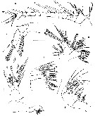 issued from : G.A. Boxshall & H.S.J. Roe in Bull. Br; Mus. Hist. (Zool.), 1980, 38 (1). [p.31, Fig.12]. Female: A, A1; B, A2; C, Md; D, Mx1; E, Mx2; F, Mxp. Scale bars 0.100 mm.
|
 issued from : G.A. Boxshall & H.S.J. Roe in Bull. Br; Mus. Hist. (Zool.), 1980, 38 (1). [p.32, Fig.13]. Female: A-D, P1 to P4. Scale bars 0.100 mm.
|
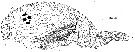 issued from : G.A. Boxshall in Phil. Trans. R. Soc. Lond., 1982, B- 297 (1086). [Fig.29]. Female: Lateral view drawn from serial s.E. micrographs. The major ridges ornamenting the body surface can be seen, and the qtippled areas represent sites of muscle attachments marked externally by the pattern of surface pits. The position of pores in the integument are shown, and A1, reflexed A2 and mandibular palp are drawn in situ.
|
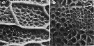 issued from : G.A. Boxshall in Phil. Trans. R. Soc. Lond., 1982, B- 297 (1086). [Plate 3, Figs.30, 31]. 30, Detail of surface ornamentation on cephalosome. The system of ridges delimits areas containing numerous surface pits. Scale bar 0.010 mm. 31, Detail of surface ornamentation on cephalosome marking the site of origin of a dorsal longitudinal trunk muscle fibre by the presence of smaller, densely packed pits. Scale bar 0.010 mm.
|
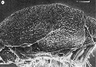 issued from : G.A. Boxshall in Phil. Trans. R. Soc. Lond., 1982, B- 297 (1086). [Plate 4, Fig.33]. 33, Lateral view of cephalosome. The area of cone organs on the side of the body can be seen. Most of the cone organs are intact, still bearing the apical globule of secretion. Scale bar 0.100 mm.
|
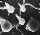 issued from : G.A. Boxshall in Phil. Trans. R. Soc. Lond., 1982, B- 297 (1086). [Plate5, Fig.35] 35, Detail from cone organ area. The cone organs visible show continuous gradation in the size of the globule of secretion. Scale bar 0.002 mm.
|
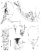
 issued from : O. Tanaka in Sympos. Ser. Mar. Biol. Ass. India, 1966, 1 (2). [p.54, Fig.3]. As Misophria cornuta. Female (from Izu Region): a, habitus (dorsal); b, forehead (frontal view); c, last thoracic segment and urosome (right lateral); d, proximal segments of urosome ( ventral); e, distal segments of urosome (ventral); f, A1; g, A2; h, Md; i, Mx1; j, Mx2; k, Mxp; l, P1; m, P2; n, P3; o, P5. Nota : Body soft-skinned, furnished with fine reticulations except the anal segment and caudal rami. A1 16-segmented, does not extend to the distal margin of the head, the proximal segments are furnished with minute spinules along the posterior margin. Proportional lengths of anterior and posterior regions of the body as 74 to 26. Head and 1st thoracic segment fused. Lateral distal corner of the 4th thoracic segment is pointed at the apex. Proportional lengths of urosmal segments anf caudal rai 19 :19 :17 :13 :12 :10 :10 = 100. The genital opening lies on the ventral distal margin. Caudal rami longer than wide (8 :6). A2 exopod 2-segmented, the distal segment with 2 setae, the remaining 5 segments have each 1 seta on the inner distal margin ; endopod 3-segmented, much longer than the exopod. Md palp large, exopod 5-segmented, endopod 2-segmented, biting gnathobase wit 7 teeth and 1 spine. Mx1 inner lobe with 9 strong spines and a smaller one and 3 setae, 2nd inner lobe with 2 setae, 3rd inner lobe with 2 setae ; endopod small with 5 setae ; exopod moderately large, with 4 terminal and 3 marginalsetae ; the outer lobe carries 5 setae. Mx2 has 5 lobes on the inner margin, 5th lobe has a strong claw and 2 feeble setae. Mxp consists of 2 large basal segments and a small 5-segmented endopod. P5 simple in structure, very small, 1-segmented, and carries a long seta on the apex and a small spine on the inner margin. P6 not observed.
| | | | | NZ: | 4 | | |
|
Carte de distribution de Benthomisophria cornuta par zones géographiques
|
| | | | | | | | | | Loc: | | | Antarct. (SW & E Pacif.), E off Azores, Japan (Izu) | | | | N: | 3 | | | | Lg.: | | | (95) F: 1,39; (96) F: 1,25; (610) F: 1,33-1,18; M: 1,37-1,06; {F: 1,18-1,39; M: 1,06-1,37} | | | | Rem.: | Prélevé à une profondeur supérieure à 4000 m.
Boxshall (1989, p.524) signale cette forme en Indien et en Antarctique (Pacif. E)
Voir aussi les remarques en anglais | | | Dernière mise à jour : 30/12/2014 | |
|
|
 Toute utilisation de ce site pour une publication sera mentionnée avec la référence suivante : Toute utilisation de ce site pour une publication sera mentionnée avec la référence suivante :
Razouls C., Desreumaux N., Kouwenberg J. et de Bovée F., 2005-2026. - Biodiversité des Copépodes planctoniques marins (morphologie, répartition géographique et données biologiques). Sorbonne Université, CNRS. Disponible sur http://copepodes.obs-banyuls.fr [Accédé le 30 mars 2026] © copyright 2005-2026 Sorbonne Université, CNRS
|
|
|
|


;)
;)
;)
;)
;)
;)
;)
;)

