|
|
 |
Fiche d'espèce de Copépode |
|
|
Misophrioida ( Ordre ) |
|
|
|
Speleophriidae ( Famille ) |
|
|
|
Expansophria ( Genre ) |
|
|
| |
Expansophria dimorpha Boxshall & Iliffe, 1987 (F,M) | |
| | | | | | | Ref.: | | | Boxshall & Iliffe, 1987 (p.230, figs.F,M); Boxshall, 1989 (p.524); Huys & Boxshall, 1991 (p.89); Vives & Shmeleva, 2010 (p.144, figs.F,M, Rem.) | 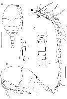 issued from : G.A. Boxshall & T.M. Iliffe in Zool. J. Linn. Soc., 1987, 91. [p.231, Fig.4]. Female (from lanzarote, Canary Islands): A, habitus (dorsal; B, idem (lateral); C, urosome (ventral); D, A1 (ventral); E, P5 (ventral). Scale bars: 0.200 mm (A); 0.100 mm (B); 0.050 mm (C, D); 0.025 mm (E). Nota: Prosome 5-segmented. Lateral margins of dorsal cephalic shield with notch (arrowed in B) at level ofrefexed antennae and mandibular palps. Cone organs not observed in area beneath reflexed appendages (see Benthomisophria palliata. 1st pedigerous somite capable of some distension as it possesses areas of folded, flexible integument anteriorly ( fig. 4B). Some ventrolateral distension also occurs to accomodate lateral cecae of midgut. Posterior margin of maxilliped-bearing somite slightly overlapping flexible area of 1st pedigerous somite (Fig.4A). Nauplius eye absent. Rostrum small, posteroventrally directed, not fused to labrum. Urosome 6-segmented. Caudal rami slightly more than twice as long as wide, armed with 6 setae as in male (Fig.7A). A 26-segmented, reaching to posterior margin of 2nd pedigerous somite. P5 uniramous and 3-segmented, positioned adjacent to ventral midline with bases touching; P6 represented by 1 seta on the plate closing off the genital opening on each side.
|
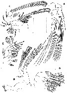 issued from : G.A. Boxshall & T.M. Iliffe in Zool. J. Linn. Soc., 1987, 91. [p.232, Fig.5]. Female: A, A2 (posterior); B, Md (posterior); C, Mx1 (anterior); D, Mx2 (posterior); E, Mxp (posterior). Scale bars = 0.050 mm.
|
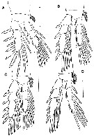 issued from : G.A. Boxshall & T.M. Iliffe in Zool. J. Linn. Soc., 1987, 91. [p.234, Fig.6]. Female: A-D, P1 to P4 (anterior views). Scale bars = 0.050 mm.
|
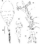 issued from : G.A. Boxshall & T.M. Iliffe in Zool. J. Linn. Soc., 1987, 91. [p.235, Fig.7]. Male: A, habitus (dorsal); B, urosome (lateral); C, A1 (ventral); D, P5 (ventral); Scale bars = 0.050 mm unless otherwise indicated. Nota: Genital somite containing 2 oval spermatophores visible through integument, on either side midline; caudal rami about twice as long as wide, armed with 1 midlateral margin seta, 1 small dorsal seta situated near the distomedial angle, 1 inner and 1 outer distal angle seta and 2 long distal setae, both of which appear to have articulated shafts. Appendages as for female except for A1 and P5 and P6. A1 23-segmented, unigeniculate on both sides with geniculation located between segments XVIII and XIX. P5 uniramous, 5-segmented, positioned adjacent to ventral midline as in female; coxa unarmed, basis with 1 lateral spine; exopod segment 1 unarmed, segment 2 with 1 medial seta, segment 3 with 2 apical spines. P6 represented by the plate closing off the genital opening, armed with 1 long and 1 short spine.
|
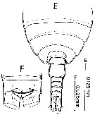 issued from : D. Jaume & G.A. Boxshall in J. Nat. Hist., 1996, 30. [p.1587, Fig.3, E-F]. Male (from Lanzarote): E, habitus, cephalosome excluded (dorsal); F, anal somite and caudal rami (dorsal).
|
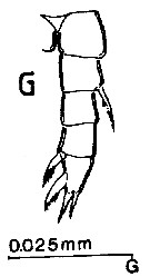 issued from : D. Jaume & G.A. Boxshall in J. Nat. Hist., 1996, 30. [p.1589, Fig.4, G]. Male: G, P5.
| | | | | NZ: | 1 | | |
|
Carte de distribution de Expansophria dimorpha par zones géographiques
|
| | | | Loc: | | | Canary Islands (Jameos del Agua, Lanzarotecave) | | | | N: | 1 | | | | Lg.: | | | (611) F: 0,58; 0,557; M: 0,51-0,45; {F: 0,56-0,58; M: 0,45-0,51} | | | | Rem.: | anchialine cave.
According to Boxshall & Iliffe (1987, p.236) the arrangement of the anterior part of the 1st pedigerous somite allows some limited distension to take place during feeding but the system is not as well developed as in Benthomisophria palliata which is profoundly modified for an opportunistic gorging strategy. Viewed from the side (Fig. 4B) it is apparent that limited ventrolateral distension of the 1st pedigerous somite is also possible. The narrow lip of integument along the posterior margin of the maxilliped-bearing somite may represent a vestige of the typical misophrioid carapace. The specific name is based on the sexual dimorphism exhibited by this species in structure of P5.
| | | Dernière mise à jour : 16/09/2021 | |
|
|
 Toute utilisation de ce site pour une publication sera mentionnée avec la référence suivante : Toute utilisation de ce site pour une publication sera mentionnée avec la référence suivante :
Razouls C., Desreumaux N., Kouwenberg J. et de Bovée F., 2005-2026. - Biodiversité des Copépodes planctoniques marins (morphologie, répartition géographique et données biologiques). Sorbonne Université, CNRS. Disponible sur http://copepodes.obs-banyuls.fr [Accédé le 26 mars 2026] © copyright 2005-2026 Sorbonne Université, CNRS
|
|
|
|


;)
;)
;)
;)
;)
;)
