|
|
 |
Fiche d'espèce de Copépode |
|
|
Misophrioida ( Ordre ) |
|
|
|
Speleophriidae ( Famille ) |
|
|
|
Expansophria ( Genre ) |
|
|
| |
Expansophria galapagensis Boxshall & Iliffe, 1990 (F,M) | |
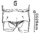
| | | | | | | Syn.: | Expansophria n.sp. Boxshall, 1989 (p.524) | | | | Ref.: | | | Boxshall & Iliffe, 1990 (p.595, figs.F,M); Huys & Boxshall, 1991 (p.89); Boxshall & Halsey, 2004 (p.223: fig.M) | 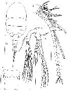 issued from : G.A. Boxshall & T.M. Iliffe in J. Nat. Hist., 1990, 24. [p.596, Fig.1]. Female (from Galapagos Islands): A, habitus (dorsal); B, genital and 1st abdominal somites (ventral); C, A1; D, A2. Scale bars 0.050 mm unless otherwise stated. Nota: Prosome 5-segmented; integument of anterior part of 1st pedigerous somite thin and highly folded, capable of expansion dorsally and laterally. Rostrum broad, posteroventrally directed, free from labrum. Surface of prosome smooth. Urosome 6-segmented; genital and 1st abdominal somites separated by complete suture line but not articulating. Caudal rami just over twice as long as wide. A1 26-segmented. A2 with sympod partly subdivided into coxa and basis, both unarmed; endopod 2-segmented and exopod 8-segmented. P6 represented by plate overlying the genital opening, armed with a long seta.
|
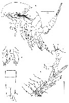
 issued from : G.A. Boxshall & T.M. Iliffe in J. Nat. Hist., 1990, 24. [p.597, Fig.2]. Female: A, labrum and rostrum; B, Md; C, Mx1; D, Mx2; E, Mxp. All scale bars 0.050 mm.
|
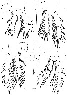 ssued from : G.A. Boxshall & T.M. Iliffe in J. Nat. Hist., 1990, 24. [p.599, Fig.3]. Female: A-D, P1 to P4; E, P5. Scale bars 0.050 mm except E (0.025 mm). P5 uniramous, positioned adjacent to ventral midline, intercoxal sclerite absent; comprising unarmed coxa, basis bearing outer seta, and a 2-segmented exopod
|
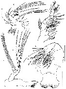
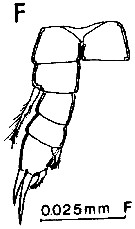
 ssued from : G.A. Boxshall & T.M. Iliffe in J. Nat. Hist., 1990, 24. [p.600, Fig.4]; Male: A, habitus (lateral); B, urosome (ventral); C, A1. Scale bars 0.050 mm exceptt A (0.100 mm). Nota: Genital somite wider than long. Anal somite with row of minute denticles ventrally along posterior margin. Caudal rami and appendages as for female except A1, P5 and P6. A1 24-segmented, geniculate on both sides with geniculation located between segments XIX and XX.
|
 issued from : D. Jaume & G.A. Boxshall in J. Nat. Hist., 1996, 30. [p.1587, Fig.3, G]. Female (from Galapagos Is.): G, anal somite and caudal rami (dorsal).
|
 issued from : D. Jaume & G.A. Boxshall in J. Nat. Hist., 1996, 30. [p.1589, Fig.4, F]. Male: F, P5.
| | | | | NZ: | 1 | | |
|
Carte de distribution de Expansophria galapagensis par zones géographiques
|
| | | | Loc: | | | Galapagos Islands (Santa Cruz Island & Totuga Bay: caves) | | | | N: | 1 | | | | Lg.: | | | (612) F: 0,699-0,624; {F: 0,62-0,70} | | | | Rem.: | From caves.
After Boxshall & Iliffe (1990, p.602) the extensive folding of the integument allows distension of the prosome to take place. The midgut in some specimens was full and visible through the body wall. It has large lateral caeca and is capable of significant swelling. This arrangement was interpreted as an adaptation to an opportunistic gorging strategy (see Boxshall & Iliffe, 1987); The species can readily be distinguished fro E. apoda which lacks P5 in the adult female. It resembles E. dimorpha from the Canaries, very closely. Each of its three species exists on only one oceanic island, one (E dimorpha in the Atlantic, the other two at opposite sides of the Pacific; other anchialine taxa have similar distributions (see Kornicker and Iliffe, 1989 a, b). A fourth has been found in Mediterranean Sea (NW Sardinia) by Jaume & Boxshall (1996a, p.1588. | | | Dernière mise à jour : 05/01/2015 | |
|
|
 Toute utilisation de ce site pour une publication sera mentionnée avec la référence suivante : Toute utilisation de ce site pour une publication sera mentionnée avec la référence suivante :
Razouls C., Desreumaux N., Kouwenberg J. et de Bovée F., 2005-2026. - Biodiversité des Copépodes planctoniques marins (morphologie, répartition géographique et données biologiques). Sorbonne Université, CNRS. Disponible sur http://copepodes.obs-banyuls.fr [Accédé le 18 juin 2026] © copyright 2005-2026 Sorbonne Université, CNRS
|
|
|
|


;)
;)
;)
;)
;)
;)
