|
|
 |
Fiche d'espèce de Copépode |
|
|
Misophrioida ( Ordre ) |
|
|
|
Misophriidae ( Famille ) |
|
|
|
Misophriopsis ( Genre ) |
|
|
| |
Misophriopsis dichotoma Boxshall, 1983 (F) | |
| | | | | | | Ref.: | | | Boxshall, 1983 (p.113, Descr.F, figs.F); Ohtsuka & al., 1992 a (p.871); Martinez Arbizu & Jaume, 1999 (p.109); Humes, 1999 (p.976) | 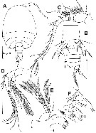 issued from : G.A. Boxshall in Bull. Br. Mus. nat. Hist. (Zool.), 1983, 44 (2). [p.116, Fig.9]. Female (from 34°57'N, 32°55'W): A, habitus (dorsal); B, urosome (ventral); C, A1 (dorsal); D, A2 (anterior); E, Md (posterior); F, detail of mandibular gnathobase. Scale bars 0.100 mm unless otherwise indicated. Nota: Prosome apparently 4-segmented but with 1st free thoracic somite entirely concealed beneath a carapace-like extension from the posterior margin of the maxilliped-bearing somite. Nauplius eye absent. Rostrum small, ventrally directed with its apex adjacent to, but not fused to, the labrum. Cone organs present in lateral areas on either side of cephalosome (see structure and function in Benthomisophria palliata). Urosome 6-segmented. Caudal rami wider than long, armed with 2 long distal margin setae, 1 medium length seta at both inner and outer distal angles, another on the dorsal surface near bases of distal setae, and 1 short lateral seta. A1 18-segmented. A2 with endopod 3-segmented; exopod 6-segmented. md with well developed gnathobase bearing distally 4 mukticusped blades, several strong spines and a small subapical patch of pinnules; mabibular palp comprising basis, 2-segmented endopod and an indistinctly 5-segmented exopod.
|
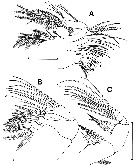 issued from : G.A. Boxshall in Bull. Br. Mus. nat. Hist. (Zool.), 1983, 44 (2). [p.118, Fig.10]. Female: A, Mx1 (posterior); B, Mx2 (anterior); C, Mxp (anterior). Scale bars 0.100 mm. Nota: Mx1 gnathobase with 7 distal margin spines, 2 hirsute setae and 3 naked setae subapically on the posterior surface, and 2 plumose setae on a spinulate swelling on the anterior surface; endite 1 short and slightly furrowed on its posterior surface, armed with 6 apical plumose setae ; endite 2 long, with 3 apical plumose setae; outer lobe rudimentary represented by 8 plumose setae on outer surface of segment; maxillulary palp biramous with 2-segmented endopod and 1-segmented exopod; segment 1 of endopod fused to basis, with junction marked by 2 subapical setae; endopod segment 1 with 4 plumose setae at inner distal angle, segment 2 with 3 naked setae arising proximal to the midpoint of the inner margin, 3 similar setae arising subapically on same margin, and 5 setae on distal margin; exopod with 9 plumose inner and distal margin setae of varying lengths and with fringes of long pinnules proximally. Mx2 6-segmented; segment 1 with 5 plumose setae on proximal endite and 3 on distal endite; segment 2 with 3 plumose setae on both proximal and distal endites; segment 3 produced medially into a curved claw armed with 2 naked setae near its base; segments 4 to 6 with a total of 7 naked setae. Mxp 7-segmented
|
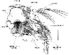 issued from : G.A. Boxshall in Bull. Br. Mus. nat. Hist. (Zool.), 1983, 44 (2). [p.120, Fig.12]. Female: ventral view of mouthparts of the left side (with A1, left paragnath and mandibular gnathobase removed). Scale bar 0.100 mm. Nota: Labrum large, posteriorly directed but not fused with rostrum; armed with 2 large medially directed spinous processes on its posterior margin.
|
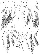 issued from : G.A. Boxshall in Bull. Br. Mus. nat. Hist. (Zool.), 1983, 44 (2). [p.119, Fig.11]. Female: A-D, P1 to P4 (anterior views); E, P5 (anteroventral); F, P6 (ventral). Scale bars 0.100 mm unless otherwise indicated. Nota: P5 biramous, comprising unsegmented protopod, 2-segmented exopod and 1-segmented endopod. P6 with transverse intercoxal sclerite joining members of leg pair reduced to a slender bar; leg comprising an outer process with a long apical seta, a median spine and an inner spinous process.
|
 issued from : P. Martinez Arbizu & D. Jaume in Helgol. Mar. Res., 1999, 53. [p.109, Table 2]. Diagnostic features for identification. exp. 2 = exopod segment 2; exp. 3 = exopod segment 3.
| | | | | NZ: | 1 | | |
|
Carte de distribution de Misophriopsis dichotoma par zones géographiques
|
| | | | Loc: | | | SW off Azores | | | | N: | 1 | | | | Lg.: | | | (606) F: 0,9; {F: 0,90} | | | | Rem.: | hyperbenthique (3000 m).
Voir aussi les remarques en anglais | | | Dernière mise à jour : 20/01/2015 | |
|
|
 Toute utilisation de ce site pour une publication sera mentionnée avec la référence suivante : Toute utilisation de ce site pour une publication sera mentionnée avec la référence suivante :
Razouls C., Desreumaux N., Kouwenberg J. et de Bovée F., 2005-2026. - Biodiversité des Copépodes planctoniques marins (morphologie, répartition géographique et données biologiques). Sorbonne Université, CNRS. Disponible sur http://copepodes.obs-banyuls.fr [Accédé le 30 mars 2026] © copyright 2005-2026 Sorbonne Université, CNRS
|
|
|
|


;)
;)
;)
;)
;)
