|
|
 |
Fiche d'espèce de Copépode |
|
|
Monstrilloida ( Ordre ) |
|
|
|
Monstrillidae ( Famille ) |
|
|
|
Cymbasoma ( Genre ) |
|
|
| |
Cymbasoma bullatum (A. Scott, 1909) (M) | |
| | | | | | | Syn.: | Thaumaleus bullatus A. Scott, 1909 (p.240, figs.M);
Monstrilla leucopsis : Wilson, 1950 (p.267);
Cymbasoma bullatus : Sekiguchi, 1981 (p.13, figs.M); Chihara & Murano, 1997 (p.1002, Pl.234: M) | | | | Ref.: | | | Suarez-Morales, 2001 a (p.58, Redescr.M, figs.M); Suarez-Morales, 2007 a (p.26, figs.M, Rem.); 2011 (p.3, fig.M, p.10); Grygier & Suarez-Morales, 2018 (p.503: Rem.). | 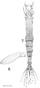 Issued from : A. Scott in The Copepoda of the Siboga Expedition Part I. Siboga-Expeditie XXIX a., 1909. [Pl. LVIII, Figs.9-10]. As Thaumaleus bullatus. Male (from off Laiwui, Paternoster Islands) ): 7, habitus (dorsal); 8, P5. Nota: Cephalic segment considerably inflated at the distal end.. It is distinctly shorter than the posterior part of the body. The frontal margin is broad and rounded with a distinct protuberance in the middle Abdomen 3-segmented. The 2nd segment is shorter and the anal segment is longer than the others.The dial end of the anal segment is expanded. Caudal rami short and broad; each ramus with 4 apical setae distinctly swollen at the base. A1 5-segmented, prehensile, equal to three-fifths of the length of the cephalic segment. P5 rudimentary and without setae.
|
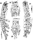 Issued from : E. Suarez-Morales in Bull. Inst. R. Sci. Nat. Belgique, Biologie, 2001, 71. [p.59, Figs.20-23]. Male (from lectotype: coll. Siboga-Expedition, St. 142, Paternoster Islands): 20-21, habitus (lateral and dorsal, respectively); 22, caudal rami (ventral view); 23, sale (dorsal view). Nota: Cephalothorax representing almost 47% of total body length. Oral papilla protruding from ventral surface, located at almost 23% of way back.Dorsal ocelli present, close to anteriormost end of head, pigment cups relatively large, separated by a distance less than an eye diameter; poorly developed, almost unpigmented, rounded in dorsal view. No sensilla observed on anterior part of cephalic region. 1st pedigerous thoracic somite and cephalon fused. Urosome consisting of 4 somites: 5th pedigerous with no appendages, genital somite (genital complex) and 2 free somites (the last is the anal somite). Anal somite being the largest of the urosome, representing almost 40% of the urosome. Caudal rami subquadrate, approximately as long as wide, with 3 terminal and 1 subterminal setae; basal parts of 2 outer setae with rounded swelling (more evident in dorsal view).
|
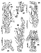 Issued from : E. Suarez-Morales in Bull. Inst. R. Sci. Nat. Belgique, Biologie, 2001, 71. [p.60, Figs.24-29]. Male (paralectotype): 24, right A1 (ventral view: marked using setal nomenclature); 25, right A1 (dorsal view); 26, right A1 (dorsal view); 27, anterior margin of head showing ventral processed (arrowed) (lateral view); 28, 4th pedigerous somite plus urosome, showing genital complex (ventral view); 29, genital complex (ventral view). Nota: Anterior region of head between antennular bases and oral papilla with one pair of horn-like processes (arrowed in fig.27). A1 length of lectotype close to 27% of total body length, 67% as long as cephalothorax. A1 5-segmented, length ratio of segments from 1st to 5th: 15.6 : 23.1 : 13.7 : 26.8 : 20.8 = 100 P5 absent from 5th pedigerous somite. Genital complex represented by cylindrical structure, moderately elongated in lateral view. Pair of relatively long subterminal digitiform genital lappets present on distal 1/3 of genital structure, both appearing elongated, strongly divergent, distally mammiliform; lappets reaching half way of preanal somite (fig.20). Rounded process present at common basal joint of lappets.
|
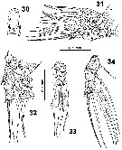 Issued from : E. Suarez-Morales in Bull. Inst. R. Sci. Nat. Belgique, Biologie, 2001, 71. [p.62, Figs.30-34]. Male: 30, intercoxal plate from P4; 31, P3; 32, P1; 33, endopod of P4; 34, exopod of P4.
|
 Issued from : E. Suarez-Morales in Bull. Inst. R. Sci. Nat. Belgique, Biologie, 2001, 71. [p.61]. Male: Armature of swimming legs. Number of setae: Arabic numerals; number of spines: Roman numerals.
|
 Issued from : E. Suarez-Morales in Zootaxa, 2007, 1662. [p.27]. Male (from 0°24'37''S, 127°36'32''E): Armature formula from swimming legs P1 to P4.
|
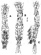 Issued from : E. Suarez-Morales in Zootaxa, 2007, 1662. [p.29, Fig.1]. Male (from 0°24'37''S, 127°36'32''E): A-C, habitus (ventral, dorsal and lateral, respectively).
|
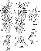 Issued from : E. Suarez-Morales in Zootaxa, 2007, 1662. [p.30, Fig.2]. Male: A, left A1 (dorsal, showing setal nomenclature of Grygier & Ohtsuka, 1995 for segments 1 to 4 and Huys & al., 2007 , for the distal segment); B, right A1 (dorsal, labeled as in A); C, cephalic region (ventral, showing protuberance and adjacent cuticular process); D, cephalic process (lateral view); E, genital lappets (ventral); F, urosome and caudal rami (lateral view, caudal setae cut short).
|
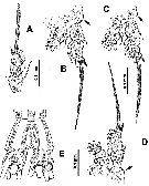 Issued from : E. Suarez-Morales in Zootaxa, 2007, 1662. [p.31, Fig.3]. Male: A, exopod of P1; B, P2; C, P3; D, P4; E, caudal rami (ventral). Sockets of outer basipodal setae arrowed.
| | | | | Ref. compl.: | | | Grygier, 1995 a (p.22, 49, 61) | | | | NZ: | 1 | | |
|
Carte de distribution de Cymbasoma bullatum par zones géographiques
|
| | | | Loc: | | | Indonesia-Malaysia (off Laiwui, Paternoster Is.), Philippines, Japan (Honshu).
Type locality: Paternoster Islands (off Laiwui). | | | | N: | 4 | | | | Lg.: | | | (5) M: 1,7; (726) M: 2,1-1,4; (987) M *: 1,62-1,74) ; {M: 1,40-2,10}
* : total body length (measured in dorsal view from anterior end of cephalothorx to posterior margin of anal somite. | | | | Rem.: | For Suarez-Morales (2007 a, p.32) several differences are apparent between the japanese specimens and C. bullatum. All these differences indicate that the japanese's specimens belong to a different, probably undescribed species of Cymbasoma.
After Suarez-Morales (2010, p.66), the female of M. leucopsis from the Philippines (Wilson, 1950 at the Caldera Bay anchorage is (after inspection of the specimen) a male assignable to Cymbasoma bullatum. The mediterranean records of this species cannot be verified (Rose, 1933; Isaac, 1975: as M. conjunctiva; Djordjevic, 1963). | | | Dernière mise à jour : 15/02/2020 | |
|
|
 Toute utilisation de ce site pour une publication sera mentionnée avec la référence suivante : Toute utilisation de ce site pour une publication sera mentionnée avec la référence suivante :
Razouls C., Desreumaux N., Kouwenberg J. et de Bovée F., 2005-2025. - Biodiversité des Copépodes planctoniques marins (morphologie, répartition géographique et données biologiques). Sorbonne Université, CNRS. Disponible sur http://copepodes.obs-banyuls.fr [Accédé le 04 juin 2025] © copyright 2005-2025 Sorbonne Université, CNRS
|
|
 |
 |


