|
|
 |
Fiche d'espèce de Copépode |
|
|
Monstrilloida ( Ordre ) |
|
|
|
Monstrillidae ( Famille ) |
|
|
|
Cymbasoma ( Genre ) |
|
|
| |
Cymbasoma morii (Tokioka, 1949) (F,M) | |
| | | | | | | Syn.: | Haemocera morii Tokioka, 1949 (p.71, figs.F); Rose, 1956 (p.463), Yamazi, 1958 (p.156, Rem.); Furuhashi, 1965; Razouls, 1982 (p.770);
Cymbasoma longispinosum : Martin Thompson, 1973 (1976) (p.617, figs.F,M, Rem.: Kerala form) | | | | Ref.: | | | Sekiguchi, 1982 (p.28, figs.F); Grygier, 1994 a (p.23, Redescr.F, figs.F); 1995 a (p.72); Chihara & Murano, 1997 (p.1002, Pl.234: F); Suarez-Morales & Palomares-Garcia, 1999 (p.197, tab.1); Suarez-Morales, 2002 (p.95: tab.1); Üstün & al., 2014 (p.1402, 1408 Table I); Grygier & Suarez-Morales, 2018 (p.502: Rem.). | 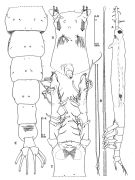 issued from : M.J. Grygier in Hydrobiologia, 1994, 292/293. [p.25, Fig.1]. Female: A, habitus (left lateral view)), most setae omitted; B, genital spines; C, anterior end of body and A1 (dorsal), long setae cut short (cf.Figg.3B); D, anterior end of body and bases of A1 (ventral); E, rear of cephalothorax (thoracomeres III-VI) and urosome (dorsal, showing pores and sensilla, furcal setae cut short (cf. Fig.3A)
|
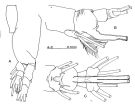 issued from : M.J. Grygier in Hydrobiologia, 1994, 292/293. [p.26, Fig.2]. Female: A, left P1 (anterior view); B-C, thoracomere VI and urosome (left and ventral views, respectively), setae and genital spines cut short.
|
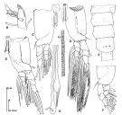 issued from : M.J. Grygier in Hydrobiologia, 1994, 292/293. [p.27, Fig.3]. Female (detailsof females from Sesoko Island, Okinawa): A, distribution of pores and sensilla on rear of cephalothorax and thoracomeres III-VI; B, tip of left A1 (dorsal view); C, left P1 (posterior view); D, left P2 (anterior), with representative setulation; E, right P3 (posterior); F, right P4 (anterior), protopod curt short; G, part of genital spine with frets (intervening striations exaggerated); H, tips of genital spines (including aberrant bifid one).
|
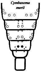 issued from : M.J. Grygier & S. Ohtsuka in Zool. J. Linnean Soc., 2008, 152. [p.499, Fig.29]. Female: Dorsal and lateral pore and pit seta patterns, from rear of cephalothorax through genital compound somite. Symbols: dots (three sizes) = pores; larger circles = pits of pit setae; smaller circles = structures of uncertain type observed by light microscopy . Pattern based on SEM and light microscopical examination.
|
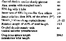 Issued from : Suarez-Morales in Mitt. Mus. Nat.kd. Berl., Zool. Reihe, 2002, 78. [p.95, Table 1]. Comparison of main taxonomic features in species of Cymbasoma with a cephalothorax relative length equal or over 60%, females only: C. morii (Tokioka, 1849). Mouth position way back anterior part of forehead in percentage of the cephalothorax length. Compare with C. bowmani, C. boxshalli, C. longispinosum, C. nicolettae, C. quintanarooense, C. striatus, C. thompsoni, C. tumorifrons, C. sp. (from Brazil), C. chelemense.
|
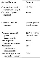 Issued from : F. Üstün, T. Terbiyik Kurt & E. Suarez-Morales in Crustaceana, 2014, 87 (11-12). [p.1403, Table I]. Taxonomic features and morphometry of females of Cymbasoma morii.
|
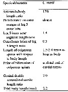 Issued from : F. Üstün, T. Terbiyik Kurt & E. Suarez-Morales in Crustaceana, 2014, 87 (11-12). [p.1404, Table I (continued)]. Taxonomic features and morphometry of females of Cymbasoma morii.
| | | | | NZ: | 3 | | |
|
Carte de distribution de Cymbasoma morii par zones géographiques
|
| | | | | | | | | | Loc: | | | Japan, Tanabe Bay, off Okinawa, Viet-Nam (Nha-Trang), W India (Vizhinjam) | | | | N: | 4 | | | | Lg.: | | | (723) F: 2,9; (783) F: 1,93-3,18; M: 1,21-1,72; (866) F: 1,5-2,9; (868) F: 1,6; {F: 1,50-2,90; M: 1,21-1,72} | | | | Rem.: | According to Yamazi (1958, p.156) the species is very rare, in summer, in the Tanabe Bay (Japan).
This form belongs to Cymbasoma longispinosum species-group. | | | Dernière mise à jour : 15/02/2020 | |
|
|
 Toute utilisation de ce site pour une publication sera mentionnée avec la référence suivante : Toute utilisation de ce site pour une publication sera mentionnée avec la référence suivante :
Razouls C., Desreumaux N., Kouwenberg J. et de Bovée F., 2005-2026. - Biodiversité des Copépodes planctoniques marins (morphologie, répartition géographique et données biologiques). Sorbonne Université, CNRS. Disponible sur http://copepodes.obs-banyuls.fr [Accédé le 26 mars 2026] © copyright 2005-2026 Sorbonne Université, CNRS
|
|
|
|


;)
;)
;)
;)
;)
;)
;)

