|
|
 |
Fiche d'espèce de Copépode |
|
|
Monstrilloida ( Ordre ) |
|
|
|
Monstrillidae ( Famille ) |
|
|
|
Cymbasoma ( Genre ) |
|
|
| |
Cymbasoma tumorifrons Suarez-Morales, 1999 (F) | |
| | | | | | | Syn.: | No Thaumaleus tumorifrons Isaac, 1974; 1975 (p.3, 7, 9, figs.F,M);
No Cymbasoma tumorifrons : Suarez-Morales & Alvarez-Silva, 2001 (p.184, figs.F, Rem.) | | | | Ref.: | | | Suarez-Morales, 1999 (p.67, Redescr. M, figs.M, Rem.); Suarez-Morales, 2002 (p.90, 95, Redescr.F, figs.F); Suarez-Morales & Morales-Ramirez, 2003 (p.210: Table 2); Vives & Shmeleva, 2010 (p.169, figs.F,M, Rem.); Grygier & Suarez-Morales, 2018 (p497, 501: Rem.) |  issued from : E. Suarez-Morales & C. Alvarez-Silva in Pacific Sc., 2001, 55 (2). [p.188, Table1]. Comparison of Type Specimen of Cymbasoma tumorifrons (Isaac) with the Mexican Specimen C. guerrerense
|
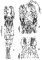
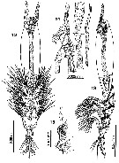 Issued from : Suarez-Morales in Mitt. Mus. Nat.kd. Berl., Zool. Reihe, 2002, 78. [p.92, Figs.12-15]. Female (from 43°5'N, 6°0'E): 12-13, habitus (ventral and lateral, respectively); 14, 5th pedigerous and genital somites (lateral); 15, oral papilla and adjacent cuticular processes (lateral).
|
 Issued from : Suarez-Morales in Mitt. Mus. Nat.kd. Berl., Zool. Reihe, 2002, 78. [p.94, Figs.16-21]. Female: 16, right A1 (dorsal view); 17, 5th pedigerous and genital somites; 18, terminal exopodal segment of P1; 19, same of P3; 20, anal somite and caudal rami (dorsal); 21, distal part of ovigerous spines (ventral view).
|
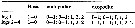 Issued from : Suarez-Morales in Mitt. Mus. Nat.kd. Berl., Zool. Reihe, 2002, 78. [p.93]. Female: Setae (Arabic numerals) and spines (Roman numerals) formula of swimming legs P1 to P4.
|
 Issued from : Suarez-Morales in Mitt. Mus. Nat.kd. Berl., Zool. Reihe, 2002, 78. [p.95, Table 1]. Comparison of main taxonomic features in species of Cymbasoma with a cephalothorax relative length equal or over 60%, females only: C. tumorifrons (Isaac, 1975). Mouth position way back anterior part of forehead in percentage of the cephalothorax length. Compare with C. bowmani, C. boxshalli, C. longispinosum, C. morii, C. nicolettae, C. quintanarooense, C. striatus, C. thompsoni, C. sp. (from Brazil), C. chelemense.
|
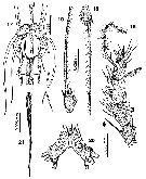
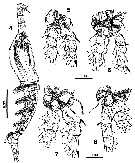
 Issued from : E. Suarez-Morales in Arthropoda Selecta, 1999, 8 (1). [p.68, Figs.1-3]. Male (from Aegean Sea): 1, habitus (dorsal); 2, forhead and A1 (dorsal); 3, genital lappets (ventral). Scale bars in mm. Nota: - Oral papilla located 27% of way back along ventral surface of cephalothorax.
- A1 relatively short and strong, 5-segmented, representing almost 39% of total body length, and ca. 80% as long as cephalothorax.
- The genital lappets are different in in the specimens examined when compared with Isaac's drawings. They were depicted as having an extremely slender distal end, with seta-like terminal projection. These structures have a terminal rounded chitinous process. Howeyver, it remains clear that this is probably one of the most acute male genital lappets in the genus.
- P5 absent (as usual in males).
- Urosome 4-segmented.
- Ratio of lengths of genital doube-somite and following two free somites 31.7 : 23.8 : 44.3 = 100.
- Anal somite with a medial constriction, apparently vestige of intersegmental division.
- Caudal rami quadrate, with terminal margin as wide as proximal one; approximately 1.1 times longer than wide, bearing 4 well-developed terminal setae.
|
 Issued from : E. Suarez-Morales in Arthropoda Selecta, 1999, 8 (1). [p.70, Figs.4-8]. Male: 4, habitus (lateral); 5, P1; 6, P2; 7, P3; 8, P4 (posterior). Scale bars in mm.
|
 Issued from : E. Suarez-Morales in Arthropoda Selecta, 1999, 8 (1). [p.69]. Male: Setae (Arabic numerals) and spines (Roman numerals) formula of swimming legs P1 to P4.
|
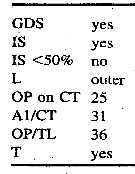 Issued from : E. Suarez-Morales & A. Morales-Ramirez in Proc. Biol. Soc. Washington, 2003, 116 (1). [p.210, Table 2]. Selected taxonomic features in Cymbasoma tumorifrons. GDS = anterior half genital double-somite globose; IS = inner seta of P5 smallest; IS <50% = inner seta <50% as long as others; L = longest seta on P5; OP on CT = position of oral papilla on cephalothorax; AI/CT = relative length (as a percent) of A1 to cephalothorax; = relative length (as a percent) of position of oral papilla anteriorly on cephalothorax; T = transverse striations on ''neck'' area. Compare with species of Cymbasoma with uniramous P5 armed with 3 setae: C. concepcionae, C. reticulatum, C. bowmani, C. quintanarooense, C. boxshalli, C. bali, C. frondipes, C. striatum, C. claparedi.
| | | | | Ref. compl.: | | | Grygier, 1995 a (p.77) | | | | NZ: | 1 | | |
|
Carte de distribution de Cymbasoma tumorifrons par zones géographiques
|
| | | | | | | Loc: | | | Medit. (Toulon) | | | | N: | 4 | | | | Lg.: | | | (736) F: 1,03-0,76; M: 0,95-0,69; (829) M: 0,93-0,7; (876) F: 0,76-1,03; {F: 0,76-1,03; M: 0,69-0,95} | | | | Rem.: | For Suarez-Morales & Alvarez-Silva (2001, p.187) the main difference between the type specimens and the Mexican specimen is the size; this is not a factor with strong taxonomic value, hence, the size of the Mexican female of this species merely increases the known range of size variation for C. tumorifrons; the slight differences between the Mediterranean and Mexican specimens are not sufficient to warrant erection of a new taxon (see Table 1). Isaac (1974) assumed that female and male belonged to a single species; however, this may have been misleading because the only way to link males and females of one monstrilloid species is to find them in the same host or verify that they share distinctive diagnostic characters ; morphologic characters that may link both sexes include striation of the head ((see in Suarez-Morales & Escamilla, 1997) and the general structure of the eyes; another feature whose value to link sexes is the branching pattern of Grygier & Ohtsuka's (1995) distal setae of the distal segment . A1 (setae b1-b-3); these are distaly branched in male C. tumorifrons (Suarez-Morales, 1999) and unbranched in the assigned female (Isaac, 1974). Althrough the Mexican female seems to represent C. tumorifrons, there is the possibility that it belongs to another species. In 2009, Suarez-Morales & Morales-Ramirez (2009, p.1264) ceate Cymbasoma guerrerense species.
After Suarez-Morales & al. (2017, p.1817) this new species was originally described as C. tumorifrons by Isaac (1974) from Emborios Bay (Aegean Sea). He determined that the males and females collected belonged to the same species, based on its co-occurrence in the same sample. In the redescription of the males, Suarez-Morales (1999) argued that the name, published by Isaac (1974) was, nomenclaturally unavailable and it was explicity deemed as a nomen nudum there. Cymbasoma tumorifrons have to be attributed to Suarez-Morales (1999). This name, however, exclusively refers to the male type specimens, thus excluding the females mentioned by Isaac (1974). The adult females designated as allotype and paratypes by Isaac (1974) are not part of the type specimens of C. tumorifrons, and, thus, represent an undescribed species. A redescription of these female specimens was published by Suarez-Morales (2002), who art that time attributed these females as C. tumorifrons based on Isaac's type specimens, plus a female found in Toulon Bay. A comparison of the original illustrations by Isaac (1974) and Suarez-Morales (2002) redescription of these female specimens from the Aegean Sea and Toulon Bay allowed us to determine that our specimen from Trieste clearly belongs to the C. mediterranea.
Cf Table 2 from Lian & Tan (2019) in C. cheni. | | | Dernière mise à jour : 12/02/2020 | |
|
|
 Toute utilisation de ce site pour une publication sera mentionnée avec la référence suivante : Toute utilisation de ce site pour une publication sera mentionnée avec la référence suivante :
Razouls C., Desreumaux N., Kouwenberg J. et de Bovée F., 2005-2026. - Biodiversité des Copépodes planctoniques marins (morphologie, répartition géographique et données biologiques). Sorbonne Université, CNRS. Disponible sur http://copepodes.obs-banyuls.fr [Accédé le 28 mai 2026] © copyright 2005-2026 Sorbonne Université, CNRS
|
|
|
|


;)
;)
;)
;)
;)
;)

