|
|
 |
Fiche d'espèce de Copépode |
|
|
Monstrilloida ( Ordre ) |
|
|
|
Monstrillidae ( Famille ) |
|
|
|
Maemonstrilla ( Genre ) |
|
|
| |
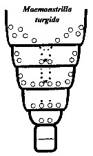
Maemonstrilla turgida (A. Scott, 1909) (F) | |
| | | | | | | Syn.: | Monstrilla turgida A. Scott, 1909 (p.239, Descr.F, figs.F); Martin Thompson & Meiyappan, 1977 (1980) (p.207, figs.F, Rem.); Grygier, 1995 a (p.77); Suarez-Morales & Dias, 2001 (p.226: Rem.)
? Monstrilla sp. Krishnaswamy, 1953 (p.75, figs.) | | | | Ref.: | | | Grygier & Ohtsuka, 2008 (p.492, figs.F, Rem.: comb. nov.) | 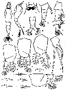 issued from : P.K. Martin Thompson & M.M. Meiyappan in Indian J. Fish., 1977 (1980), 24 (1-2). [p.207, Fig.1]. As Monstrilla turgida. Female (from Minicoy Island, Indian Ocean): a-b, habitus (dorsal and lateral, respectively); c, forehead (dorsal, enlarged); d-e, urosome (dorsal and lateral, respectively; enlarged); f, A1; g, P1; h, P2; i, P3; j, P4; k, P5. Nota: - Ratio of cephalothorax : rest of body 49.21 : 50.79. - Urosome 3-segmented, markedly short, about 1/7 total length. - Genital segment sub-quadrangular in shape and transversed at its mid-length by a distinct suture; longer than the combined length of following two segments; - A pair of ovigerous spines present on ventral side which is about one-and-half times length of urosome; - 3rd urosomal segment broader distally. - Caudal rami longer than broad (ratio 58.82:41.18, each with 1 outer marginal seta and 5 sub-equal apical setae. - Mouth placed near anterior region of cephalosome. - Eyes well developed. - Proportional lengths of various segments of body (caudal rami included) 49.21; 9.95; 12.05; 7.85; 6.28; 5.24; 2.09; 2.62; 4.71 = 100. - A1 4-segmented, attains about 1/3 length of cephalosome, last segment longest, but shorter than combined length of first three segments; proportional lengths 23.66; 17.74; 16.67; 41.93 = 100.
|
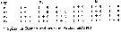 issued from : P.K. Martin Thompson & M.M. Meiyappan in Indian J. Fish., 1977 (1980), 24 (1-2). [p.208]. As Monstrilla turgida. Female: Setae and spines on three segments of exopodite (Re) and endopodite (Ri).
|
 issued from : P.K. Martin Thompson & M.M. Meiyappan in Indian J. Fish., 1977 (1980), 24 (1-2). [p.209]. As Monstrilla turgidaCharacters comparison between specimens fro Pacific Ocean and Minicoy Island.
|
 issued from : M.J. Grygier & S. Ohtsuka in Zool. J. Linnean Soc., 2008, 152. [p.494, Fig.24]. Female (from Ryukyu Islands): A, habitus (ventral); B, left A1 (ventral; setules omitted; setal designations after Grygier & Ohtsuka, 1995, fig.6); C, urosome (ventral; ovigerous spines and most setae cut short, some representative setules shown on furcal setae, copulatory pore arrowed). Scale bars = 0.500 mm in A; 0.0100 mm in B, C. Nota female: - Bulbous cephalothorax much the same as in M. hyottoko, but considerably larger and with relatively longer ovigerous spines. - Measurements taken as defined in figure 1 (see M. polka): Length in lateral view (sum of lengths of cephalothorax, metasome and urosome) 49.4-54.8, 26.1-30.4 and 17.2-20.8 %, respectively (this sum exceeding body length as seen in dorsal view by 2.4-8.7 %. - Height and greatest width of cephalothorax respectively, 40.9-57.9 and 41.9-57.9 of cephalothorax length; in several specimens height and width equal. - A1 with non-articulating boundary between 3rd and 4th segments, suggested by constriction; 1st and 2nd segments distinct. Length 41.6-51.3 % that of cephalothorax. - Width of incorporated pediger 68.4-88.5 % of greatest width, widths of succeeding three free pedigers and genital compound somite relative to that of incorporated pediger 76.1-89.1, 64.8-77.2, 53.8-63.5 and 30.7-40.3 %, respectively. - Ovigerous spines 45.1-56.1 % as long as body in dorsal view (7 pairs measured). - Cuticular reticulation completely absent, but cephalothorax with faint but dense circular striae; scattered minute dots also seen by phase contrast. - Oral papilla long, straight, conical, directed ventrally. Pair of closely spaced pores just in front of it.; another pair, further apart, at level of posterior bases of A1. 2 more pairs of pores, even further apart, behind oral papilla. - 3 or 4 small scars behind base of each A1, middle one(s) smallest, anterior and posterior ones surrounded by radiating ridges. - Pair of hair-like sensilla on forehead; dorsal to them, symmetrical pattern of about 9 small pores and dorsolateral pair of larger pores; apparently no wrinkled pit that of M. hyottoko.
- 3 cups of naupliar eye large, equal in size.
- A1 with non-articulating boundary between 3rd and 4th segments, suggested by constriction; 1st and 2nd segments distinct. Full setation probably present, but 1 or 2 setae missing on each antennule observed.
- Spines extending to halfway between P1 and oral papilla, slender and cylindrical with taperd tips (figs.13B, 24A).
- Caudal rami with ventral pore and 6 setae distributed as in M. hyottoko; short dorsal seta usually simple, but sometimes with distal setules; other( setae biserially plumose and medial one longest.
|
 issued from : M.J. Grygier & S. Ohtsuka in Zool. J. Linnean Soc., 2008, 152. [p.462, Fig.1, A, B]. Measurements taken from specimens in species of Maemonstrilla: Lengths in lateral view from example M. polka (A) and in dorsal view (example M. turgida (B).
|
 issued from : M.J. Grygier & S. Ohtsuka in Zool. J. Linnean Soc., 2008, 152. [p.497, Fig.27]. Female: A, P1 (posterior); B, intercoxal sclerite of P1 (anterior); C, right P2 (anterior); D, left P3 (posterior); E, outer apical exopodal seta of left P3; F, right P4 (anterior); G, right P5 (medial view); H, compound genital somite (lateral, anterior to left) showing prominence at anterior base of ovigerous spines (latter cut short). In A, C, D, F and G, setules omitted, some setae cut short. Scale bars = 0.200 mm (A-D, F); 0.100 mm in E, G, H. Nota: Full plesiomorphic setation of legs P1-P4 present as outlined by Huys & Boxshall (1991), including inner seta of 1st endopodal segment and weak inner seta of 1st exopodal segment (fig.27A, C, D, F). Outer basis seta short and hair-like in P1, 2 and 4, longer and plumose in P3 but shorter than exopod; pore anterior to this seta in each leg. Tiny pore on anterior face of 3rd segment of each ramus. Outer spiniform setae of exopod simple, that on 3rd segment a little longer than that on 1st. Outer side of outer apical exopodal seta lined with closely adjacent, bluntly conical denticles (figs. 25G, 27E). - P5 with wide exopodal lobe bearing 2 apical setae and 21 subapical outer seta; narrower but longer endopodal lobe with 1 apical seta; all setae biserially plumose, outer 2 exopodal setae longer than others (figs.24C, 27G). - Round copulotary pore on midline within crescent-shaped slot at posterior base of conical process bearing ovigerous spines (fig.24C).
|
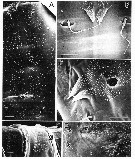
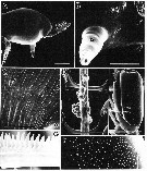 issued from : M.J. Grygier & S. Ohtsuka in Zool. J. Linnean Soc., 2008, 152. [p.495, Fig.25]. SEM. Female: A, cephalothorax (lateral); B, oral papilla (o), scars, pores (arrows) and base of A1 (a); C, striations of cephalothorax and 2 left-hand pores (arrows) behind oral papilla; D, ovigerous spines with attached eggs overlying two intercoxal sclerites; E, protopod of P4 (lateral), showing pathches of denticles, arrows bounding region enlarged in F; F, detail of E; G, detail of outer apical exopodal seta of P2. Scale bars = 0.200 mm (B, D, E); 0.005 mm in C; 0.010 mm (F, G).
|
 issued from : M.J. Grygier & S. Ohtsuka in Zool. J. Linnean Soc., 2008, 152. [p.496, Fig.26]. SEM. Female: A, dorsal spinulation and pores (arrows) of 1st free pediger (anterior at top); B, spiniform scales and pit setae at centre rear of dorsum of free pediger 1 (posterior at bottom); C, pore, spiniform scales, and pit setae at centre rear of dorsum of free pediger 2 (posterior at bottom left); D, right dorsal spinulation, pore and pit setae of free pediger 3 (anterior at right); E, spinulation of genital compound somite (g, letter placed on this segment's dorsal suture and penultimate segment of urosome (dorsolateral), anterior at right. Scale bars = 0.010 mm.
|
 issued from : M.J. Grygier & S. Ohtsuka in Zool. J. Linnean Soc., 2008, 152. [p.499, Fig.29]. Female: Dorsal and lateral pore and pit seta patterns, from rear of cephalothorax through genital compound somite. Symbols: dots (three sizes) = pores; larger circles = pits of pit setae; arrowheads = spiniform scales. Pattern based on SEM and light microscopical examination.
|
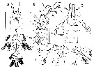
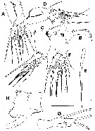
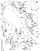 issued from : M.J. Grygier & S. Ohtsuka in Zool. J. Linnean Soc., 2008, 152. [p.498, Fig.28]. Female (Syntype, 'Siboga' Expedition, Sta. 142): A, habitus (lateral); B, right A1 (ventral view; setal designation after Grygier & Ohtsuka, 1995, fig.6), dotted 4v3-seta missing, but drawn in based on its presence in left antennule; C, right P2 (endopod); D, loose, broken-off exopod of P2, 3 or 4; E, rami of left P3 (above) and P4 (below (anterior view); F, basis and rami of right P3 (anterior view); G, rami of right P4 (anterior view); H, P5 and genital compound somite (lateral)I, telson* and caudal rami (lateral). Scale bars = 0.500 mm (A); 0.100 mm (B); 0.200 mm in C-I. * Telson: Terminal part of the body having its mesoderm derived directly from the teloblasts after budding of the metamers has ceased. Represented by the anal somite, terminal 'segment' of the body, bearing the anus either terminally or dorsally, somitic complex incorporating abdominal somite plus telson (Huys & Boxshall, 1991, Appendix 3, p.455, 459).
|
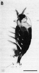 issued from : M.J. Grygier & S. Ohtsuka in Zool. J. Linnean Soc., 2008, 152. [p.477, Fig.13, B]. B, ovigerous female (neotype from Sesoko Island). photomicrographs of preserved specimen. Scale bar = 0.500 mm. Nota For the authors the unusual direction of the ovigerous spines suggests that species of Maemonstrilla practise subthoracic brooding. Although it is conceivable that the spines point forward only after the final moult and bend backwards to the 'normal' position when the eggs are deposited, several exemples refute this. A few eggs (about 0.023 mm in diameter) were found attached to the anteriorly pointing spines of two specimens (see fig.25D), and there were two large clusters (part of a single larger original mass ?) on the spines of another specimen of this species. A contrario all three specimens of M. polka bore very large egg masses held beneath and matching in length, the cephalothorax and metasome. M/ okame also were carrying eggs beneath the thorax.
|
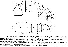
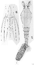 Issued from : A. Scott in The Copepoda of the Siboga Expedition Part I. Siboga-Expeditie XXIX a., 1909. [Pl. LVIII, Figs.5-6]. As Monstrilla turgida. Female (from off Laiwui, coast of Obi Major): 5, habitus (dorsal); 6, P5. Nota: Cephalic segment greatly inflated just behind the frontal margin. It is slightly longer than the posterior part of the body. Frontal margin broad and boldly rounded. Abdomen 3-segmented. Genital segment sub-quadrangular in shape and traversed near the middle by a distinct suture. It is one and half times longer than the combined length of the next two segments. The segment is nearly as long as the united length of the next two segments and caudal rami. The 2nd and 3rd segments sub-equal in length. Caudal rami slightly longer than broad and about one and a half times longer than the anal segment; each ramus with 1 outer marginal seta and 5 apical setae. The apical setae sub-equal in length. A1 4-segmented and equal to one-third of the length of the cephalic segment. P5 moderately long and rather slender. The apex bi-lobed; inner lobe narrower than outer lobe and extends beyond the apex of it. It is furnished with 1 seta. The outer lobe with 3 setae.
|
 Issued from : M.J. Grygier & S. Ohtsuka in Zool. J. Linnean Soc., 2008, 152. [p.492]. Maemonstrilla turgida Female (from Rtukyuan specimens): - Cephalothorax non-reticulated, but with faint encircling striations. Cuticle appearing smooth by light microscopy, but patches of minute spinules revealed by SEM dorsally on metasomal pedigers and all urosomal segments except telson, and on outer face of coxa of P1-P4. - Pair of spinulose, spine-like scales posteriorly near dorsal midline on both 1st and 2nd free pedigers. - Outer basis seta of P3 shorter than exopod. - Inner seta present on 1st segment of each ramus of P1-P4. - P5 bilobed, its exopodal lobe with 3 setae and endopodal lobe with 1 seta (perhaps up to 2 setae). - No ventral protrusion of posterior part of genital compound.
| | | | | NZ: | 3 | | |
|
Carte de distribution de Maemonstrilla turgida par zones géographiques
|
| | | | | | | | | | Loc: | | | Indian (Lagoon Minicoy Is.), Indonesia-Malaysia (Ceram Sea, coast of Orbi Major), Souhth China Sea, S Japan (Sesoko Islands, Ishigaki Is.)
Type locality: 0°24' S, 127°36' E. | | | | N: | 3 | | | | Lg.: | | | (5) F: 2; (781) F: 2,42-2,26; (1077) * F: 1,38-2,21; {F: 2,00-2,42}
* sum of lengths of cephalothorax, metasome and urosome in lateral view. | | | | Rem.: | After Martin Thompson & Meiyappan (1977) the slight variations noted in the specimens described from Minicoy compared to the type material may be due to the geographical variations in the species.
For Grygier & Ohtsuka (2008, p.500) the unidentified Monstrilla figured by Krishnaswamy (1953) from Kundugal Channel (India) seems to be a female of Maemonstrilla turgida or a similar species.
Grygier & Ohtsuka judged that M. pustulata Suarez-Morales & Dias, 2001 from Brazil shows 'some affinities' to M. turgida, i.e. a similar cephalothoracic shape and a bilobed female P5 with 3 + 1 setation; but they notedseveral differences, especially the opposed direction of the ovigerous spines, and did not press a case for a phyletic connection. | | | Dernière mise à jour : 24/04/2020 | |
|
|
 Toute utilisation de ce site pour une publication sera mentionnée avec la référence suivante : Toute utilisation de ce site pour une publication sera mentionnée avec la référence suivante :
Razouls C., Desreumaux N., Kouwenberg J. et de Bovée F., 2005-2025. - Biodiversité des Copépodes planctoniques marins (morphologie, répartition géographique et données biologiques). Sorbonne Université, CNRS. Disponible sur http://copepodes.obs-banyuls.fr [Accédé le 17 août 2025] © copyright 2005-2025 Sorbonne Université, CNRS
|
|
|
|


;)
;)
;)
;)
;)
;)
;)
;)
;)
;)

