Issued from : C.C. Davis
in Trans. am. microsc. Soc., 1949, 68 (3). [p.253, Fig. 1-9]. As
Monstrilla reticulata.
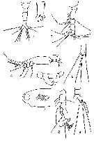
Female (from Biscayne Bay, Florida): 1, urosome (dorsal view); 3, left A1; 4, habitus (lateral view); 6, P5; 7, portion of the cephalic segment showing reticulation (lateral view).
Male (from Biscayne Bay): 2, genital segment showing genital protuberance and spermatophore (ventral view); 5, urosome (lateral view); 8, A1 (ventral view); 9, urosome, posterior portion with furcal ramus.
Nota: 1 female found off Coconut Grove (March 10, 1948); all others individuals obtained off Chicken Key in Biscayne Bay: 1 ripe female (December 13, 1947; 1 female (January 16, 1948); 2 females and 1 male (March 30, 1948); 1 female, 1 ripe, 2 females carrying eggs and 1 male (April 6, 1948); 1 female (April 13, 1948); 1 female (April 27, 1948); 1 female (May 4, 1948; 2 females and 1 male (June 1, 1948); 12 females and 1 male 'June 8, 1948).
Female:
- Colorless, transparent or translucent for the most paert, however, the distal 3/4 of A1, the area around the eyes, that between the segments of the metasome, ventral surface of urosome posterior to ovigerous spines, furcal rami, were blackish-brown, while eyes themselves reddish-black.
- Cephalic segment approximately same length as the remainder body, and its surface is finely reticulate (reticulation sometimes difficult to ascertain).
- Oral papilla very prominent, located about 1/5 of the distance from the anterior margin.
- 2 dorsal eyes and 1 ventral, prominent.
- On the anterior margin of the cephalic segment, between bases of A1, frontal organ prominent.
- Cephalic segment very clear and transparent in all except 2 specimens, in these two the entire cephalic segment filled with a large number of eggs.
- The four thoracic segments (2nd, 3rd, 4th and 5th) posterior to the cephalic segment subequal in length, but progressively narrower posteriorly.
- Genital segment about twice as long as the segments that precede and follow it, and it is relatively slightly swollen ventrally, on the anterior half of the segment. It bears the ventral ovigerous spines, closely appressed or slightly fused together near their bases.
- Ovigerous spines extend only slightly beyond the furcal rami.
Anal segment very short, and fused with the furcal rami dorsally and ventrally, being clearly set off only laterally.
- A1 consist of 4 clear segments. Antennal seta not well developed, and none of them dichotomously branched.
- P5 short and thick, with an inner lobe bearing a single terminal seta, and an outer lobe bearing 3 terminal setae; the two inner of these arise very close together.
- Furcal rami somewhat longer than broad. Setae very fine, the largest is borne on the outer margin, while terminally there is a group of 3, borne close together near the inner corner, and a 4th on the outer corner.
Male:
-Cephalic segment opaque and without obvious reticulation
- Genital segment with genital protuberance, very prominent, wkith relatively small, unornamented genital lappets.
- There are 3 segments between the genital segment and the furcal rami, with as in the female, the anal segment very short and fused dorsally and ventrally with the furcal rami.
- Furcal rami bear 1 strong seta on outer margin and 3 terminal setae, outermost of which is weaker and shorter than the rest.
A1: 5-segmented, geniculate, with a rather strong hooked spine and 3 dichotomously branced setae on terminal segment.
- P5 lacking.


;)
;)

 Toute utilisation de ce site pour une publication sera mentionnée avec la référence suivante :
