|
|
 |
Fiche d'espèce de Copépode |
|
|
Cyclopoida ( Ordre ) |
|
|
|
Oncaeidae ( Famille ) |
|
|
|
Oncaea ( Genre ) |
|
|
| |
Oncaea waldemari Bersano & Boxshall, 1994 (F,M) | |
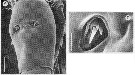
| | | | | | | Syn.: | Oncaea media minor: Malt, 1982 a | | | | Ref.: | | | Bersano & Boxshall, 1996 ["1994"] (p.30, figs.F,M); Boxshall, 1998 (p.227); Böttger-Schnack, 2001 (p.57, 70, figs.F,M, juv.5, Rem.); Böttger-Schnack & al., 2004 (p.1130, tab.1, Rem.); Wi & al., 2009 (p.110, figs.F, Rem.); Vives & Shmeleva, 2010 (p.326, figs.F,M, Rem.); Böttger-Schnack & Machida, 2011 (p.111, Table 1, 2, fig.2, 3, DNA sequences, phylogeny); Böttger-Schnack & Schnack, 2013 (p.4: Table 1, Rem.: s.str-Group) | 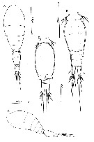 issued from : J.G.F. Bersano & G.A. Boxshall in Nauplius, Rio Grande, 1994, 2. [p.31, Fig.1]. Female (from 31°40'-33°45'S, 51°00'-52°20'W): A, habitus (dorsal); B, urosome (dorsal); C, habitus (lateral). Nota : Ratio Prosome/urosome length 1.7 : 1. Entire surface of dorsal cephalic shield pitted and ridged ; ornamented with sensillae and integumental pores of varying sizes. Rostrum with pair of lateral sensillae. Proportional lengths (%) of urosome somites and caudal rami 12 :49 :6 :8 :10 :15. Genital double-somite 1.4 times longer than wide, greatest width anterior to mid-level, lateral margins smoothly rounded ; ornamented with 3 pairs of integumental pores on dorsal surface, 2 anterior to gonopores, 1 posterior. Paired gonopores located about 35 % of distance along genital double-somite ; each gonopore closed by operculum formed by sixth leg, armed with 3 spinous processes. Anal somite 2/3 as long as caudal rami ; about 1.5 times wider than long, ornamented with pair of small and pair of large integumental pores dorsally, to each side of anal cleft, and pairs of pores on lateral and ventral surfaces. Anal operculum weakly developed, ornamented with minute denticles along free posterior margin ; pair of sensilla located lateral to operculum. Caudal rami 2.6 times longer than wide ; lateral seta (II) located about 38 % of distance along ramus ; dorsal seta VII just shorter than innermost distal seta (V), outermost seta III spiniform, distal major setae IV and V plumose, seta V about 27 % longer than seta IV. P5 with exopodal segment fused to somite, not separate basally, forming short exopodal lobe. Outer protopodal seta short, located on surface of somite dorsal to origin of exopodal lobe. Exopodal lobe with 2 apical setae. P6 represented by plate closing gonopores ; armed with 3 spinous processes.
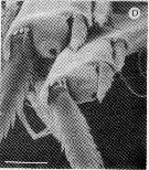
Male: D, urosome (ventral). Urosome 6 segmented comprising 5th pedigerous somite, large genital somite, plus 4 free abdominal somites. Genital somite nearly twice as long as wide, with evenly rounded convex lateral margins. 1st to 3rd abdominal somites all very short, subequal in length. Anal somite about twice as wide as long; ornamented with pair of sensillae and 3 pairs of integumental pores (as in female). Caudal rami about 1.2 times longer than anal somite; about 1.7 times longer than greatest width. Setation similar to that of female but with lateral seta (II) located just proximal to mid point of lateral margin. Appendages as for female except A1, Mxp and P6. P5 as for female. P6 represented by large, unarmed opercular plates (= genital lappets) closing off paired gonopores at posterior margin of genital somite (see Fig.1 D)
|
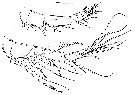 issued from : J.G.F. Bersano & G.A. Boxshall in Nauplius, Rio Grande, 1994, 2. [p.32, Fig.2]. Female: A, A1. Nota : A1 6-segmented ; relative lengths of segments (%) measured along posterior non-setigerous margin 8 : 26 :41 :10 :4 :11. Setation formula 3, 8, 4, 2 +1 aesthetasc, 2 +1 aesthetasc, 6 + aesthetasc. Aesthetasc on segment 4 originating in large depression in surface. Male: B, A1 (with long setation elements drawn cut off). Nota: A1 4-segmented, apical segment representing fused distal 3 segments of female. Relative lengths of segments (measured along posterior non-setigerous margin) 7: 25:42:26. Segmental setation pattern: 3, 7, 5, 11 + 3 aesthetascs
|
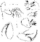 issued from : J.G.F. Bersano & G.A. Boxshall in Nauplius, Rio Grande, 1994, 2. [p.33, Fig.3]. Female: A, A2; B, Md (with inset detail of distal margin elements); C, Mx1; D, Mx2; E, Mxp (syncoxa not shown). Male: F, Mxp (syncoxa not shown). Appendages male as for female except Mxp. Nota : A2 3-segmented, comprising long coxobasis plus 2-segmented endopod ; 1st endopodal segment about 1.4 times longer than apical segment. Apical segment double, comprising fused 2nd and 3rd endopodal segments. Coxobasis bearing single spiny seta at inner distal angle, unornamented. 1st endopodal segment unarmed, ornamented with row of tiny denticles along inner margin and 2 denticles plus 2 integumental pores near apex of convex outer margin. Apical segment with proximal group of 4 short, spiniform setae on inner margin and distal group of 5 strong claw-like elements and 2 slender setae. Labrum with bilobed free posterior margin, ornamented with row of denticles along convex sections of posterior margin. Md consisting of coxa bearing 5 distal elements on gnathobase : dorsal spinulate seta, slender spinulate seta, multicusped blade, simple blade, unilaterally setulate seta. Mx1 indistinctly bilobed, with 7 setae. Inner lobe (derived from praecoxal arthrite) bearing 1 medial and 2 distal elements ; outer lobe (derived from palp) bearing 4 elements, increasing in length from inner to outer. Segment of Mx1 with entirely smooth integument. Ornamentation of setation elements sa figured. Mx2 comprising stout syncoxa and distal basis. Syncoxa unarmed, surface pitted ornamented with spinule row and 2 integumental pores. Surface of syncoxal integumentfinely pitted. Basis drawn out into spinulate distal process, carrying 2nd spinulate spine and 2 slender seta-like elements. Mxp 4-segmented. Syncoxa small, unarmed. Basis robust armed with 2 long inner margin setae, each ornamented with spinules ; distal seta slightly longer than proximal ; surface of basis pitted, irregular row of large spinules present near inner margin of basis. 1st endopodal segment small, free and unarmed. Distal claw incorporating distal part of endopod ; armed with short seta proximally on inner margin ; ornamented with comb-like row of spinules along most of curved concave margin. Mxp male more robust than in female; comprising unarmed syncoxa, powerful basis armed with 2 short setae on inner margin and ornamented with 2 rows of rounded spinules along inner margin and a row of pointed spinules in distal half of inner margin; distal claw about as long as basis, claw incorporating endopodal segment proximally, armed with short spinulate inner spine.
|
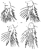 issued from : J.G.F. Bersano & G.A. Boxshall in Nauplius, Rio Grande, 1994, 2. [p.34, Fig.4]. Female & Male: A, P1; B, P2; C, P3; D, P4. Nota : Swimming legs 1 to 4 joined by intercoxal sclerites, biramous, with 3-segmented rami. Exopods with all outer margin spines bilaterally serrate ; terminal spines serrate on outer margin plumose on inner margin ; all inner margin setae plumose. Terminal spines just shorter than third segment in all legs. Endopods with inner margin setae plumose, distal spines serrate an douter margin spines serrate an douter margin spines serrate (P2) or naked (P3 and P4). Lateral margins of all endopodal segment ornamented with row of pinnules ; P2 and P3 endopods with conical terminal process present between distal margin spines ; apex of process with large integumental pore (see Fig.6D). Tip of P4 lacking any projection between distal spines. P5 with exopodal segment fused to somite, not separate basally, forming short exopodal lobe. Outer protopodal seta short, located on surface of somite dorsal to origin of exopodal lobe. Exopodal lobe with 2 apical setae.
|
 issued from : J.G.F. Bersano & G.A. Boxshall in Nauplius, Rio Grande, 1994, 2. [p.34]. Formula of swimming legs P1 to P4 female and male (A rabic numerals = setae; Roman numerals = spines).
|
 issued from : J.G.F. Bersano & G.A. Boxshall in Nauplius, Rio Grande, 1994, 2. [p.36, Fig.5 A, D]. Female: A, genital double-somite (dorsal view); D, sixth leg closing genital aperture (dorsal). Scale bars: 0.020 mm (A); 0.005 mm (D)
|
 issued from : J.G.F. Bersano & G.A. Boxshall in Nauplius, Rio Grande, 1994, 2. [p.36, Fig.5 C, E]. Female: C, anal somite (dorsal view showing anal operculum and associated sensillae pores; E, anal somite and caudal rami (ventral). Scale bars: 0.010 mm (C); 0.10 mm (E)
|
 issued from : J.G.F. Bersano & G.A. Boxshall in Nauplius, Rio Grande, 1994, 2. [p.37, Fig.6 C]. Female: 4th segment of A1, showing large origin aesthetasc. Scale bar: 0.005 mm.
|
 issued from : J.G.F. Bersano & G.A. Boxshall in Nauplius, Rio Grande, 1994, 2. [p.37, Fig.6 A, B]. Female: A, Mxp, ventrolateral view showing basal setae and free endopodal segment (arrowed); B, Mx1 and Mx2, lateral view showing maxillule partly hidden beneath anterior labrum.
|
 issued from : J.G.F. Bersano & G.A. Boxshall in Nauplius, Rio Grande, 1994, 2. [p.37, Fig.6 D]. Conical terminal processes on endopods of P2 and P3, showing apical pores. Scale bar: 0.005 mm
|
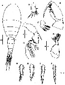 issued from : J.H. Wi, Y.H. Yoon & H.Y. Soh in Ocean Sci. J., 2009, 44 (2). [p.111, Fig.11]. Female (from East Sea of Korea): A, habitus (dorsal); B, A2; C, Md; D, Mx1; E, Mx2; F, Mxp; G, endopodite 3 of P1; H, endopodite 3 of P2; I, endopodite 3 of P3; J, endopodite 3 of P4. Scale bars in micrometers.
| | | | | Ref. compl.: | | | Böttger-Schnack & al., 2001 (p.1029, tab.1); Krsinic & al., 2007 (p.528); McKinnon & al., 2008 (p.844: Tab.1, p.846: Tab.II); Miyashita & al., 2009 (p.815, Tabl.II); Nishibe & al., 2009 (p.491, Table 1: seasonal abundance); Böttger-Schnack & Schnack, 2009 (p.131, Table 3, 4); Vidjak & al., 2012 (p.243, Rem.: p.253); Krsinic & Grbec, 2012 (p.57, 63: abundance); Tachibana & al., 2013 (p.545, Table 1, seasonal change 2006-2008); Mazzocchi & al., 2014 (p.64, Table 3, 4, occurrence); Benedetti & al., 2016 (p.159, Table I, fig.1, functional characters); Vidjak & al., 2016 (p.629, Rem.); Yebra & al., 2019 (p.1, Table 4: length, width, F,M) | | | | NZ: | 6 | | |
|
Carte de distribution de Oncaea waldemari par zones géographiques
|
| | | | | | | | | | Loc: | | | S Brazil, English Channel (Plymouth), Medit. (Alboran Sea, Balearic Basin, Adriatic Sea, Sibenik Harbour, Ionian Sea, Lebanon Basin), Red Sea (S), G. of Aden, Strait of bab al Mandab, N Arabian Sea, W Australia (North West Cape), Japan, Tosa Bay, Tokyo Bay, E Korea | | | | N: | 21 | | | | Lg.: | | | (248) F: 0,58-0,49; M: 0,41-0,36; (819) F: 0,42-0,50; M: 0,34-0,36; (1072)* F: 0,517-0,528; (1225) F: 0,49-0,76; M: 0,37-0,58; {F: 0,42-0,76; M: 0,34-0,58}
*: Body length in lateral aspect. | | | | Rem.: | L'année de publication de la description originale de cette espèce par Bersano & Bosxshall est 1996, alors que l'année données dans le journal est 1994. Alors la citation correcte pour cette espèce est :
Oncaea waldemari Bersano & Boxshall 1996 ["1994"]
(Pour explication, voir la note de Ruth Böttger-Schnack pour l'espèce O. waldemari dans le site du WoRMS.)
Voir aussi les remarques en anglais | | | Dernière mise à jour : 26/04/2019 | |
|
|
 Toute utilisation de ce site pour une publication sera mentionnée avec la référence suivante : Toute utilisation de ce site pour une publication sera mentionnée avec la référence suivante :
Razouls C., Desreumaux N., Kouwenberg J. et de Bovée F., 2005-2026. - Biodiversité des Copépodes planctoniques marins (morphologie, répartition géographique et données biologiques). Sorbonne Université, CNRS. Disponible sur http://copepodes.obs-banyuls.fr [Accédé le 21 juin 2026] © copyright 2005-2026 Sorbonne Université, CNRS
|
|
|
|


;)
;)
;)
;)
;)
;)
;)
;)
;)
;)

