|
|
 |
Fiche d'espèce de Copépode |
|
|
Calanoida ( Ordre ) |
|
|
|
Eucalanoidea ( Superfamille ) |
|
|
|
Eucalanidae ( Famille ) |
|
|
|
Subeucalanus ( Genre ) |
|
|
| |
Subeucalanus flemingeri Prusova, Al-Yamani & Al-Mutairi, 2001 (F,M) | |
| | | | | | | Ref.: | | | Prusova & al., 2001 (p.47, Descr. F, M, figs.F, M); Al-Yamani & Prusova, 2003 (p.38, figs.F,M); Al-Yamani & al., 2011 (p.29, 30, Figs.F,M) | 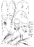 Issued from : I.Yu. Prusova, F. Al-Yamani & H. Al-Mutairi in Zool. Inst., St. Petersbourg, 2001, 10 (1). [p.48, Figs. 1-11]. Female (from 29°25'N, 48°30'E): 1, habitus (dorsal); 2, distal part of the second inner seta of the left caudal ramus; 3, habitus (right lateral); 4, cephalon and rostrum (ventral); 5, cephalon (right lateral); 6, urosome (dorsal); 7, urosome (right lateral); 8 a-e, seminal receptacles (right lateral view); 9, A1 segments 1-9; 10, A1 segments 10-17; 11, A1 segments 18-23. Scale bars = 0.1 mm. 1-7, 9-11: holotype; 8 a-e: paratypes. Nota: Head and 1st pediger segment completely fused, pedigers 4 and 5 partly fused. - Rostrum elongated and ending in fine filaments. - 2nd inner seta on left caudal ramus greatly elongated. - A1 23-segmented, extending beyond tips of caudal rami by last 5-6 segments, with stout plumose setae on last segments and all the other setae filiform.
|
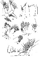 Issued from : I.Yu. Prusova, F. Al-Yamani & H. Al-Mutairi in Zool. Inst., St. Petersbourg, 2001, 10 (1). [p.49, Figs. 12-19]. Female : 12, A2; 13, Md palp; 14, Md gnathobase; 15, Mx1; 16, inner lobe 1 of Mx1 (another view); 17, inner lobe 1 of Mx2 (detached); 18, Mx2 (without inner lobe 1); 19, Mxp. Scale bars = 0.1 mm. 12-15, 17-19: holotype; 16: paratype. Nota: A2 exopod 7-segmented, exopodal segment 1 with 4 setae, exopodal segments 2 to 6 with 1 seta each and exopodal segment 7 with 3 terminal setae, the two inner ones much elongated. Endopod 2-segmented, endopodal segment 1 with 2 setae, endopodal segment 2 with 2 sets of 9 medial and 7 terminal setae. - Md with 3 setae on inner margin of basis; exopod 5-segmented, with 1 seta at each of the indistinctly separated exopodal segments 1-4 and 2 terminal setae at exopodal segment 5. Endopod 2-segmented, 1st segment with 2 setae, 2nd segment with 4 setae. Masticatory edge of gnathobase with 2 rows of medium-sezed teeth. - Inner lobe 1 (= arthrite) of Mx1 with 9 well developed and 4 small apical spines; inner lobe 2 with 4 long, thin distal setae; outer lobe 1 of Mx1 with 6 long and stout and 3 short and thin setae; basis with 5 setae; exopod with 5 setae; endopod 3-segmented, endopodal segments 1 and 2 with 4 setae each, endopodal segment 3 with 5 apical setae. - Coxa of Mxp with a posterior seta close to proximal end and 3 groups of setae along medial margin, with 2, 3, and 3 setae respectively in order from proximal; basis with a distal group of 3 setae; endopod 6-segmented, with 2, 3, 4, 4, 4, and 4 setae; 2 inner setae at distal segment stout and elongated.
|
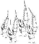 Issued from : I.Yu. Prusova, F. Al-Yamani & H. Al-Mutairi in Zool. Inst., St. Petersbourg, 2001, 10 (1). [p.50, Figs. 20-231]. Female (paratype): 20, P1; 21, P2; 22, P3; 24, P4. Scale bar = 0.1 mm.
|
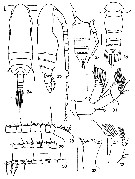 Issued from : I.Yu. Prusova, F. Al-Yamani & H. Al-Mutairi in Zool. Inst., St. Petersbourg, 2001, 10 (1). [p.52, Figs. 24-33]. Male: 24, habitus (dorsal); 25, same (right lateral); 26, urosome (dorsal); ; 27, urosome and P5 (right lateral); 28, A1 segments 1-10; 29, A1 segments 11-15; 30, A1 segments 16-23; 31, A2; 32, Md palp; 33, Md gnathobase. Scale bars = 0.1 mm. Nota: Prosome nearly 4.1 times as long as urosome and 3.1 times as long as wide - head and pediger 1 fused. - Urosome of 5 somites. -Genital segment slightly asymmetrical, with genital aperture on the left side. - A1 23-segmented, extending beyond end of urosome by last 5-6 segments; all segments with fine setules and aesthetascs, mostly 2 aesthetascs on each segment. - A2 more robust than that of female; setae on coxa, basis and endopodal segment 1 far smaller than in female; segmentation and setation of exopod as in female. - Md palp more robust than in female, with 3 setae on inner margin; segmentation and setation of exopod and endopod as in female.
|
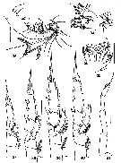 Issued from : I.Yu. Prusova, F. Al-Yamani & H. Al-Mutairi in Zool. Inst., St. Petersbourg, 2001, 10 (1). [p.53, Figs. 34-41]. Male: 34, Mx1; 35, Mx2; 36, Mxp; 37, P1; 38, P2; 39, P3; 40, P4; 41, P5. Scale bars = 0.1 mm. Nota: Mx1 and Mx2 as in female. - Mxp shorter than in female; endopod 6-segmented, with 2, 3, 3, 4, 3, and 5 setae; larger setae of endopodal segments 2-6 curved; 2 of 5 distal setae stout, greatly elongated and heavily plumosed. - P5 uniramous, 4-segmented, ending with distal spine nearly as long as the last segment; distal segment with fine hairs at outer edge.
|
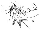 Issued from : V.N. Andronov in Russian Acad. Sci. P.P. Shirshov Inst. Oceanol. Atlantic Branch, Kaliningrad, 2014. [p.89, Fig.23: 5]. Subeucalanus flemingeri after Prusova & al., 2001. Mxp male.
| | | | | Ref. compl.: | | | Goetze & Ohman, 2010 (p.2110, Table 1) | | | | NZ: | 1 | | |
|
Carte de distribution de Subeucalanus flemingeri par zones géographiques
|
| | | | | | | Loc: | | | Indian (Arabian Gulf, Kuwait) | | | | N: | 1 | | | | Lg.: | | | (1195) F: 2,01-2,14; M: 1,97-2,14; {F: 2,01-2,14; M: 1,97-2,14} | | | | Rem.: | For Prusova & al. (2001, p.51) this species is very similar to wem>S. pileatus and S. subcrassus in the body shape, but distinguished from them by the more rounded forehead in dorsal and lateral views, and by the shape and arrangement of seminal receptacles. S. flemingeri differs from S. subcrassus in its smaller size and the narrower genital segment in dorsal view. The male is distinguished from S. pileatus and S. subcrassus by the cask-shaped second urosomal segment, shape and proportion, of P5 segments including distal spine in order from proximal is in S. flemingeri 30 : 42 : 27 : 25 : 26; in S. pileatus 18 : 17 : 15 : 13 : 11; and in S. subcrassus 28 : 27 : 15 : 20 : 25; and distal segment of P5 covered with hairs (according to Giesbrecht, 1892, these segments are naked in S. pileatus and S. subcrassus. | | | Dernière mise à jour : 04/05/2016 | |
|
|
 Toute utilisation de ce site pour une publication sera mentionnée avec la référence suivante : Toute utilisation de ce site pour une publication sera mentionnée avec la référence suivante :
Razouls C., Desreumaux N., Kouwenberg J. et de Bovée F., 2005-2025. - Biodiversité des Copépodes planctoniques marins (morphologie, répartition géographique et données biologiques). Sorbonne Université, CNRS. Disponible sur http://copepodes.obs-banyuls.fr [Accédé le 26 décembre 2025] © copyright 2005-2025 Sorbonne Université, CNRS
|
|
 |
 |


