|
|
 |
Fiche d'espèce de Copépode |
|
|
Calanoida ( Ordre ) |
|
|
|
Diaptomoidea ( Superfamille ) |
|
|
|
Pseudodiaptomidae ( Famille ) |
|
|
|
Pseudodiaptomus ( Genre ) |
|
|
| |
Pseudodiaptomus andamanensis Pillai, 1980 (F,M) | |
| | | | | | | Ref.: | | | Pillai, 1976 (1980) (p.256, figs.F,M) | 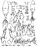 issued from : P.P. Pillai in J. mar. biol. Ass. India, 1976 (1980), 18 (2). [p.257, Fig.3]. Female (from Port Blair, Andaman Sea): a, habitus (dorsal); b, urosome (dorsal); c, anal segment and caudal rami (enlarged); d, urosome (lateral, right side); e, urosome (lateral, left side); f, rostrum; g, A1 (distal segments, enlarged); h, Md (masticatory edge); i, distal enlargment of seta on Mxp; j, P1; k, P2; l, P5.Head and 1st thoracic segment separate, 4th and 5th fused. Posterior angles of last thoracic segment asymmetically into outwardly directed spines, left spine reaching to posterior 2/3 of the genital segment being longest. Rostrum with 2 long filaments, strongly developed. Urosomal segments and caudal rami proportions 35:17:14:12:22 = 100. Genital segment asymmetrical and with lateral swellings; anteriorly at right lateral margin it is produced externally into a recurved peg-like structure; when viewed dorsally, the left lateral margin appears uneven; ventrally genital segment has a prominent genital boss; genital pore paired, is guarded anteriorly by two sets of fine spines and with a median genital groove; at the posterior margin ventrally a cluster of 5-6 small needle-like spines present. Caudal rami symmetrical, length: width ratio = 2.8:1. A1 21-segmented, reaches the anterior margin of genital segment. Male: m, habitus (dorsal); n, urosome (dorsal); o, A1; p, P5. Nota: Urosomal segments with caudal rami proportions 13:18: 18:19 = 100. A1 19-segmented.
|
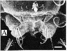
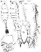 issued from : T.C. Walter, S. Ohtsuka, S. Putchakarn, K. Pinkaew & S. Chullasorn in Proc. Biol. Soc. Washington, 2002, 115 (3). [p.659, Fig.5]. Female (from Ko Aew Is. & Phuket Is., Andaman Sea): A-B, habitus (dorsal and lateral, respectively); C, genital double-somite (ventral); D, P5 (posterior); E, A1; F, antennular segment 19 specialized seta. Scale bars: 1.0 mm (A-B); 0.1 mm (C-E); 0.05 mm (F).
|
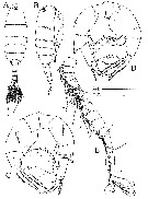 issued from : T.C. Walter, S. Ohtsuka, S. Putchakarn, K. Pinkaew & S. Chullasorn in Proc. Biol. Soc. Washington, 2002, 115 (3). [p.660, Fig.6]. Male: A-B, habirus (dorsal and lateral, respectively); C, P5 (posterior); D, P5 (anterior); E, A1. Scale bars: 1.0 mm (A-B); 0.1 mm (C-E).
|
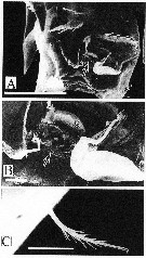 issued from : T.C. Walter, S. Ohtsuka, S. Putchakarn, K. Pinkaew & S. Chullasorn in Proc. Biol. Soc. Washington, 2002, 115 (3). [p.661, Fig.7]. Female: A, genital double somite (ventral view); genital atrium indicated by large arrow, left hair-sensillum by small arrow. B, genital double somite (ventral view, magnified); g = genital operculum; genital atrium indicated by large rarrow; spermatophore remnant to the right of arrow. C, genital double somite, ventral view of left hair-sensillum (magnification of A, indicated by small arrow). Scale bars: 0.1 mm (A-B); 0.01 mm (C).
|
 issued from : T.C. Walter, S. Ohtsuka, S. Putchakarn, K. Pinkaew & S. Chullasorn in Proc. Biol. Soc. Washington, 2002, 115 (3). [p.662, Fig.8, A]. Female: A, labrum (ventral view). Scale bar 0.1 mm.
| | | | | Ref. compl.: | | | Dussart & Defaye, 1983 (p.35); Madhupratap & Haridas, 1986 (p.105, tab.2); Walter, 1986 (p.131); 1986 a (p.503); 1987 (p.367); Maiphae & Sa-ardrit, 2011 (p.641, Table 2) | | | | NZ: | 1 | | |
|
Carte de distribution de Pseudodiaptomus andamanensis par zones géographiques
|
| | | | | | | Loc: | | | Andaman Is. (Marine Bay, Port Blair) | | | | N: | 2 | | | | Lg.: | | | (1005) F: 2,16-2,20; M: 1,83-1,88; (1061) F: 2,19; M: 1,80; {F: 2,16-2,20; M: 1,80-1,88} | | | | Rem.: | coastal.
Incomplete data.
Voir aussi les remarques en anglais | | | Dernière mise à jour : 29/03/2012 | |
|
|
 Toute utilisation de ce site pour une publication sera mentionnée avec la référence suivante : Toute utilisation de ce site pour une publication sera mentionnée avec la référence suivante :
Razouls C., Desreumaux N., Kouwenberg J. et de Bovée F., 2005-2025. - Biodiversité des Copépodes planctoniques marins (morphologie, répartition géographique et données biologiques). Sorbonne Université, CNRS. Disponible sur http://copepodes.obs-banyuls.fr [Accédé le 05 juillet 2025] © copyright 2005-2025 Sorbonne Université, CNRS
|
|
|
|


;)
;)
;)
;)
;)

