|
|
 |
Fiche d'espèce de Copépode |
|
|
Calanoida ( Ordre ) |
|
|
|
Diaptomoidea ( Superfamille ) |
|
|
|
Pseudodiaptomidae ( Famille ) |
|
|
|
Pseudodiaptomus ( Genre ) |
|
|
| |
Pseudodiaptomus culebrensis Marsh, 1913 (F,M) | |
| | | | | | | Syn.: | no Pseudodiaptomus culebrensis : Collado & al., 1984 | | | | Ref.: | | | Marsh, 1913 (p.4, figs.F,M); 1933 (p.33, figs.F,M, Rem.); Wright, 1936 (p.14, 16, fig.M); 1937 a (p.162, fig.M); Dussart & Defaye, 1983 (p.32); Grindley, 1984 (p.226); Walter, 1986 (p.131); 1986 a (p.503, fig.1); 1987 (p.366); 1989 (p.601, figs.F,M; Rem.) | 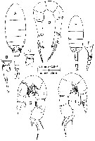 issued from : T.C. Walter in Bull. Mar. Sc., 1989, 45 (3). [p.602, Fig.6]. Female: A, habitus (dorsal); B, last thoracic segments and urosome (lateral right side); C, last thoracic segments and genital segment (lateral left side); D, P5 (anterior view). Male: E, habitus (dorsal); F, last thoracic segments and urosome (lateral left side); G, P5 (posterior view); H, idem (anterior view).
|
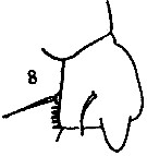 issued from : S. Wright in Ann. Acad. Brasileira Sci., 1937, 9 (2). [Pl.1, Fig.8]. After Marsh, 1933. Male: exopodite 1 of right P5 (posterior view).
|
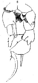 issued from : S. Wright in Annaes. Acad. bras. Cienc., VIII (1), 1936. [Pl.III, Fig.6 (no 7 as in the legend)]. Male: 6, P5 (redrawn from Marsh, 1933)
|
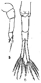 Issued from : C.D. Marsh in Proc. U.S. natn. Mus., 1933, 82 (18) (2959). [Pl. 17, figs.5-6]. Female (from Rio Culebra, Panama): 5, P5; 6, urosome. Nota: Caudal rami asymmetrical, about 7 times as long as wide and ciliate on the inner margins.
|
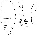 Issued from : C.D. Marsh in Proc. U.S. natn. Mus., 1933, 82 (18) (2959). [Pl. 18, figs.1-3]. Female:1, cephalothorax (dorsal); 3, A2. Nota: The segmentation of the exopod of A2 was indistinct in the material examined; in figure two central segments are shown. In others only one can be distinguished. The dorsal surface of the last two segments of the thorax is armed with hairs. A1 22-segmented, exceeding the cephalothorax. In all specimens examined the rifgt egg sac was atrophied, the left contained about 8 eggs. Male: 2, urosome (dorsal).
|
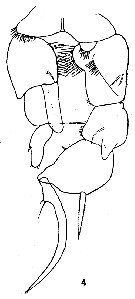 Issued from : C.D. Marsh in Proc. U.S. natn. Mus., 1933, 82 (18) (2959). [Pl. 17, fig.4]. Male: P5.
| | | | | Ref. compl.: | | | Suarez-Morales & Gasca, 1998 a (p.111); Alvarez-Silva & al., 2005 (p.39); Alvarez-Silva & Torres-Alvarado, 2013 (p.241, Table1: seasonal abundance) | | | | NZ: | 2 | | |
|
Carte de distribution de Pseudodiaptomus culebrensis par zones géographiques
|
| | | 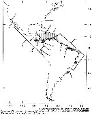 issued from : T.C. Walter in Bull. Mar. Sc., 1989, 45 (3). [p.625, Fig.18]. issued from : T.C. Walter in Bull. Mar. Sc., 1989, 45 (3). [p.625, Fig.18].
Map of American continents showing the zoogeography of Pseudodiaptomus species. |
| | | | Loc: | | | NE Pacif. (Central America: Panama), W Mexico, G. of California, Coyuca lagoon (Guerrero), Zihuatanejo Bay | | | | N: | 4 | | | | Lg.: | | | (950) F: 1,15-1,27; M: 0,83-0,93; (1148) F: 1,22; M: 0,88; {F: 1,15-1,27; M: 0,83-0,93} | | | | Rem.: | brackish.
Incomplete data.
Voir aussi les remarques en anglais | | | Dernière mise à jour : 14/10/2014 | |
|
|
 Toute utilisation de ce site pour une publication sera mentionnée avec la référence suivante : Toute utilisation de ce site pour une publication sera mentionnée avec la référence suivante :
Razouls C., Desreumaux N., Kouwenberg J. et de Bovée F., 2005-2026. - Biodiversité des Copépodes planctoniques marins (morphologie, répartition géographique et données biologiques). Sorbonne Université, CNRS. Disponible sur http://copepodes.obs-banyuls.fr [Accédé le 14 juin 2026] © copyright 2005-2026 Sorbonne Université, CNRS
|
|
|
|




;)
;)
;)
;)
;)
;)
;)