|
|
 |
Fiche d'espèce de Copépode |
|
|
Calanoida ( Ordre ) |
|
|
|
Diaptomoidea ( Superfamille ) |
|
|
|
Pseudodiaptomidae ( Famille ) |
|
|
|
Pseudodiaptomus ( Genre ) |
|
|
| |
Pseudodiaptomus hickmani Sewell, 1912 (F,M) | |
| | | | | | | Syn.: | no Pseudodiaptomus hickmani : Dakin & Colefax, 1940 (p.89, figs.F,M) | | | | Ref.: | | | Sewell, 1912 (p.364, figs.F,M); 1924 (p.786); 1932 (p.235); Marsh, 1933 (p.37, figs.F,M); Silas, 1972 (p.648); Ranga Reddy & Radhakrishna, 1982 (p.262, Redescr.F,M, figs.F,M) | 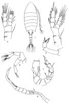 Issued from : R.B.S. Sewell in Rec. Indian Mus., 1912, 7. [Pl.XXII, Figs.1-7]. Female (from Bay of Bengal): 1, habitus (dorsal); 2, A2; 3, P1; 4, P2; 5, P5. Nota: Abdomen and caudal rami contained twice in the length of the cephalothorax. Head and 1st thoracic segment separate, 4th and 5th separate.Rostrum bfid with long and delicate spines. Abdomen 4-segmented, the first three of which are armed posteriorly with a row of triangular teeth extending across the dorsal surface. Proportional lengths of urosomites and furca as 10:4:6:4:7. Caudal ramus 3 times as long as wide. Genital operculum produced posteriorly in a single spine on the right side; On the right side of the segment is a blunt spinous process and anterior to the genital orifice is a curved row of needle-shaped spines, which terminates laterally above mentioned process; dorsally the segment is armed on each side
Male: 6, right A1; 7, P5.
Nota: Abdomen 5-segmented, of which the 2nd, 3rd and 4th armed posteriorly with a complete ring of triangular teeth. Proportional lengths of urosomites and furca as 2:5:5:5:3:5. The grasping A1 21-segmented; the knee-joint occurs between the 18th and 19th, the 13th to 17th segments are swollen.
|
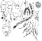 Issued from : Y. Ranga Reddy & Y. Radhakrishna in Hydrobiologia, 1982, 87. [p.263, Plate III, Figs. 1-11]. Female (from 16)32'-16°47'N, 81°4'-81°22'E): 1, habitus (dorsal); 2, postrior part of prosome and urosome with spermatophore (lateral); 3, posterior segment of prosome and genital segment (lateral); 4, segment genital (ventral); 5, caudal ramus; 6 & 6a, A1 and its modified seta; 7, P1; 8, P2; 9, P4; 10, P5; 11, egg sac. Nota: Rostrum strongly developed with 2 long acute spines overreaching the 2nd segment of A1. - Prosme twice the length of urosome. - Head and 1st pedigerous segment separate, 4th and 5th pedigeous segments fused. - Posterolateral corners of the 5th pedigerous segment rounded with a sharp, posterolaterally-directed spine; a few short hairs occur on the margin internal to the spine. - Urosome 4-segmented. - Genital segment slightly asymmetrical, proximally swollen with a short, strong spine on the right side; the armature of this segment is more complex than described by Sewell. - Urosomites together with the caudal rami are in the proportion of 35 : 17 : 19 : 16 : 25. - Caudal rami symmetrical, somewhat divergent, 23.3 times as long as broad with long coarse hairs on their inner margins. The five caudal setae are jointed; the middle seta the longest; a small sensory bristle lies between the inner 2 setae. - A1 21-segmented, reaching a little beyond the posterior margin of prosome. - The structural details of A2 and oral parts quite similar to those of P. binghami. Sewell'sremark that A2 of P. hickmanidiffer from others in possessing a row of spine-like processes on the margin of the last segment of the endopodite appears to be not well-founded as this character is shared by other species. - P1- P4 are identical with P. binghami but for the spine pattern of the basipodite segments. In P1, 1 or 2 strong spines , present on the outer margin of coxa, are absent in other species. - P5 asymmetrical, the left leg, particularly its 2 exopoditesegments, being more slender than their counterparts of the right leg. Each leg consists of 4 segments, 2 each of basipodite and exopodite. Coxa naked. Basis bears 1 seta on the posterior face. Exopodite 1 slightly over twice as long as broad with a small spine at the distal outer corner. Exopodite 2 has 1 spine at about its distal corner, and the segment proper ends in an out-curved, claw-like process serrated on both margins. Between this and the outer spine is a large spine with a lobe-like serrated process at its base on the inner side. - The ovigerous female bears a single large, compact efgg sac, furrowed in the middle owing to the compression of urosome, with 24-38 individual eggs.
|
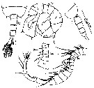 Issued from : Y. Ranga Reddy & Y. Radhakrishna in Hydrobiologia, 1982, 87. [p.263, Plate III, Figs. 12-15]. Male: 12, posterior part of prosome and urosome (dorsal); 13, same (lateral); 14 & 14a, right A1 and its segments 9-14; 15, P5 (posterior view). Nota: Prosome as in the female except that the right spine of the 5th pedigerous segment is comparatively larger than its left counterpart and both are backwardly directed. - Urosome 5-segmented; the posterodorsal margin of segments 2-4 with short spines; the 4 midorsal spines of 4th segment distinctly larger. - Genital segment bears at its distal right corner a few small spines arranged in a short transverse row. In the lateral view from the left side, the 2nd urosomite, in addition, shows a few minute spines arranged in 2 short transverse rows. - Caudal rami and setae as in the female. - All appendages except for the right A1 and P5 similar to those of the female. - Right A1 21-segmented, with 3 segments distal to the geniculation; segments 13-17 swollen; segment 3 with a long jointed aesthetasc besides 2 setae; segments 10-13 with a spine each, the spine on segment 10 characteristically bifid and hoohed; segment 17 with an unarmed tooth plate, overlapping the proximal part of the succeeding segment. The tooth plate on segment 18 with long, straight spine-like teeth in the proximal 2/3. Segment 19 has 2 unarmed 'tooth plates'. - P5 asymmetrical, biramoius; basipodite 2-segmented, exopodite 3-segmented in the right leg and 2-segmented in the left. Right leg with coxa naked; basis quite larger than coxa and bears on its outer aspect a Y-shaped spinous process (i.e. endopodite), the two arms of which are nearly of equal length; basis has near the distal outer margin a few spinules arranged in a short transverse row, and 1 short seta at about the centre of the segment on the posterior face. Exopodite 1 has on the outer distal angle a Y-shaped spinous process, the outer arm of which has a short subapical spine; the segment also has a row of short blunt spines on its proximal inner margin. Exopodite 2 longer than broad, having on its distal outer region a short thick spine with serrated margins; near this spine lie a few minute spines on the outer margin; at the middle of the inner margin of the same segment lie a short blunt spinous projection and 1 seta. Exopodite 3 bent inward, 'sickle-shaped', apically drawn out into a slender spinous process, and having on the proximal inner side a pair of short setae, one of which lies on a small lobe; the inner margin of the segment, distal to the lobe, serrated. Left leg with coxa and basis as in the right leg; the endopodite long, unjointed and 'fringed'. Exopodite 1 has a small spine at its distal corner. Exopodite 2 narrow at its base and expands distally into a large thin plate, whose ornamentation can be best understood from the figure. - Colour: Light brown in preserved specimen. A few orange red spots at the base of Mxp and on the ventral part of the anal segment.
| | | | | Ref. compl.: | | | Sewell, 1948 (p.323); Dussart & Defaye, 1983 (p.31); Sarkar & al., 1986 (p.178); Madhupratap & Haridas, 1986 (p.105, tab.2); Walter, 1986 (p.133); 1986 a (p.503); 1987 (p.367); Ramaiah & al., 1996 (p.3) | | | | NZ: | 1 | | |
|
Carte de distribution de Pseudodiaptomus hickmani par zones géographiques
|
| | | | | | | Loc: | | | India, Lake Kolleru, at Kolletikota, Chilka Lake, Salt Lakes, Calcutta, Hooghly estuary, Diamond Harbour, Burma, Hinze Basin | | | | N: | 5 | | | | Lg.: | | | (80) F: 1,26-1,37; (81) F: 1,37; M: 1,3; (1193) F: 1,31-1,42; M: 1,10-1,19; {F: 1,26-1,42; M: 1,10-1,30} | | | | Rem.: | limnique & saumâtre, estuaire.
Pour Sewell (1912) certaines identifications dans lindien comme P. serricaudatus pourraient appartenir à cette nouvelle espèce.
Voir aussi les remarques en anglais | | | Dernière mise à jour : 02/03/2016 | |
|
|
 Toute utilisation de ce site pour une publication sera mentionnée avec la référence suivante : Toute utilisation de ce site pour une publication sera mentionnée avec la référence suivante :
Razouls C., Desreumaux N., Kouwenberg J. et de Bovée F., 2005-2026. - Biodiversité des Copépodes planctoniques marins (morphologie, répartition géographique et données biologiques). Sorbonne Université, CNRS. Disponible sur http://copepodes.obs-banyuls.fr [Accédé le 07 mai 2026] © copyright 2005-2026 Sorbonne Université, CNRS
|
|
|
|


;)
;)
;)

