|
|
 |
Fiche d'espèce de Copépode |
|
|
Calanoida ( Ordre ) |
|
|
|
Diaptomoidea ( Superfamille ) |
|
|
|
Pseudodiaptomidae ( Famille ) |
|
|
|
Pseudodiaptomus ( Genre ) |
|
|
| |
Pseudodiaptomus ishigakiensis Nishida, 1985 (F,M) | |
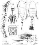
| | | | | | | Ref.: | | | Nishida, 1985 (p.128, figs.F,M, p.132-133 Rem.: historical account); Chihara & Murano, 1997 (p.893, Pl.165: F,M); Boxshall & Halsey, 2004 (p.173, figs.F,M) |  issued from : S. Nishida in Publ. Seto Mar. Biol. Lab., 1985, 30 (1/3). [p.129, Fig.2]. Female (from Kabira Bay, Ishigaki Is.): a, habitus (dorsal); b, idem (lateral left side); c, genital segment (ventral); d, anal segment and caudal rami (dorsal); e, right A1 (ventral); f, A2. CL = cephalothorax length (dorsal measure). Nota : Rostrum with 2 long filaments. Head and 1st pedigerous somite separate, 4th and 5th fused. Proportional lengths of urosomal segments and caudal ramus 32.2 : 14.8 :18.4 : 11.6 : 23.0. 1st to 3rd urosomal segments each with row of triangular spinules on dorsoposterior margin ; size of spinules increasing from first to third segment. Genital segment produced ventrally, slightly asymmetrical in dorsal view ; dorsal surface with a few medial, transverse rows of very fine spinules ; ventral surface with transverse row of spinule anterior to genital area, a pair of fine setae near posterior margin. Lateral outer margin of caudal ramus divided into 4 portions (proportions of anterior to posterior portions 2 :2 :1 :1) by 2 small notches at base of lateral seta. A1 22-segmented ; segments 6-7 incompletely segmented, the former with short spine ; segment 20 having specialized seta with small teeth on medial margin. A2 : Exopod with 7 terminal and 8 subterminal setae, and lateral fringe of fine hairs. Endopod 4-segmented (3rd segment inconspicuous and looking like membrane connecting 2nd and 4th segments.
|
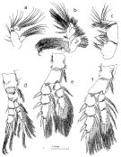
 issued from : S. Nishida in Publ. Seto Mar. Biol. Lab., 1985, 30 (1/3). [p.130, Fig.3]. Female: a, Md; b, Mx1; c, Mxp; d, P1; e, P2; f, P3. Nota : Md : basal segment of palp with 4 inner marginal setae ; exopod inconspicuously 2-segmented, each segment with 5 and 8 setae, respectively : endopod with 6 setae, segmentation incomplete. Mx1 : Arthrite (inner lobe of praevoxa) with 9 strong and 2 finer spines ; anterior surface with row and patch of spinules. 2nd and 3rd inner lobes each with 4 terminal setae. Outer lobe with 7 long and 2 short setae. Exopod with 10 marginal setae. Endopod incompletely 3-segmented ; 1st to 3rd segments with 4, 4, 7 setae, respectively. Mxp 6-segmented ; 2nd basal segment large, 4 distal segments small.
|
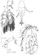
 issued from : S. Nishida in Publ. Seto Mar. Biol. Lab., 1985, 30 (1/3). [p.131, Fig.4, a-e]. Female: a, P4; b, P5. P5 uniramous, 4-segmented and symmetrical ; 2nd segment, unlike like P. marinus Sato, 1913 (Grindley & Grice, 1969), without spinules on outer distal margin ; 3rd segment length 3.2 times width, with outer distal seta ; 4th segment with outer distal seta ; inner distal part produced distally into curved spiniform process with serrate membrane on both margins ; terminal spine with teeth on distal half of inner margin, and short serrate spine near its base. Male: c, habitus (dorsal); d, right A1 (ventral view); e, P5. Proportional lengths of abdominal segments and caudal ramus 9.0 : 20.2 : 17.9 : 17.6 : 15.7 : 19.6 ; 2nd to 4th abdominal segments each with row of triangular spinules on whole posterior margin ; the size of spinules increasing from 2nd to 4th segment. Right A1 21-segmented. P5 biramous, with 2 basal segments, 2-segmented exopod and 1-segmented endopod. 1st basal segment with row of spinules on anterior surface. 2nd basal segment with medial outer seta and distal outer row of spinules. 1st segment of right exopod with row of spinules on inner margin, on distal end with V-shaped bifurcated spine without spinule in the fork ; a little proximally to its base with short thick spinule. 2nd exopodal segment with long, straight naked spine on outer distal margin, and short thick spinule on posterior surface near medial point of inner margin ; terminal hook with 1 inner and 1 outer midmarginal setae. Right endopod bifurcate ; inner ramus slender and pointed, with fine seta near distal end ; outer ramus thick and with 5 blunt distal spinules, one with teeth on tip. 1st segment of left exopod with short naked spine on outer distal corner. 2nd segment with short terminal spine, and thick long outer spine with serrate membrane on inner margin ; outer margin between these spines fringed with tiny spinules ; inner margin with 3 fine setae distally, and row of setules proximally ; 1 fine seta on medial posterior surface. Left endopod with patch of spinules on posterior surface near distal end, row of setules on distal inner margin, and serrate membrane on distal outer margin.
| | | | | Ref. compl.: | | | Oka & Saisho, 1994 (p.51, abundance); Ohtsuka & al., 1995 (p.159); Walter, 1986 (p.133); 1987 (p.367); Sakaguchi & al., 2011 (p.18, Table 1, 2, occurrences); Ohtsuka & Nishida, 2017 (p.565, 578, Rem., Table 22.1) | | | | NZ: | 1 | | |
|
Carte de distribution de Pseudodiaptomus ishigakiensis par zones géographiques
|
| | | | Loc: | | | SW Japan (Kabira Bay: Ishigaki Is., Nabsei islads, Kyushu: Sumiyo Bay) | | | | N: | 3 | | | | Lg.: | | | (866) F: 1,2-1,29; M: 1,01-1,05; {F: 1,2-1,29; M: 1,01-1,05} | | | | Rem.: | saumâtre – marine.
Voir aussi les remarques en anglais | | | Dernière mise à jour : 11/05/2019 | |
|
|
 Toute utilisation de ce site pour une publication sera mentionnée avec la référence suivante : Toute utilisation de ce site pour une publication sera mentionnée avec la référence suivante :
Razouls C., Desreumaux N., Kouwenberg J. et de Bovée F., 2005-2025. - Biodiversité des Copépodes planctoniques marins (morphologie, répartition géographique et données biologiques). Sorbonne Université, CNRS. Disponible sur http://copepodes.obs-banyuls.fr [Accédé le 31 décembre 2025] © copyright 2005-2025 Sorbonne Université, CNRS
|
|
 |
 |


