|
|
 |
Fiche d'espèce de Copépode |
|
|
Calanoida ( Ordre ) |
|
|
|
Epacteriscoidea ( Superfamille ) |
|
|
|
Ridgewayiidae ( Famille ) |
|
|
|
Stargatia ( Genre ) |
|
|
| |
Stargatia palmeri Fosshagen & Iliffe, 2003 (F,M) | |
| | | | | | | Ref.: | | | Fosshagen & Iliffe, 2003 (p.17,18, figs.M) | 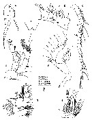 issued from : A. Fosshagen & T.M. Iliffe in Sarsia, 2003, 88. [p.19, Fig.2]. Male (from Bahamas): A, habitus (lateral); B, rostrum (ventral view); C, right A1; D, right A1 (segments XI-XV); E, A2; F, Md; G, Mx1; H, Mx2. Nota: Right A1 22-segmented with 2 areas of geniculation, typical one between segments 18 and 19 (XX-XXI), and complex proximal one with several modifications from segments 7 to 13 (IX-XV); segment 2 (II-IV) compound; segments 8-13 (X-XV) condensed, appearing to have flexure joint between XI and XII, and between XV and XVI. A2: Exopod 9-segmented; distal compound segment of endopod with distinct inner lobe bearing 9 setae and elongate terminal lobe with 7 setae. Md: Coxa reduced in size; gnathobase with pointed teeth, 2 ventral ones longest, isolated and deeply incised; basis with 2 inner setae; endopod enlarged and highly modified, 1st segment with 3 elements, 2 setae and 1 distal long pointed process with filament, 2nd segment with coarsely serrated inner margin, with 10 elements, 9 of which are pointed processes gradually tapering into long thin filament, and 1 ordinary seta at tip of endopod; exopod indistinctly 4-segmented with 6 setae, proximal seta strong and curved. Mx1: praecoxal arthrite with 15 elements; coxal epipodite with 9 setae, 2 proximal ones modified into short, smooth and flattened setae, ponted at tip; coxal endite and 1st basal endite each with 4 setae, 2nd basal endite with 5 modified setae; endopod elongated, distinctly 3-segmented with 5, 5 and 7 elements, respectively, most of which with bulbous base ending in filament; exopod with 11 setae. Mx2: endopod indistinctly 3-segmented, with 8 setae; long setae bearing widely spaced setules.
|
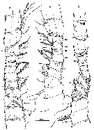 issued from : A. Fosshagen & T.M. Iliffe in Sarsia, 2003, 88. [p.20, Fig.3]. Male: right A1 (segments VII-XVII and the proximal flexure zone); A (dorsal view); B (ventral view); C (posterior view; arrows point to flexure joints between segments XI and XII, and between XV and XVI; only muscles that are associated with the joints are drawn). Figure drawn by R. Huys.
|
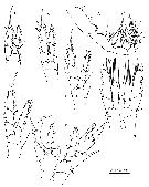 issued from : A. Fosshagen & T.M. Iliffe in Sarsia, 2003, 88. [p.21, Fig.4]. Male: A, Mxp; B, P1; C, P2; D, P3; E, P4; F, P5 (posterior). Copepodid male stage V: G, P5 (anterior).
|
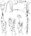 issued from : A. Fosshagen & T.M. Iliffe in Sarsia, 2003, 88. [p.22, Fig.5]. Female: A-B, habitus (dorsal and lateral, respectively); C, urosome (lateral); D, A1; E, P2; F, P5. Nota: A1 26-segmented. Urosome 4-segmented. Caudal rami as in male but with seta V on left side longer, about double length of corresponding seta on right side. Copepodid female stage V: G, P5.
| | | | | NZ: | 1 | | |
|
Carte de distribution de Stargatia palmeri par zones géographiques
|
| | | | Loc: | | | Bahamas (Stargate Blue Hole: cave) | | | | N: | 1 | | | | Lg.: | | | (883) M: 1,49-1,58; {M: 1,49-1,56} | | | | Rem.: | anchialine cave | | | Dernière mise à jour : 01/02/2015 | |
|
|
 Toute utilisation de ce site pour une publication sera mentionnée avec la référence suivante : Toute utilisation de ce site pour une publication sera mentionnée avec la référence suivante :
Razouls C., Desreumaux N., Kouwenberg J. et de Bovée F., 2005-2025. - Biodiversité des Copépodes planctoniques marins (morphologie, répartition géographique et données biologiques). Sorbonne Université, CNRS. Disponible sur http://copepodes.obs-banyuls.fr [Accédé le 06 juillet 2025] © copyright 2005-2025 Sorbonne Université, CNRS
|
|
|
|




;)
;)
;)
;)