|
|
 |
Fiche d'espèce de Copépode |
|
|
Calanoida ( Ordre ) |
|
|
|
Clausocalanoidea ( Superfamille ) |
|
|
|
Scolecitrichidae ( Famille ) |
|
|
|
Macandrewella ( Genre ) |
|
|
| |
Macandrewella omorii Ohtsuka, Nishida & Nakaguchi, 2002 (F,M) | |
| | | | | | | Ref.: | | | Ohtsuka & al., 2002 (p.545, figs.F,M) | 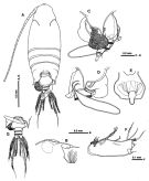 issued from : S. Ohtsuka, S. Nishida & K. Nakaguchi inJ. Nat. Hist., 2002, 36 [p.547, Fig.11]. Female (from off Tokashiki Shima Is.): A, habitus (dorsal); B, rostrum and cuticular lens (lateral); C, prosomal end and urosome with spermatophore (lateral left side); D, idem (lateral right side); E, genital double-somite (ventral); F, syncoxa of Mxp; G, prosomal ends and urosome with spermatophore (dorsal). Nota: Cuticular lens present at base of rostrum. Posterior ends of prosome slightly asymmetrical. 2nd and 3rd urosomites with striated posterior margin. Anal somite small. A1 23-segmented. Endopod of Mx2 with 3 worm-like and 5 brush-lke sensory setae. Mxp with syncoxa having of long setules proximally. P5 absent.
|
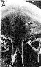 issued from : S. Ohtsuka, S. Nishida & K. Nakaguchi inJ. Nat. Hist., 2002, 36 [p.548, Fig.12 A]. Female (SEM micrograph): A, rostrum and cuticular lens indicated by arrow (frontal view). Scale = 0.1 mm.
|
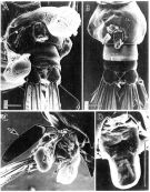 issued from : S. Ohtsuka, S. Nishida & K. Nakaguchi inJ. Nat. Hist., 2002, 36 [p.549, Fig.13]. Female (SEM micrographs): A, urosome with spermatophore (ventral); B, idem (ventral, spermatophore removed); C, idem (lateral right side, spermatophore proper and linguiform genital operculum indicated by large and small arrows, respectively); D, genital operculum. Scales = 0.01 mm.
|
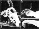 issued from : S. Ohtsuka, S. Nishida & K. Nakaguchi inJ. Nat. Hist., 2002, 36 [p.548, Fig.12 C-E]. Female (SEM micrographs): c, sensory setae on endopod of Mx2 (D: brush-like; E: worm-like; closed and open circles, respectively. Scales = 0.01 mm (C); 0.001 mm (D, E).
|
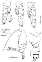 issued from : S. Ohtsuka, S. Nishida & K. Nakaguchi inJ. Nat. Hist., 2002, 36 [p.550, Fig.14]. Female: A, right P2 (anterior, coxa omitted); B, 2nd endopod segment of left P2 (anterior); C, P3 (anterior, coxa omitted); D, P4 (anterior, coxa omitted). Male: E, habitus (lateral left side); F, cuticular lens (lateral); G, right posterior corner of prosome (lateral right side); H, prosomal end and urosome (lateral left side).
|
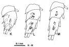 issued from : S. Ohtsuka, S. Nishida & K. Nakaguchi inJ. Nat. Hist., 2002, 36 [p.541, Fig.8]. Male: K, endopod of P2 (anterior); L, endopod of P3 (anterior); M, endopod of P4 (anterior).
|
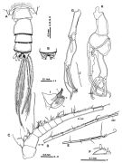 issued from : S. Ohtsuka, S. Nishida & K. Nakaguchi inJ. Nat. Hist., 2002, 36 [p.551, Fig.15]. Male: A, prosomal ends and urosome (dorsal); B, anal somite and caudal rami (ventral); C, A1 (segments I to X-XV); D, A1 (segments XVI-XVII to XXII); E, A1 (segments XXIII to XXVII-XXVIII); F, A1 (compound segment XXVII-XXVIII); G, left P5; H, right P5; I, terminal portion of left exopod of P5; J, inner margin of left endopod of P5. Nota: 5th pediger asymmetrical, with right process slightly larger than left; 2nd urosomite with left side more swollen laterally than right side.
|
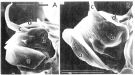 issued from : S. Ohtsuka, S. Nishida & K. Nakaguchi inJ. Nat. Hist., 2002, 36 [p.552, Fig.16]. Male (SEM micrographs): A-B, terminal portion of left exopod of P5 (elements indicated by \"a-d\"). Scales = 0.05 mm.
| | | | | NZ: | 1 | | |
|
Carte de distribution de Macandrewella omorii par zones géographiques
|
| | | | | | | Loc: | | | SW Okinawa (off Kume Shima Is., off Tokashiki Shima Is.) | | | | N: | 1 | | | | Lg.: | | | (903) F: 3,32-3,54; M: 3,38-3,62; 3,53-4,05; {F: 3,32-3,54; M: 3,38-4,05} | | | | Rem.: | hyperbenthique.
Voir aussi les remarques en anglais | | | Dernière mise à jour : 06/04/2016 | |
|
|
 Toute utilisation de ce site pour une publication sera mentionnée avec la référence suivante : Toute utilisation de ce site pour une publication sera mentionnée avec la référence suivante :
Razouls C., Desreumaux N., Kouwenberg J. et de Bovée F., 2005-2026. - Biodiversité des Copépodes planctoniques marins (morphologie, répartition géographique et données biologiques). Sorbonne Université, CNRS. Disponible sur http://copepodes.obs-banyuls.fr [Accédé le 17 juin 2026] © copyright 2005-2026 Sorbonne Université, CNRS
|
|
|
|




;)
;)
;)
;)
;)
;)
;)
;)