|
|
 |
Fiche d'espèce de Copépode |
|
|
Calanoida ( Ordre ) |
|
|
|
Diaptomoidea ( Superfamille ) |
|
|
|
Temoridae ( Famille ) |
|
|
|
Eurytemora ( Genre ) |
|
|
| |
Eurytemora herdmani Thompson & Scott, 1897 (F,M) | |
| | | | | | | Ref.: | | | in Herdman, Thompson & Scott, 1897 (p.78, figs.F,M); Giesbrecht & Schmeil, 1898 (p.103; Bremen, 1906 (p.100, fig.116); Sharpe, 1910 (p.410, figs.F); Willey, 1920 a (p.12); Gurney, 1931 a (p.185); C. Wilson, 1932 (p.112, fig.75 a, b)Mori, 1937 (1964) (p.66, figs.M, juv.F); Brodsky, 1950 (1967) (p.283, figs.F,M); Heron, 1964 (p.204, Rem.F,M, figs.F,M); Faber, 1966 (p.191, 195, figs.N); Shih & al., 1971 (p.47, 208); Katona, 1971 (p.5, 16, comparison); Heron & Damkaer, 1976 (p.129, Table 2); Kos, 1976 (Vol. II, figs.F,M), Rem.; 1977 a (p.23, Rem.F,M, figs.F,M); Robins & McLaren, 1982 (p.529, nuclear DNA contents); McLaren & Marcogliese, 1983 (p.721, cell nucleus); Sévigny & Odense, 1985 (p.455, enzymatic system); Kos, 1984 (1985) (p.228, Rem.); Dussart & Defaye, 1983 (p.48); Sazhina, 1985 (p.53, figs.N); Ohtsuka & al., 1996 b (p.153, figs.F: mouth parts, gut contents); Chihara & Murano, 1997 (p.917, Pl.181: F,M); G. Harding, 2004 (p.18, figs.F,M); Boxshall & Halsey, 2004 (p.208: fig.F); Dodson & al., 2010 (p.653, table 1, 2, fig.4, 5, 6, key Female: p.146); Kos, 2016 (p.80, figs.F, M, Rem.) | 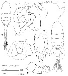 issued from : G.A. Heron in Crustaceana, 1964, 7 (3). [p.205, Figs.12-18]. Female (from NW Alaska): 12, habitus (dorsal); 13, genital segment (ventral); 14, Md (cutting edge of gnathobade); 15, P5. Nota: A1 24-segmented, reaching to metasomal segment 6. Caudal rami with dorsal surface hirsutee; inner margings with long fine hairs. Male: 16, habitus (dorsal); 17, right A1 (segments 8-14 with detail of spines): 18, P5 (posterior). Nota: Right A1 21-segmented, segments 13 to 17 enlarged
|
 issued from : G. Harding in Key to the adullt pelagic calanoid copepods found over the continental shelf of the Canadian Atlantic coast. Bedford Inst. Oceanogr., Dartmouth, Nova Scotia, 2004. [p.18]. Female & Male. L = left leg; R = right leg.
|
 Issued from : S. Ohtsuka, M. Shimozu, A. Tanimura, M. Fukuchi, H. Hattori, H. Sasaki & O. Matsuda in Proc. NIPR Symp. Polar Biol., 1996, 9. [p.158, Fig.4, C-D]. Eurytemora herdmani Female (from Bering Sea, Chukchi Sea): Mandibular cutting edge, ventralmost tooth (arrowed). Scale bar = 10 µm.
|
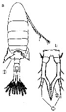 Issued from : M. Chihara & M. Murano in An Illustrated Guide to Marine Plankton in Japan, 1997. [p.919, Pl. 181, fig.284 a-b]. Female: a, habitus (dorsal); b, P5. Note characteristics numbered 1, 2, 3 into circles.
|
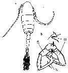 Issued from : M. Chihara & M. Murano in An Illustrated Guide to Marine Plankton in Japan, 1997. [p.919, Pl. 181, fig.284 a-b]. Male: a, habitus (dorsal); b, P5. Note characteristics numbered 1, 2, 3 into circles.
|
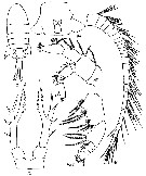 Issued from : M.S. Kos in Zoological Institut RAS, St. Petersburg, 2016, 179, [p.80, Fig. 40]. Eurytemora herdmani female (from the Japan Sea ).
|
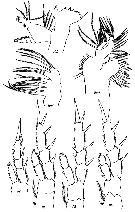 Issued from : M.S. Kos in Zoological Institut RAS, St. Petersburg, 2016, 179, [p.81, Fig. 41]. Eurytemora herdmani female (from the Japan Sea ).
|
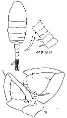 Issued from : M.S. Kos in Zoological Institut RAS, St. Petersburg, 2016, 179, [p.82, Fig. 42]. Eurytemora herdmani male (from the Japan Sea).
|
 Issued from : S.I. Dodson, D.A. Skelly & C.E. Lee in Hydrobiologia, 2010, 653. [p.141, Table 2]. Descriptions of North American species of Eurytemora from Alaska.
|
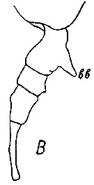 Issued from : M.S. Kos in Field guide for plankton. Zool Institute USSR Acad., Vol. II, 1976. After Kos (original). Female: B, abdomen (lateral view).
| | | | | Ref. compl.: | | | W.H. Johnson, 1933 (p.1, vertical distribution vs. light); Lacroix, 1960 (p.11, 26); M.W. Johnson, 1961 (p.311, Table 2); Marshall & Orr, 1962 (tab.3); Martin, 1965 (p.188); Faber, 1966 a (p.419, 420); Katona, 1970 (p.373, fig.1, 2, 3, generation times, reproduction, sex ratios, survival vs. temperature); Itoh, 1970 (tab.1); Conover & Mayzaud, 1975 (p.151, fig.5, abundance); McLaren, 1976 a (p.200, heritability: size, age of maturity, mortality, sex ratio); Mackas & Bohrer, 1976 (p.77, fig.1, gut contents); Conover, 1978 (p.66, 69, feeding); Poulet, 1978 (p.1126, grazing); Mayzaud & Poulet, 1978 (p.1144, feeding); Poulet & Marsot, 1978 (p.1403, chemosensory-grazing); 1980 (p.198, Fig.2, 6, 7, 8, Table 3, feeding); Gagnon & Lacroix, 1981 (p.401, fig.5, Table 1, tidal effect); McLaren & Corkett, 1981 (p.77, growth-temperature, production, P/B quotient); Robins & McLaren, 1982 (p.529); Citarella, 1982 (p.791, 798: listing), frequency, Tableau II, V); Poulet & Ouellet, 1982 (p.341, chemosensory, feeding); Gagnon & Lacroix, 1982 (p.9, fig.4, Table 1); 1983 (p.fig.2, tidal estuary); McLaren & Marcogliese, 1983 (p.721, body size vs. nucleus counts); Jacoby & Youngbluth, 1983 (p.84, Table 4, Rem: mating); Tremblay & Anderson, 1984 (p.6); De Ladurantaye & al., 1984 (p.21, tab.I, fig.3, 4, 5, advective processes in fjord); George V.S., 1985 (p.145, demography); Citarella, 1989 (p.123, abundance); Huntley & Lopez, 1992 (p.201, Table A1, egg-adult weight, temperature-dependent production); Mayzaud & al., 1992 (p.197, enzyme activity/food concentration); Escribano & McLaren, 1992 (p.77, food-temperature-length-weigt); Mauchline, 1998 (tab.33, 45, 47, 48, 51); Manning & Bucklin, 2005 (p.233, Table 1); Teegarden & al., 2008 (p.33, Rem.: feeding and toxicity); DFO, 2009 (p.1, fig. 13, seasonal variability); Hopcroft & al., 2010 (p.27, Table 1, 2, fig.9); Turner & al., 2011 (p.1066, Table II, abundance 1998-2008); Matsuno & al., 2011 (p.1349, Table 1, abundance vs years); Matsuno & al., 2012 (Table 2); Lasley-Rasher & Yen, 2012 (p.433, mating vs predator); Gusmao & al., 2013 (p.279, Table 4, sex ratio, fig.3: sex ratio vs temperature); Ohashi & al., 2013 (p.44, Table 1, Rem.); Coyle & al., 2014 (p.97, table 3); Ohtsuka & Nishida, 2017 (p.565, Table 22.1). | | | 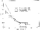 issued from : S.K. Katona in Helgol. Meersunters, 1970, 20. [p.376, Fig.1]. issued from : S.K. Katona in Helgol. Meersunters, 1970, 20. [p.376, Fig.1].
Generation times (± 2 SD) in 20 p.1000 salinity sea water at different temperatures.
W.H.O.I.: culture started with females taken from Oyster Pond, a fresh brackish pond near Woods Hole (Massachussets)
Hamble: Culture from the Hamble River at Southampton (England)
For E. herdmani from Nahant (Massachussets). |
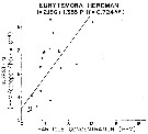 Issued from : R.J. Conover in Rapp. P.-v. Réun. Cons. int. Explor. Mer, 1978, 173. [p.69, Fig.54]. Issued from : R.J. Conover in Rapp. P.-v. Réun. Cons. int. Explor. Mer, 1978, 173. [p.69, Fig.54].
Regression between rate of ingestion (I) for Eurytemora herdmani from Bedford Basin, Nova Scotia (Canada) and particle concentration in the same environment. |
| | | | Loc: | | | N America (Rimouski, Saguenay fjord, upper St. Lawrence estuary, G. of St. Lawrence, Narragansett Bay, G. of Maine, Damariscotta River estuary, Rhode Island, Woods Hole, Bay of Fundy), Massachusetts Bay, Passamaquoddy Bay, Halifax (Nova Scotia), Shediac Bay, Bedford Basin, Northumberland Strait, Baie-des Chaleurs, Hudson Bay, W Alaska, Chukchi Sea, Beaufort Sea, E Siberia, S Kurils, S Korea, Japan (Akkeshi Bay N Japan, Bering Sea (inner shelf, western neritic) | | | | N: | 66 | | | | Lg.: | | | (22) F: 1,6-1,3; M: 1,5-1,2; (91) M: 1,5-1,15; (280) F: 1,39-0,73; (566) F: 1,6-1; M: 1,75-0,87; (571) F: 1,36-1,05; M: 1,46-1,16; (573) F: 1,6; M: 1,6; (866) F: 1,5-1,6; M: 1,28-1,53; (1232): F: 1,05-1,60; M: 1,16-1,50; {F: 0,73-1,60; M: 0,87-1,75}
In culture (Katona, 1970), at 2°C: F = 1.105 mm ±0.057; M = 1.197 mm ±0.033 and at 21.5°C F = 1.097 mm ±0.037; M = 1.073 mm ±0.023.
At 14°C females show a linear and significant size increase with decreasing salinity from 33 p.1000 (0.964 mm ±0.049) to 20 p.1000 (1.127 mm ± 0.022) to 15 p.1000 (1.227 mm ±0.036). The trend was apparent but less pronounced in males. These data suggest that E. herdmani is more stenothermal than E. affinis, and also that E. herdmani may be best adapted to salinities somewhat below full-strength sea water. | | | | Rem.: | saumâtre, marine (littorale)
Voir aussi les remarques en anglais | | | Dernière mise à jour : 19/06/2023 | |
|
|
 Toute utilisation de ce site pour une publication sera mentionnée avec la référence suivante : Toute utilisation de ce site pour une publication sera mentionnée avec la référence suivante :
Razouls C., Desreumaux N., Kouwenberg J. et de Bovée F., 2005-2026. - Biodiversité des Copépodes planctoniques marins (morphologie, répartition géographique et données biologiques). Sorbonne Université, CNRS. Disponible sur http://copepodes.obs-banyuls.fr [Accédé le 05 février 2026] © copyright 2005-2026 Sorbonne Université, CNRS
|
|
 |
 |


