|
|
 |
Fiche d'espèce de Copépode |
|
|
Calanoida ( Ordre ) |
|
|
|
Arietelloidea ( Superfamille ) |
|
|
|
Arietellidae ( Famille ) |
|
|
|
Protoparamisophria ( Genre ) |
|
|
| |
Protoparamisophria biforaminis Ohtsuka, Nishida & Machida, 2005 (F) | |
| | | | | | | Ref.: | | | Ohtsuka & al., 2005 (p.2492, Descr.F, figs.F) | 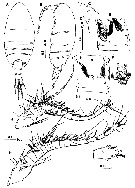 issued from : S. Ohtsuka, S. Nishida & R.J. Machida in J. Nat. Hist., 2005, 39 (27). [p.2493, Fig.4]. Female (from Sulu Sea): A-B, habitus (dorsal and lateral, respectively); C, rostrum (lateral); D, idem (anterior); E, genital double-somite (left lateral; gonopore and copulatory pore indicated by large and small arrowhead, respectively); F, idem (ventral; spematophore remnant omitted; symbols as in E); G, spermatophore remnant on genital double-somite; H, left A1; I, terminal segments of left A1; J, right A1. Scale bars in mm. Nota: Rostrum narrowly pointed with 2 fine filaments (1 missing in figure). Urosome 4-segmented. Genital double-somite asymmetrical, with longitudinal ridge on left side and paired, small papillae dorsolaterally; ventral surface with paired gonopores anteriorly (indicated by large arrowheads) and paired copulatory pores (indicated by small arrowheads) at mid-length; seminal receptacles asymmetrically arranged, packed with sperm; spematophore remnant tinged brownish, covering copulatory pores, and having paired, bulbous, well-chitinized ducts, each connected to copulatory pore. Caudal rami symmetrical, fringed with long setules along inner margin. A1 asymmetrical, with left about 1.3 times as long as right; 1st (I-III) to 8 (X) segments fringed with long setules along posterior margin (only scars remaining on some segments in fig.4H, I); left A1 22-segmented. Right A1 with same fusion pattern and armature as left.
|
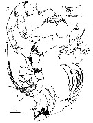 issued from : S. Ohtsuka, S. Nishida & R.J. Machida in J. Nat. Hist., 2005, 39 (27). [p.2494, Fig.5]. Female: A, A2; B, Md (mandibular cutting edge); C, Md (mandibular palp); D, Mx1 (slightly streched); E, Mx2; F, Mxp. Scale bars in mm. Nota: A2: basis with inner seta at inner distal corner; endopod 2-segmented, 1st segment with seta at distal 1/3 length and patch of minute spinules distally; 2nd segment with 1 naked, 1 serrate and 1 plumose setae subterminally, 1 short and 5 long setae terminally, and patch of minute spinules along outer margin; exopod indistinctly 7-segmented, with setal formula 0, 0, 1, 1,2, 1, 3 (1 vestigial). Md without patch of long setules on gnathobase, cutting edge with 2 patches of minute spinules and 3 teeth, dorsalmost bicuspid; mandibular palp with ebdopod rudimentary, 1-segmented, with 1 naked, short and 1 plumose, long setae terminally; exopod 5-segmented, with setal formula 1, 1, 1, 1, 2; terminal setae well developed. Mx1 with 5 naked spines and 1 process on praecoxal arthrite; coxal endite with short seta; coxal epipodite bearing 8 setae; basal endite with minute seta; endopod 1-segmented, bulbous, with 3 unequal setae, gradually increasing in size distally; exopod 1-segmented, lamellar, bearing 3 terminal setae. Mx2 stout; 1st praecoxal endite with 2 unequal spinulose setae and vestigial element; 2nd praecoxal to 2nd coxal endites each bearing 2 spinulose setae; basis relatively elongate; basal spine relatively long and naked; endopod 4-segmented, with setal formula 1, 3, 2, 2, all bearing longitudinal row of fine spinules. Mxp with syncoxa bearing 1 middle and 2 subterminal serrate setae, and terminal patch of short spinules and subterminal patch of long ones; basis shorter than syncoxa, with 2 subterminal serrate setae, patch of long setules midway, and longitudinal patch of spinules; 1st endopodal segment almost separate from basis, with serrate seta; 2nd to 5th segments bearing 4, 4, 3, and 3 setae, respectively; 6th segment with setae a and b not reduced.
|
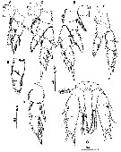 issued from : S. Ohtsuka, S. Nishida & R.J. Machida in J. Nat. Hist., 2005, 39 (27). [p.2495, Fig.6]. Female: A, P1 (anterior); B, endopod of P1 (anterior); C, exopod of P1 (anterior); D, P2 (anterior); E, P3 (anterior); F, P4 (posterior); G, P5 (posterior). Scale bars in mm. P5 slightly asymmetrical; coxae and intercoxal sclerite only partly coalescent; basis bearing thick, plumose seta at outer distal corner (left seta missing); endopod represented by acutely pointed process with 1 terminal and 1 subterminal plumose setae; exopod indistinctly 3-segmented, with suture visible on both sides; 1st and 2nd exopodal segments each bearing serrate spine, near base of which 2 processes present; 3rd exopodal segment with proximal spiniform seta, subterminal serrate spine, and 2 terminal serrate spines.
|
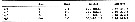 issued from : S. Ohtsuka, S. Nishida & R.J. Machida in J. Nat. Hist., 2005, 39 (27). [p.2497]. Female: Armature formula of swimming legs P1 to P4. Roman numerals = spines; Arabic numerals = setae.
| | | | | NZ: | 1 | | |
|
Carte de distribution de Protoparamisophria biforaminis par zones géographiques
|
| | | | Loc: | | | Sulu Sea (central)
Type locality: 08°57.31'N, 120°11.31'E. | | | | N: | 1 | | | | Lg.: | | | (983) F: 2,28; {F: 2,28} | | | | Rem.: | hyperbenthic (1516 m).
For Ohtsuka & al. (2005, p.2497), the asymmetrical A1 and stout spines on the legs strongly suggest that it is a truly hyperbenthic calanoid (see in Bradford-Grieve, 2002). | | | Dernière mise à jour : 29/01/2015 | |
|
|
 Toute utilisation de ce site pour une publication sera mentionnée avec la référence suivante : Toute utilisation de ce site pour une publication sera mentionnée avec la référence suivante :
Razouls C., Desreumaux N., Kouwenberg J. et de Bovée F., 2005-2026. - Biodiversité des Copépodes planctoniques marins (morphologie, répartition géographique et données biologiques). Sorbonne Université, CNRS. Disponible sur http://copepodes.obs-banyuls.fr [Accédé le 31 mars 2026] © copyright 2005-2026 Sorbonne Université, CNRS
|
|
|
|


;)
;)
;)
