|
|
 |
Fiche d'espèce de Copépode |
|
|
Calanoida ( Ordre ) |
|
|
|
Arietelloidea ( Superfamille ) |
|
|
|
Arietellidae ( Famille ) |
|
|
|
Paraugaptiloides ( Genre ) |
|
|
| |
Paraugaptiloides mirandipes Ohtsuka, Nishida & Machida, 2005 (M) | |
| | | | | | | Ref.: | | | Ohtsuka & al., 2005 (p.2498, Descr.M, figs.M) | 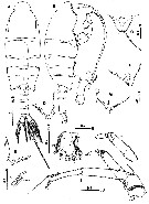 issued from : S. Ohtsuka, S. Nishida & R.J. Machida in J. Nat. Hist., 2005, 39 (27). [p.2499, Fig.7]. Male (from Sulu Sea): A-B, habitus (dorsal and lateral, respectively); C, rostrum (anterior; right rostral filament missing); D, F, right distal corner of prosome (dorsal and lateral, respectively); E, G, left distal corner of prosome (dorsal and lateral, respectively); H, left caudal ramus (dorsal); I, A2 exopod (setae omitted); K, terminal portion of A2 endopod. Scale bars in mm. Nota : Body relatively weakly chitinized expcept for P5. Prosome about 2.3 times as long as urosome. Cephalosome produced posterolaterally to cover anterior part of 1st pediger. Rostrum produced posteriorly, with pair of long filaments. 5th pediger produced posteriorly at each side into irregular processes furnished with glandular openings and hair-like sensillae ; posterior margin of processes fringed with fine setules. large spermatophore visible, extending from 1st pediger to genital somite. Urosome 5-segmented.Genital somite with gonopore opening at right posterolateral corner. 2nd and 3rd urosomites almost equal in length. Anal somite relatively conspicuous. Caudal rami symmetrical, fringed with long setules on both sides ; rudimentary seta I present, seta V longest. A2 with unarmed coxa ; basis with inner seta terminally; endopod 2-segmented, 1st segment unarmed, 2nd segment with 3 inner setae of unequal length and 1 reduced and 5 well-developed terminal setae; exopod indistinctly 7-segmented, setal formula 0, 1, 1, 1, 1, 1, 3 (1 vestigial).
|
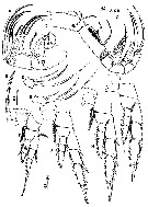
 issued from : S. Ohtsuka, S. Nishida & R.J. Machida in J. Nat. Hist., 2005, 39 (27). [p.2500, Fig.8]. Male: A, 1st to 12th segments of left geniculate A1; B, 13th to 19th segments of left A1; C, proximal spiniform element on A1 segment XIX; D, distal spiniform element on left geniculate A1 segment XIX; E, anterior spiniform element on left geniculate A1 segment XX; Fterminal segments of left geniculate A1; G, Md; H, Mx1. Scale bars in mm. Nota : Left A1 geniculate, 19 segmented, with 1st (I-IV) and 2nd (V) posteriorly fringed with fine setules ; 1st to 5th (VIII) segments each with square plate-like structure posterodistally ; geniculation positioned between segments 17 (XX) and 18 (XXI-XXIII) ; large anterodistal process originating from compound segment XXIV-XXV, reaching beyond antennulary tip ; terminal three segments corresponding to ancestral XXIV-XXVIII incompletely fused with fusion lines visible clearly. Right A1 23-segmented, reaching to anal somite seta terminally ; Md with 2 mono- and 1 tricuspid teeth and 2 patches of setules along cutting edge ; patch of long setules present near base of palp ; endopod absent, but possibly represented by round prominence ; exopod 5-segmented, setal formula 1, 1, 1, 1, 2. Mx1 with praecoxal arthrite bearing 5 serrate spines and 1 process ; coxal endite with 1 serrate seta ; coxal epipodite with 8 setae (2 of which relatively short) ; basis with rows of long setules and vestigial seta ; endopod 1-segmented, bulbous, with 2 serrate unequal setae ; exopod 1-segmented, lobate, with 3 long setae terminally.
|
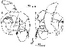
 issued from : S. Ohtsuka, S. Nishida & R.J. Machida in J. Nat. Hist., 2005, 39 (27). [p.2501, Fig.9]. Male: A, Mx2; B, basal spine of Mx2; C, Mxp (some setae on endopod omitted); D, 4th endopodal segment of Mxp; E, 5th endopodal segment of Mxp; F, P2 (anterior); G, P2 (anterior); H, P3 (anterior); I, P4 (anterior). Scale bars in mm. Nota : Mx2 with 1st praecoxal endite bearing 2 setae and 1 rudimentary element ; 2nd praecoxal to 2nd coxal endites each with 2 serrate setae ; basis bearing long spine with longitudinal row of fine spinules ; endopod indistinctly 4-segmented, all setae on which well developed, setal formula 1, 3, 2, 2. Mxp with syncoxa bearing 1 middle and 2 subterminal serrate setae and subterminal patch of minute spinules ; basis as long as syncoxa, with 2 serrate setae and subterminal patch of fine spinules ; 1st endopodal segment nearly incorporated into basis, with serrate seta ; 2nd to 5th endopodal segments bearing 4, 4, 3 and 3 setae, respectively ; 5th endopodal segment slightly longer than preceding segment ; terminal endopodal segment with setae a and b rudimentary.
|
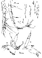
 issued from : S. Ohtsuka, S. Nishida & R.J. Machida in J. Nat. Hist., 2005, 39 (27). [p.2502, Fig.10]. Male: A, P5 (anterior); B, idem (posterior); C, tip of proximal element on 3rd exopodal segment of left P5. Scale bars in mm. Nota : P5 highly complex, enlarged, heavily chitinized. Coxae and intercoxal sclerite completely fused. Lef leg with basis bearing plumose subterminal seta on posterior surface ; endopod distinctly 2-segmented, located near inner distal corner of basis, 1st segment diamond-shaped, 2nd segment originating from inner midpoint of preceding segment ; exopod 3-segmented, 1st segment elongate, with minute element at outer distal corner ; 2nd segment massive , expanded outwards ; 3rd segment strongly curved, bearing proximal short seta, middle curved spine with spatulate tip (fig.10C), and terminal bifurcate process. Right leg with basis bearing plumose seta on posterior surface ; endopod 1-segmented with broad base along inner margin of basis, furnished with fine setules subterminally on posterior surface ; exopod indistinctly 3-segmented, 1st segment curved inwards at mid-length, carrying 2 round processes on anterior surface and triangula router distal process with fine seta ; 2nd segment extremely elongated, as long as basis and 1st exopodal segment combined, inserted on inner subterminal corner of preceding segment, and bearing short seta at mid-length of inner margin, distal patch of fine setules and subterminal round process with minute spinule at tip ; 3rd segment lamellar with 3 minute setae along distal margin.
Unidentified ciliates were attached near the distal patch of fine setules of the 2nd exopodal segment of the right P5.
| | | | | NZ: | 1 | | |
|
Carte de distribution de Paraugaptiloides mirandipes par zones géographiques
|
| | | | Loc: | | | Indonesia-Malaysia: Sulu Sea (central)
Locality type: 08°52.66'N, 120°25.28'E. | | | | N: | 1 | | | | Lg.: | | | (983) M: 5,26; {M: 5,26} | | | | Rem.: | hyperbenthic (2430-2450 m). | | | Dernière mise à jour : 28/01/2015 | |
|
|
 Toute utilisation de ce site pour une publication sera mentionnée avec la référence suivante : Toute utilisation de ce site pour une publication sera mentionnée avec la référence suivante :
Razouls C., Desreumaux N., Kouwenberg J. et de Bovée F., 2005-2026. - Biodiversité des Copépodes planctoniques marins (morphologie, répartition géographique et données biologiques). Sorbonne Université, CNRS. Disponible sur http://copepodes.obs-banyuls.fr [Accédé le 17 juin 2026] © copyright 2005-2026 Sorbonne Université, CNRS
|
|
|
|


;)
;)
;)
;)
