|
|
 |
Fiche d'espèce de Copépode |
|
|
Calanoida ( Ordre ) |
|
|
|
Clausocalanoidea ( Superfamille ) |
|
|
|
Stephidae ( Famille ) |
|
|
|
Stephos ( Genre ) |
|
|
| |
Stephos vivesi Jaume, Boxshall & Gràcia, 2008 (F,M) | |
| | | | | | | Ref.: | | | Jaume & al., 2008 (p.504, figs.M, F); S.Y. Moon & al., 2015 (p.31: Rem.) | 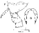 issued from : D. Jaume, G.A. Boxshall & F. Gràcia in Systematics and Biodiversity, 2008, 6 (4). [p.506, Fig.2, F, G]. Female (from Majorca, Balearic Islands): F, last prosomie with attached P5, genital double-somite and 1st free abdominal somite (left lateral); G, P5 (anterior). Nota : P5 symmetrical, uniramous, 3-segmented with proximal segment fused to intercoxal sclerite. 1st and 2nd segments unarmed ; 3rd segment longer than previous 2 combined, with tapering distal half ; tiny seta and pore positioned submarginally on anterior surface about midway along segment, seta close to lateral margin, pore close to medial margin ; 2 rows of denticles along tapering portion of segment.
|
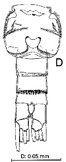 issued from : D. Jaume, G.A. Boxshall & F. Gràcia in Systematics and Biodiversity, 2008, 6 (4). [p.507, Fig.3, D]. Female: D, urosome (ventral). Nota : Urosome 4-segmented. Genital double-somite adorned with several rows of ventrolateral and dorsolateral rows of spinules.
|
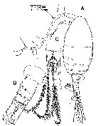
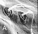
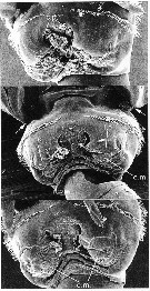 issued from : D. Jaume, G.A. Boxshall & F. Gràcia in Systematics and Biodiversity, 2008, 6 (4). [p.511, Fig.8]. Females: SEM micrographs ventral views of the genital double-somite in different specimens showing sequence of stages of extrusion of an unidentified material. Arrows indicate the tiny terminal spinule on the inner margin of the last prosomite; c.p. = copulatory pores; e;M. = extruded material; l.o. = left genital operculum; r.o. = right genital operculum. Nota: Reproductive system showing paired arrangement, with paired genital opercular plates located ventrally, each partially concealing copulatory pore, placed ventromedially. Genital opercular plates capable of retraction.
|
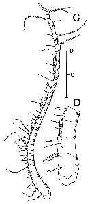 issued from : D. Jaume, G.A. Boxshall & F. Gràcia in Systematics and Biodiversity, 2008, 6 (4). [p.508, Fig.4, C-D]. Female: C, left A1 (ventral); D, detail of proximal part of latter. Scale bar: 0.05 mm.
|
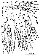
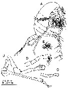 issued from : D. Jaume, G.A. Boxshall & F. Gràcia in Systematics and Biodiversity, 2008, 6 (4). [p.506, Fig.2, A-E]. Male: A, body (lateral); B, P5 (anterior); C, detail of distal segment of right leg (lateral); D, detail of distal segment of left leg (medial); E, detail of distal segment of left leg (ventral = anterior). Nota: P5 strongly asymmetrical, uniramous and filiform, both longer than urosome. Left leg 5-segmented, segments 2 and 3 each with rounded outgrowth on medial margin; 5th segment short and rounded, reflexed posteriorly, with row of 8 unequal long hyaline lamellae on distal margin and patch of short, stout spinules on posterior surface. Right leg 4-segmented; segment 3 very elongate, (segment longer than urosome), straight and slender except slightly expanded proximal part, with small pointed process proximally on lateral margin; segment 4 roughly spatulate, with 2 rounded outgrowths proximally on anterior surface, tiny spine proximally on lateral margin, and finely denticulated distomedial margin.
|
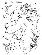
 issued from : D. Jaume, G.A. Boxshall & F. Gràcia in Systematics and Biodiversity, 2008, 6 (4). [p.507, Fig.3, A-C]. Male: A, body (dorsal); B, urosome (dorsal); C, detail of caudal rami (ventral). Nota : Long sensilla present on each side of 3rd pedigerous siomite. Somites 4 and 5 completely fused, with posterolateral corners rounded and slightly asymmetrical in dorsal aspect, extending posteriorly slightly further on right side. Urosome 5-segmented, with all somites except anal somite about same length. Genital somite slightly produced laterally on left side, with gonopore opening posterolaterally on left side ventral surface ; ornamentation of somite reduced to cluster of few short spinules anterodorsally on right margin of somite. Posterior hyaline frill of 4th urosomite transformed progressively into row of long spinules ventrally and ventrolaterally (as in female). Anal somite short, with scarcely developed, hyaline operculum. Caudal rami symmetrical, 1.7 times longer than wide, with 4 distal setae and 1 reduced seta (seta VII) on outgrowth midway along medial margin ; no trace of seta I.. Distal setae short (just longer than urosome) and stiff, spinulose, distolateral seta (seta III) slightly shorter than rest. Internal tissue of distal setae finely corrugated.
|
 issued from : D. Jaume, G.A. Boxshall & F. Gràcia in Systematics and Biodiversity, 2008, 6 (4). [p.508, Fig.4, A-B]. Male: A, forehead (ventral), showing rostrum, proximal segment of A1 and labrum; B, right A1 (ventral). Nota : Rostrum hardly developed, rounded, with two bifid sensillae frontally. A1 long and symmetrical, extending beyond posterior margin of prosome, non-geniculate, 23-segmented with failure to express articulations between segments I-IV (Segment 1 corresponding to compound ancestral segment I-IV ; although vestige of articulation between segments II and III expressed dorsally), X-XI and XXVII-XXVIII.
|
 issued from : D. Jaume, G.A. Boxshall & F. Gràcia in Systematics and Biodiversity, 2008, 6 (4). [p.509, Fig.5]. Male: A, right A2 (lateral); B, Md (mandibular gnathobase); C, Md (mandibular palp); D, right Mx1 (anterior); E, right Mx2 (with disarticulated free ebdopod; lateral); F, left Mxp (anteromedial). Nota : A2 exopod indistinctly 7-segmented, with intersegmental articulation between segments 2 and 3 not completely expressed. Endopod 2-segmented ; distal segment expanded subdistally into medial lobe bearing 8 setae, and with distal portion crowned with 7 setae, and transverse row of long spinules subdistally on lateral margin. Mandibular gnathobase with row of spinules subdistally on ventral margin. Cutting edge comprising 8 teeth plus dorsal spinulose seta ; ventralmost tooth largest, smooth and unicuspid ; 3 adjacent teeth with row of 3 small denticles, unicuspid ; 4 dorsal teeth reduced, tricuspidate, with transverse comb of long spinules basally. Mandibular palp biramous ; basis with 4 setae on inner margin, proximalmost seta inipinnate, others smooth ; exopod indistinctly 5-segmented, setal formula 1, 1, 1, 1, 2. Endopod 2-segmented, proximal with 4 setae on distomedial angle, distal segment with 10 setae. Mx1 with praecoxal arthrite carrying 8 marginal spines plus 4 stiff setae on posterior surface ; 2 combs of short spinules located transversely on ventral margin of arthrite. Coxal epipodite with 9 setae ; coxal endite with 3 stiff setae. Basis with cluster of 6-7 denticles on anterior sutrface ; proximal basal endite with 4 setae ; distal basal endite indistinct, with 5 setae ; no trace of basal exite. Exopod with 11 marginal setae, and row of setules along distal portion of medial margin. Endopod not articulated to basis, indistinctly 3-segmented, setal formula 4, 4, 7. Mx2 indistinctly 6-segmented, comprising partially coalesced praecoxa and coxa, allobasis and 3-segmented endopod. Armature of praecoxal and coxal endites 5, 3, 3, 3, respectively. Basal endite with 4 setae, 1 stouter than rest ; endopodal endite with 1 seta on tip. Free endopod setal formula 1, 1, 3, respectively. Integument of praecoxa ornamented with patch of denticles on posterior margin. Praecoxal and coxal endites with cluster of long spinules subdistally on lateral surface ; distal coxal endite with additional row of denticles proximally on medial surface. Mxp 7-segmented with syncoxa, basoendopod and free 5-segmented endopod. Syncoxa ornamented with patch of short denticles and 2 rows of spinules on posterior surface. Basoendopod with 3+2 setae, medial row of hyaline lamellae submarginally on anterior surface of segment. Free endopod setal formula 4, 4, 3, 3 + 1, 3 +1.
|
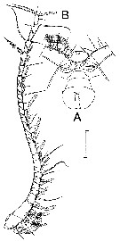
 issued from : D. Jaume, G.A. Boxshall & F. Gràcia in Systematics and Biodiversity, 2008, 6 (4). [p.510, Fig.6]. Male: A, left P1 (anterior); B, detail of ornamentation on posterior side of inner basal seta of latter; C, P2 (anterior); D, P3 (anterior); E, P4 (posterior). Nota : Swimming legs 1-4 progressively larger towards posterior. Most setae on all legs with internal tissue finely corrugated.
|
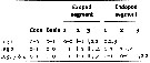 issued from : D. Jaume, G.A. Boxshall & F. Gràcia in Systematics and Biodiversity, 2008, 6 (4). [p.509]. Male: Armature of the swimming legs P1 to P4. (Roman numerals = spines; Arabic numerals = setae).
|
 issued from : D. Jaume, G.A. Boxshall & F. Gràcia in Systematics and Biodiversity, 2008, 6 (4). [p.511, Fig.7, A. Female (SEM micrographs): A, detail of frontal sensillae. Nota: Note bifid sensillae as in male description.
| | | | | NZ: | 1 | | |
|
Carte de distribution de Stephos vivesi par zones géographiques
|
| | | | | | | Loc: | | | W Medit. (Balearic Islands, Majorca: Cova des Coll) | | | | N: | 1 | | | | Lg.: | | | (1017) F: 0,44-0,45; M: 0.44-0.45; {F: 0,44-0,45} | | | | Rem.: | In Cova des Coll (E Majorca), typical mixing-zone cave excavated in an upper Miocene coral reef, with a direct connexion connection to the sea.
Jaume & al. (2008, p.511) points to the species exhibits an external configuration of the genital system for the genus: paired genital opercular plates located ventrally, each partially concealing copulatory pore, placed ventromedially; genital opercular plates capable of retraction. All other stephids for which reliable information is available possess a single genital operculum covering both genital apertures.
The species belongs to ''group IV'' (see genus Stephos).
Thie species name by the authors to honour Dr Francisco Vives from Palma de Mallorca. | | | Dernière mise à jour : 14/08/2015 | |
|
|
 Toute utilisation de ce site pour une publication sera mentionnée avec la référence suivante : Toute utilisation de ce site pour une publication sera mentionnée avec la référence suivante :
Razouls C., Desreumaux N., Kouwenberg J. et de Bovée F., 2005-2026. - Biodiversité des Copépodes planctoniques marins (morphologie, répartition géographique et données biologiques). Sorbonne Université, CNRS. Disponible sur http://copepodes.obs-banyuls.fr [Accédé le 23 juin 2026] © copyright 2005-2026 Sorbonne Université, CNRS
|
|
|
|


;)
;)
;)
;)
;)
;)
;)
;)
;)
;)

