|
|
 |
Fiche d'espèce de Copépode |
|
|
Calanoida ( Ordre ) |
|
|
|
Clausocalanoidea ( Superfamille ) |
|
|
|
Diaixidae ( Famille ) |
|
|
|
Procenognatha ( Genre ) |
|
|
| |
Procenognatha semisensata Markhaseva & Schulz, 2010 (F,M) | |
| | | | | | | Ref.: | | | Markhaseva & Schulz, 2010 (p.5, figs.F, M); Laakmann & al., 2019 (p.330, fig. 2, 4, phylogenetic relationships) | 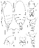 issued from : E.L. Markhaseva & K. Schulz in Proc. Zool. Inst. RAS, 2010, 314 (1). [p.6, Fig.1]. Female (from 00°45'N, 05°35'W): A-B, habitus (lateral and dorsal, respectively); C, rostrum (ventral); D, same (lateral); E, posterior prosome and urosome (dorsal); F-G, same (lateral); H, genital double-somite (ventral); . Scale bars: 0.5 mm = A-B; 0.1 mm = C-H. Holotype: A, B, G, H; paratype: C, D, F. Nota : - Cephalosome and 1st pediger fused, 4th and 5th incompletely separate. - Rostrum as a poorly developed plate with 2 filaments. - Posterior corners prolonged into rounded lobes extending to mid-length of genital double-somite. - Genital double-somite slightly asymmetrical, more prominent on the right dorsal view. - Genital double-somite and urosome somites 2 and 3 with fringe of spinules along posterior borders. - Caudal rami with 4 terminal plus 1 small dorsolateral and 1 small ventral seta each. - A1 longer than prosome, 24 free segments.
|
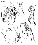 issued from : E.L. Markhaseva & K. Schulz in Proc. Zool. Inst. RAS, 2010, 314 (1). [p.7, Fig.2]. Female: A, A2 (coxa, basis, endopod); B, A2 (exopod); C, Mx2 (brush-like setae not shown); D, E, F, endopod (different limbs). Scale bars 0.1 mm Holotype: A-E; paratype: F. A2 exopod 8-segmented. Mx2 : praecoxal endite bearing 5 setae, coxal (previously considered as distal praecoxal endite) with 3 setae ; basal endites (previously considered as coxal endites) with 3 setae each ; lobe of proximal endopodal segment (previously considered as proximal basal endite) with 4 setae, all sclerotized ; endopod with 9 setae, 3 terminal setae sclerotized and 6 brush-like setae of different morphology , the longest, poorly sclerotized, seta plumose from one side,with poorly developed brush-like head, 1 short sensory seta, with poorly developed brush on top, and 4 well-developed brush-like sensory setae (1 thick and 3 thin).
|
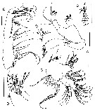 issued from : E.L. Markhaseva in Proc. Zool. Inst. RAS, 2010, 314 (1). [p.8, Fig.3]. Female: A, Md; B, Md (cutting edge); C, Mx1; D, Mx1 (praecoxal arthrite); E, Mxp. Scale bars 0.1 mm. Holotype: A, C, E; paratype: B, D. Mx1 : praecoxal arthrite with 9 terminal spines, 4 posterior setae and 1 anterir seta, proximalmost posterior seta swollen in its first half (markegd by arrow) ; coxal endite with 3 setae, coxal epipodite with 9 setae ; proximal basal endite with 4 setae, distal basal endite with 5 setae ; endopod with 12 setae , exopods with 7 setae (right limb of one of paratypes with 9 setae). Mxp : 3 setae on distal praecoxal endite, of them the distalmost with poorly developed brush-like head.
|
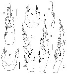 issued from : E.L. Markhaseva & K. Schulz in Proc. Zool. Inst. RAS, 2010, 314 (1). [p.10, Fig.4]. Female: A, P1; B, P1 (exopod segment 3); C, P2; D, P3; E, P4; F-G, P5. Nota: - P5 uniramous, symmetrical, 3-segmented with 1 lateral and medial spine each and 2 spine-like unarticulated and unequal extensions terminally.
|
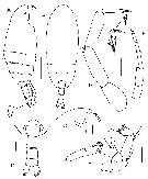 issued from : E.L. Markhaseva & K. Schulz in Proc. Zool. Inst. RAS, 2010, 314 (1). [p.11, Fig.5]. Male: A-B, habitus (lateral and dorsal, respectively); C, dorehead (lateral); D, posterior prosome and urosome (dorsal); E, A2; F, right P5; G, left P5. Scale bars: A, B = 0.5 mm; C-G = 0.1 mm. Nota: - A2, Md, Mx1, Mx2, Mxp and P2-P4 similar to those of female. - P5 biramous, exopods 3-segmented on both sides, endopods bud-like, rudimentary. Left leg longer than right ; exopod segment 2 with spinules distally, exopod segment 3 ornamented with posterior and terminal spinules. Right leg, exopodal segment 1 with lateral distal spine, segment 3 with 2 slightly unequal terminal attenuations.
|
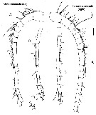 issued from : E.L. Markhaseva & K. Schulz in Proc. Zool. Inst. RAS, 2010, 314 (1). [p.12, Fig.6]. Female (paratype): A, right A1 (segments I-XVIII); B, same (segments XIX-XXVIII). A1 24-segmented, longer than prosome Male (paratype): C, left A1 (segments I-XVII); D, same (segments XVII-XXVIII incompletely separate). Scale bar 0.1 mm.
|
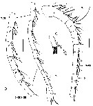 issued from : E.L. Markhaseva in Proc. Zool. Inst. RAS, 2010, 314 (1). [p.14, Fig.7]. Male (paratype): A, right A1 (segments I-XV); B, same (segments XVI-XXVIII); C, same (segments XVII-XXII, enlarged). Scale bars 0.1 mm. Nota : - A1 nearly as long as body ; left A1 23-segmented ; right A1 22 segmented, with trace of geniculation (proximal excavation on each of segments XVIII to XXIII (marked arrows) and additional medial spine-like seta present on each of XVII to XX segments. - P5biramous, exopods 3-segmented on both sides; endopods bud-like, rudimentary. Left leg longer than right; exopodal segment 2 with spinules distally; exopoal segment 3 ornamented with posterior and terminal spinules;. Right leg, exopodal segment 1 with lateral distal spine, segment 3 with 3 slightly unequal terminal attenuations.
|
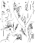 issued from : E.L. Markhaseva & K. Schulz in Proc. Zool. Inst. RAS, 2010, 314 (1). [p.15, Fig.8]. Male (paratype): A, Md (palp); B, Md (cutting edge); C, Mx1 (praecoxal arthrite and coxa); D, Mx1 (basis, endopod and exiopod); E, Mx2 (proximal praecoxal endite); F, Mx2 (endopod and distal basal endite; setae of distal basal endite not figured); G, Mxp; H, P1 (endopod); I, P1 (lateral spines of exopod segments 1-3). Scale bars 0.1 mm.. A2, Mx1, Mx2, Mxp and P1 and P2 similar to those of female.
| | | | | NZ: | 1 | | |
|
Carte de distribution de Procenognatha semisensata par zones géographiques
|
| | | | Loc: | | | Atlantic (00°45'N-00°39'S, 05°35-02°30'' W) | | | | N: | 1 | | | | Lg.: | | | (1050) F: 2,5-2,75; M: 1,95-2,3; {F: 2,50-2,75; M: 1,95-2,30} | | | | Rem.: | above the sea bed: 5050-5169 m.
Remarks from Markhaseva & Schulz (2010, p.5) :
Praecoxal arthrite of Mx1 with proximalmosst posterior seta swollen in its basal aprt (vs. Not swollen in other ‘bradfordians’) ; the latter character is an apomorphy of the genus.
Mx2 endopod with 3 sclerotized terminal setae (vs. Worm-like sensory setae in remaining ‘bradfordian genera’). Endopod with 9 setae (coposition : 3scl + 1scl /br + 5br).
The male is distinguished by the right A1 with traces of geniculation.
A high number and unique combination of plesiomorphic characters (ancestral or primitive characteristics) exhibited by Procenognatha semisensata (type species) suggest that this species is the most primitive among ‘bradfordian taxa’ : ancestral setation of A2 exopod (1, 1-1-1, 1, 1, 1, 1, 1 and 3) ; endopod with a total number of 9 setae ; presence of 3 sclerotized setae on endopod terminally (a unique plesiomorphy for ‘bradfordians’.
Syncoxa of Mxp with ancestral setation formula (1, 2, 3).
A1 male with traces of geniculation, oral parts not reduced, and biramous P5.
The new genus and Cenognatha are closely related and share number of characters. | | | Dernière mise à jour : 30/07/2019 | |
|
|
 Toute utilisation de ce site pour une publication sera mentionnée avec la référence suivante : Toute utilisation de ce site pour une publication sera mentionnée avec la référence suivante :
Razouls C., Desreumaux N., Kouwenberg J. et de Bovée F., 2005-2026. - Biodiversité des Copépodes planctoniques marins (morphologie, répartition géographique et données biologiques). Sorbonne Université, CNRS. Disponible sur http://copepodes.obs-banyuls.fr [Accédé le 28 février 2026] © copyright 2005-2026 Sorbonne Université, CNRS
|
|
 |
 |


