|
|
 |
Fiche d'espèce de Copépode |
|
|
Monstrilloida ( Ordre ) |
|
|
|
Monstrillidae ( Famille ) |
|
|
|
Maemonstrilla ( Genre ) |
|
|
| |
Maemonstrilla hyottoko Grygier & Ohtsuka, 2008 (F) | |
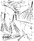
| | | | | | | Ref.: | | | Grygier & Ohtsuka, 2008 (p.463, figs.F) | 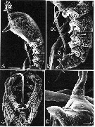 issued from : M.J. Grygier & S. Ohtsuka in Zool. J. Linnean Soc., 2008, 152. [p.464, Fig.2]. SEM . Female (from Sesoko Island): A, cephalothorax and metasome (lateral, left legs removed); B, metasome and urosome of same specimen (lateral), showing ovigerous spines (os) and leg 5 (numbered); C, metasome urosome of different specimen (ventral), showing ovigerous spines (small arrow), widely spaced legs 1-4, and low, wide intercoxal sclerites (large arrow): D, genital compound somite of specimen in A, ventrolateral view, showing base of ovigerous spines, copulatory opening (arrow), and spur-like posteroventral process. Scale bars = 0.200 mm (A); 0.100 mm (B, C); 0.020 mm in D. Nota: - Cephalothorax distinctly bulbous anteriorly; incorporated 1st pediger widening again in dorsal view behind distinct 'waist' (fig.1B). - Body length in lateral view (sum of lengths of cephalothorax, metasome and urosome (as defined in fig.1A) 49.6-52.1, 28.1-31.2 and 19.2-20.7 %, respectively. - Height and greatest width of cephalothorax 49.2-53.9 and 50.8-54.3 % of cephalothorax length respectively. - Lateral cups of naupliar eye about same size as ventral cup, about 80 µm in diameter. - P1-P4 each with 2 arcuate sclerites laterally at base of leg (fig.2B). Intercoxal sclerites low and wide (arrow) with ventrally produced outer corners (fig.2C). Protopod with longitudinal groove on anterior face. - Ovigerous spines arising from conical projection of ventral surface of anterior half of genital compound segment (fig.2B, C). Crescent-shaped slit leading to copulatory opening at posterior base of cone (arrow fig.2D); slit flanked by pair of small knobs projecting from cone. Tips of ovigerous spines found at any point between P2 and midlength of cephalothorax; spines cylindrical, smooth, but slightly thickened and wrinkled at two-thirds length, tips naked and tapered. Posteroventral protrusion of genital compound somite large, subconical, spur-like (figs.2D, 4G).
|
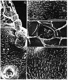 issued from : M.J. Grygier & S. Ohtsuka in Zool. J. Linnean Soc., 2008, 152. [p.465, Fig.3]. SEM . Female: A, 'face' and left A1 (a), ventral view, showing oral papilla (o), scars (s), 3 pairs of pores (arrows), and segments of A1 (1-4).; B, anterodorsal surface of cephalothorax (anterior at bottom), showing pores (small arrows) and pit-like organ (large arrow); C, detail of pit-like organ in B, D, pores (arrows) at border between narrow and wide reticular meshes in posterior half of cephalothorax, posterior to right. Scale bars = 0.050 mm (A, D); 0.100 mm in B; 0.010 mm in C. Nota: - Oral papilla very large, funnel-shaped, directly ventrally and preceded by pair of simple pores. Pair of tubular pores, with 4 spines on lip of each pore, situated further anteriorly between posterior ends of bases of A1. Cluster of 3 knob-like scars behind base of each antennule, middle knob smaller than others. - Forehead region with pair of circles formed of cuticular ridges just anterodorsal to A1 bases, smaller circle on midline just posterior to these, and pair of pores anterodorsal to large circles (pair of hair-like sensilla seen arising from these pores in some paratypes). Subcircular pit with wrinkled thin cuticle on dorsal midline of forehead, 10 µm in largest diameter (fig.3B, C). In an SEM specimen, 2 pores behind this structure, and 3 pores on each side further laterally (arrows fig.3B); numbers and positions of these pores not necessarily constant in other specimens. - 3 pairs of dorsal pores on cephalothorax at posterior boundary between narrow and broad reticular meshes (figs.3D, arrows). Arrangement of dorsal pores and dorsal and lateral pit setae from rear of cephalothorax to urosome (shown in fig. 29, fig.5A-D). - Dorsal pores on free pedigers 1-3 only. Dorsal pit setae in one widely separated pair on incorporated pediger; in 1, 2, 2 and 1 pair, respectively, on succeeding 4 free pedigers. In addition, 4 pairs of dorsolateral and lateral pit setae on incorporated pediger and 2 pairs each on 1st and 2nd free pedigers, anterior pair on each segment located more ventrally than posterior one.
|
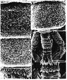 issued from : M.J. Grygier & S. Ohtsuka in Zool. J. Linnean Soc., 2008, 152. [p.467, Fig.5]. SEM . Female: A, dorsal surface of free pedigers 1 and 2; two obscured pores indicated by arrows; B, dorsal surface of free pediger 3; C, dorsal surface of urosome, including genital compound somite (g), and outer faces of legs 4; D, rear dorsal surface of free pediger 4, showing pit setae; E, dorsal surface of rear of urosome, including telson (t). Scale bars = 0.050 mm (A, B, E); 0.100 mm in C; 0.010 mm in D. Nota: - Dorsum of 1st free pediger (fig.5A) covered with sharp, posteriorly directed denticles arrayed in 6 longitudinal rows of rectangular patches, these patches numbering 3-3-7-7-3-3, respectively; those of 2nd free pediger arranged the same (fig.5A); denticle patches of 3rd free pediger fewer, 3-2-4-4-2-3 (fig.5B); and those of 4th free pediger 2-3-2-2-3-2 (fig.5C). - Dorsum of genital compound (fig.5C): anterior third with 6 rows of 3 denticle patches each; middle third with grid of cuticular ridges; posterior third with 6 rows of denticle patches arrayed 1-2-2-2-2-1, followed by band of 6 arrays of considerably larger denticles, with largest 3 or 4 denticles in each array extending beyond somite's posterior margin. - Penultimate segment (fig.5C) with transverse band of 4 denticle patches followed by 6 arrays of large denticles like those on genital compound somite. - P1-P4: Posterior faces of rami lightly reticulated (fig.5C). - Caudal rami (figs. 5-5C, 6D) lightly reticulate dorsally and with ventromedial condyle articulating to telson, anal slit visible in telson. Each ramus with ventral distal pore and 6 setae (at least 5 of them plumose), 1 arising ventrolaterally and the other 5 apically; most dorsal apical seta (seta VII of Huys & Boxshall, 1991) shorter than others, appearing smooth in some individuals but setulose in 2 specimens (perhaps setules orientated dorso-ventrally).
|
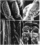 issued from : M.J. Grygier & S. Ohtsuka in Zool. J. Linnean Soc., 2008, 152. [p.468, Fig.6]. SEM . Female: A, protopods of legs 3 and 4, outer view (anterior to left); B, endopod of a swimming leg, inner view showing site of absent seta on 1st segment (arrow); C, outer apical seta (large arrow) on 3rd exopodal segment of P1 and pore (small arrow) on 3rd endopodal segment of P2 (anterior view); D, telson (t) and caudal rami (c),, ventral view), showing anal anal opening (large arrow), and ventral pores and condylar articulation (small arrow) of rami. Scale bars = 0.050 mm (A); 0.020 mm in B, D: 0.010 mm in C. Nota: - P1-P4: Pore just in front of lateral seta on basis; this seta short and hair-like in P1, P2 and P4, but much longer and plumose in P3 (fig.4D, plumosity omitted). Distal pore on anterior face of 3rd segment of each ramus (fig.6C). No inner seta on 1st exopodal or 1st endopodal segment (fig.6B). Outer spiniform seta on 1st and 3rd exopodal segment in P1-P4 simple, that of 3rd segment bigger. Sparse spinules as well as 2 rows of long setules found on natatory setae (fig.6C): 1 inner seta on 2nd segment of each ramus; 2 inner setae on 3rd segment of each ramus in P1, but 3 inner setae on exopod in P2-P4; 1 outer seta on 3rd segment of endopod, and 2 apical setae on each ramus (fig.4B-D, F). Outer apical seta of exopod with outer row of widely spaced, talon-like denticles instead setules (fig.4E,6C)
|
 issued from : M.J. Grygier & S. Ohtsuka in Zool. J. Linnean Soc., 2008, 152. [p.466, Fig.4]. Female: A, right A1 (dorsal view;, setules omitted; setal designations after Grygier & Ohtsuka, 1995, fig.6); B-D, right legs 1-3, respectively (posterior view; setules omitted); E, outer apical exopodal seta of left P3 (setules along non-toothed side omitted); F, right P4 (posterior view; setules omitted); G, 4th free pediger and genital compound somite (lateral view), showing right P5, proximal parts of ovigerous spines, and posteroventral spur. Scale bar = 0.100 mm (A, E, G); 0.020 mm (B-D, F). Nota: - A1 length 40.1-46.1 % of cephalothorax length. Clearly 4-segmented, but proximal region of 1st segment consisting of complexly folded arthropodial membrane and sclerites for muscle attachment; reticular ridges becoming lower and simpler distally. All seta elements identified by Grygier & Ohtsuka (1995) (figs.3A, 4A). Spiniform 2v-setae and 3-seta much longer than 4-setae; row of spinules present on at least 2v-seta, possibly on other spiniform spiniform setae. Outer distal b1-3 setae each branched at least twice in asymmetrically dichotomous manner; b5 seta bifid. Apical 6-setae similar to each other in size. - P1-P4: suture separating coxa and basis extending diagonally across posterior face and continuing around onto outer part of anterior face (fig.4B-D, F). Sharp, distally pointing denticles on outer faces of coxa and exopod (figs.5C, 6A), as large as denticles on posterior margin of genital, compound somite. - P5 rod-like, distally slightly clavate, with 2 apical setae (figs.4G, 2B).
|
 issued from : M.J. Grygier & S. Ohtsuka in Zool. J. Linnean Soc., 2008, 152. [p.499, Fig.29]. SEM. Female: Dorsal and lateral pore and pit seta patterns, from rear of cephalothorax through genital compound somite. Symbols: dots (three sizes) = pores; larger circles = pits of pit setae. Pattern based on SEM and light microscopical examination.
|
 Issued from : M.J. Grygier & S. Ohtsuka in Zool. J. Linnean Soc., 2008, 152. [p.493]. Key to the Ryukyu species of the Maemonstrilla hyottoko species-Group. M. hyottoko Female: 1 - Cuticle of cephalothorax, A1, lateral sides of trunk, dorsum telson, and caudal rami reticulated; outer faces of P1-P4 and dorsum of free pedigers, genital compound somite, and penultimate somite spinulose. Cephalothoracic reticulations comprising ridges with abundant or sparse spinules; simple or complex cuticular ornamentation within at least some meshes. 2 - Oral papilla large and conical, directed ventrally. Genital compound somite distinctly divided transverse ridge. 3 - P1-P4 each lacking with two low, coxal rounded lobes; outer face of coxa evenly covered with large spines. Cephalothoracic reticulations with many side branches; besides polygonal meshes anteriorly and posteriorly, wide zone of narrow meshes present behind level of oral papilla.
Compare with M. simplex, M. okame, M. polka, M. spinicoxa.
| | | | | NZ: | 1 | | |
|
Carte de distribution de Maemonstrilla hyottoko par zones géographiques
|
| | | | | | | Loc: | | | S Japan (Sesoko Island, Ishigaki Is., Myako Is.: Hirara Port)
Type locality: 26°38.2' N, 127°51.8' E. | | | | N: | 1 | | | | Lg.: | | | (1077) F: 1,15-1,60 *
*: Sum of lengths of cephalothorax, metasome and urosome, in lateral view. | | | Dernière mise à jour : 23/04/2020 | |
|
|
 Toute utilisation de ce site pour une publication sera mentionnée avec la référence suivante : Toute utilisation de ce site pour une publication sera mentionnée avec la référence suivante :
Razouls C., Desreumaux N., Kouwenberg J. et de Bovée F., 2005-2026. - Biodiversité des Copépodes planctoniques marins (morphologie, répartition géographique et données biologiques). Sorbonne Université, CNRS. Disponible sur http://copepodes.obs-banyuls.fr [Accédé le 22 juin 2026] © copyright 2005-2026 Sorbonne Université, CNRS
|
|
|
|


;)
;)
;)
;)
;)
;)

