|
|
 |
Fiche d'espèce de Copépode |
|
|
Cyclopoida ( Ordre ) |
|
|
|
Oncaeidae ( Famille ) |
|
|
|
Triconia ( Genre ) |
|
|
| |
Triconia denticula Wi, Shih & Soh, 2011 (F) | |
| | | | | | | Ref.: | | | Wi, Shih & Soh, , 2011 (p.590, Descr. F, figs.F); Soh & al., 2013 (p.119, figs.F) | 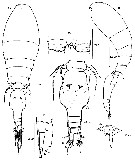 issued from : J.-H. Wi, K.-S. Shih & H.Y. Soh in Zool. Studies, 2011, 50 (5). [p.591, Fig.2]. Female (from 126°5'E, 32°00'N): A-B, habitus (dorsal and lateral, respectively); C, urosome (dorsal), caudal setae numbered using Roman numerals; D, genital aperture (right side), arrow indicating a small process; E, posteromedial part of anal somite (ventral), secretory pores indicated by an arrow; F, left caudal ramus (lateral view), serration near insertion of setae II and III, and secretory pore indicated by an arrow. Nota : Most surfaces of body densely covered with minute retractile granulations (not figured). Prosome length 1.7 times as long as urosome (including caudal rami), 1.9 times as long as urosome excluding caudal rami. 2nd pedigerous somite without conspicuous dorsoposterior rojection in lateral view.. Proportional lengths (%) of urosomites and caudal rami 11.2 : 51.9 : 9.0 : 7.8 : 11.2 : 8.9. Genital double-somite about 1.5 times as long as maximum width (in dorsal view) and 1.8 times as long as postgenital somites combined. Maximum width measured at 3/5 distance of posterior margin ; bulgy part from area of genital apertures to ¾ distance from anterior margin ; paired genital apertures about 1/3 distance from anterior margin of dorsal surface, armed with a single long spine, short process. Dorsolateral and ventral surfaces covered with minute denticles. Secretory pores on dorsal surface (fig.C). Anal somite about 1.3 times wider than long, almost same length as caudal rami, with large anal opening, with linear row of small denticles on it ; dorsal and ventral surfaces covered with small scales ; 2 secretory pores located on either side of anal opening and 4 on mid-posterior region ; posterior margin finely serrated ventolaterally. Caudal rami about 1.75 times as long as wide, with 6 elements, secretory pore located near insertion of seta II , fringed with serrations. Antero- and posterolateral setae II + III long, spiniform, and unipinnate along medial margin ; outer terminal seta IV long and plumose ; inner terminal seta V longest and plumose, 1.5 times longer than seta IV and 2.1 times longer than seta VI ; terminal accessory seta VI long and plumose ; dorsal seta VII much shorter than seta VI, plumose and bi-articulate at base. P5 consisting of long plumose seta on lateral surface of 1st urosomal somite and 1-segmented exopod with 2 unequal setae (1 stout, naked seta and 1 longer slender seta). P6 represented by operculum closing off each genital aperture ; armed with long spine and a small process on inner side of it.
|
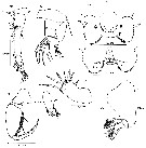 issued from : J.-H. Wi, K.-S. Shih & H.Y. Soh in Zool. Studies, 2011, 50 (5). [p.592, Fig.3]. Female: A, A1; B, A2, individual elements on lateral margin of 2nd endopodal segment numbered using Roman numerals; C, labrum (anterior), bold arrows indicating integumental pockets; D, labrum (posterior), small arrows indicating secretory pores on lobes; E, Md, individual setae designated using small letters; F, Mx1; G, Mx2; H, Mxp. Nota : A1 6-segmented, relative lengths (%) of segments measured along posterior non-setiferous margin 8.1 : 20.3 : 44.6 : 10.8 : 4.1 : 12.1. Armature formula 1- [3], 2-[8], 3-[5], 4-[3+ae], 5-[2+ae], 6-[6+(1+ae)]. A2 3-segmented. Endopod 2-segmented ; lateral armature of distal endopodal segment with1 pectinate, strong spiniform seta III and 3 naked setae (I, II and IV) ; distal armature consisting of 5 long curved setae, ornamented with row of fine spinules unilaterally, and 2 slender naked setae on distal margin. Md with 5 elements on gnathobase : 3 setae and 2 blades. Mxp 4-segmented, comprising syncoxa, basis, and 2-segmented endopod, distal endopodal segment drawn out into long curved claw ornamented with strong spinules along entire concave margin, accessory armature consisting of slender naked seta on outer proximal margin, and unipectinate spine basally fused to inner proximal corner of claw.
|
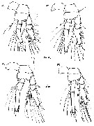 issued from : J.-H. Wi, K.-S. Shih & H.Y. Soh in Zool. Studies, 2011, 50 (5). [p.594, Fig.4]. Female: A-D, P1 to P4, respectively (posterior views). Nota : Distal endopodal segments of P2-P4 with conical process (expressed as a cone) between outer distal and terminal spines, with apical pore.
|
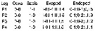 issued from : J.-H. Wi, K.-S. Shih & H.Y. Soh in Zool. Studies, 2011, 50 (5). [p.593]. Female: Armature formula of swimming legs P1-P4. (Roman numerals indicate spines, Arabic numerals indicate setae).
|
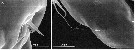 issued from : J.-H. Wi, K.-S. Shih & H.Y. Soh in Zool. Studies, 2011, 50 (5). [p.602, Fig.9, A-B]. Female (SEM photographs): A, genital aperture (right side), arrow indicating small process; B, genital double-somite (in lateroventral view) showing exopodal outer seta of P5.
| | | | | NZ: | 1 | | |
|
Carte de distribution de Triconia denticula par zones géographiques
|
| | | | | | | Loc: | | | East China Sea (S Cheju island) | | | | N: | 1 | | | | Lg.: | | | (1081)* F: 0,66-0,71; {F: 0,66-0,71}
* Body length in lateral view (in mm). | | | | Rem.: | For Wi & al. (2011, p.593) the species can be misidentified as T. rufa in dorsal view, because of the similar shape of the genital double-somite, but the species is included in the similis-subgroup due to the absence of a dorsal projection on the 2nd pedigerous somite that exists in T. rufa, which belongs to the cnifera-subgroup. | | | Dernière mise à jour : 05/12/2016 | |
|
|
 Toute utilisation de ce site pour une publication sera mentionnée avec la référence suivante : Toute utilisation de ce site pour une publication sera mentionnée avec la référence suivante :
Razouls C., Desreumaux N., Kouwenberg J. et de Bovée F., 2005-2026. - Biodiversité des Copépodes planctoniques marins (morphologie, répartition géographique et données biologiques). Sorbonne Université, CNRS. Disponible sur http://copepodes.obs-banyuls.fr [Accédé le 14 juin 2026] © copyright 2005-2026 Sorbonne Université, CNRS
|
|
|
|


;)
;)
;)

