Suarez-Morales & al., 2017 (p.1805, Descr.F, M, figs.F, M, Rem.)
Figures
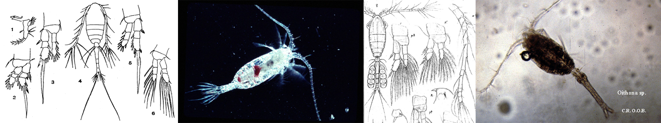
Planche 1 Issued from : E. Suarez-Morales, A. Goruppi, A. de Olazabal & V. Tirelli in J. Nat. Hist., 2017, 51 (31-32) [p.1807, Fig.4]. Female (from G. of Trieste): a-b, habitus (dorsal and ventral, respectively); c, right A1 with armature (dorsal view); d, cephalic region showing preoral ornamentation and oral papilla (lateral); e, urosome, dorsal view showing subtriangular small processes on posterior margin of genital, double-somite (arrow); f, urosome with P5 (lateral); g, same (ventral); h, P5, ventral view (outer setae cut short); i, intercoxal sclerites of P1-P4 (anterior view); j, 3rd exopodal segment of P3 showing apical elements. Scale bars: 200 µm (4, b); 50 µm (c-g); 25 µm (h-j).
Nota: - Cephalothorax representing 61.6 % of total body length (caudal rami excluded). - Midventral oral papilla located at 27.5 % of cephalothorax length. - Pair of relatively large eyes present, pigment cups well-developed, separated by one eye diameter, weakly pigmented; ventral cup slightly larger than lateral cups. - Cephalic area moderately produced, with flat forehead, frontal surface smooth, with pair of sensilla. - Cephalic cuticular ornamentation deduced. - urosome of 3 urosomites together representing 15 % of total body length (caudal rami excluded); relative lengths 34 : 44 : 22 = 100. 5th pedigerous somite with straight lateral margins; P5 inserted on distal 1/3 of somite. - Genital double-somite with postero-lateral margins with small subtriangular processes (arrowed in fig. 4e). - Anal somite about half as long as genital double-somite, ventrally produced.- Caudal ramus subquadrate, 1.1 times longer than wide, armed with 3 subequally long, sparsely setulated caudal setae; - Ovigerous spines paired, , relatively long, representing 30 % of total body length (caudal rami excluded); spines basally separated, slender, equally long, straight at their base and along shaft, both tapering distally. A1 representing about 26 % of total body length (caudal rami excluded) and 44 % of cephalothorax length; 4-segmented, suture between segments 3 and 4 absent. Relative length of distal antennulary segment 42 %. - Pedigerous somites 2-4 together accounting for 24 % of total body length in dorsal view. P1-P4 slightly increasing in size posteriorly; intercoxal sclerites subrectangular, with smooth surface, decreasing in size (fig.4 i). - P5 basally conjoined, distinctly bilobate, inner (endopodal) lobe conspicuous, unarmed, arising proximally, digitiform, reaching slightly beyond midlength of long outer lobe. Outer (exopodal) lobe elongated , slender0, armed with 2 subequally long setae on distal position.
Planche 2 Issued from : E. Suarez-Morales, A. Goruppi, A. de Olazabal & V. Tirelli in J. Nat. Hist., 2017, 51 (31-32) [p.1808, Fig.5]. Male (from Trieste): a, habitus (dorsal); b, cephalic region and right A1 showing armature (dorsal); c, cephalic region showing preoral ornamentation and oral papilla (lateral); d, same (ventral); e, urosome (dorsal); f, same, ventral view showing genital complex with diverging lappets; g, urosome (lateral); h, P1 with intercoxal sclerite; i, P3 with intercoxal sclerite. Scale bars: 200 µm (a); 50 µm (b-i).
Nota: - Cephalothorax representing 44.8 % of total body length. - Midventral oral papilla moderately developed, located at 24 % of cephalothorax length. - Cephalic region slightly protuberant bilaterally in dorsal view. - Pair of dorsal ocelli present, weakly developed; pigment cups medium-sized. Ocelli separated by the length or slighlly less than one eye diameter, faintly pigmented. Ventral cup larger than lateral cups. - Pair of sensilla between antennulary. - Forehead area as in female, produced with flat lightly striated surface. Ventral rounded protuberance between antennulary bases, smilar to that of the female. Urosome 4-segmented (5th pedigerous, genital complex, preanal and anal somites. - 5th pedigerous somite with smooth ventral and dorsal surfaces. - Genital somite slightly shorter than 5th pedigerous somite. Genital complex of type II (Suarez-Morales & McKinnon, 2014) represented by pair of divergent, digitiform genital lappets, these slightly asymmetrical, right ramus slightly narrower than left in lateral view; lappets barely reaching to midlength of long anal somite; pair of small medial thumb-like processes present at common basal joint of lappets; lappets surface smooth. Anal somite with weak constriction , about 1.2 times as long as preanal somite in dorsal and lateral views, comprising 30 % of urosome length; no suture visible on ventral or dorsal surfaces. - Caudal rami subrectangular, about 1.3 times as long as wide, about as long as anal somite. Each ramus with 3 caudal setae. - A1 relatively long, 66 % of cephalothorax length; 5-segmented, segments 1-2 and 2-3 separated, segments 3-4 fused as in female; segment 5 located distal to geniculation (as for Huys & al., 2007, setal nomenclature: elements A-E and 1, 2, 4 present). - Pedigerous somites 2-4 together accounting for 34 % of total body length (caudal rami excluded) in dorsal view. - Coxae of each pair unarmed, joined by intercoxal sclerite, slightly longer than wide, ornamented with small quadrate fields of spinules. Bases of P1-P4 separated from coxae posteriorly by oblique articulation; with outer basal setae; on P3, this seta about 7 times longer, sparselysetulated. Endopods and exopods of P1-P4 triarticulated. Outer spine on distal exopodal segment of P1-P4 about 0.3 times as long as segment. Armature formula of legs as in female.
Distribution
NZ: 1
Carte de distribution de Cymbasoma pseudobidentatum par zones géographiques
(1206)* F: 0,78; M: 0,74; [F: 0,78; M: 0,74]
* Total length from the end of anterior cephalothorax to the posterior end of anal somite.
Remarques
For Suarez-Morales & al. (2017, p.4809), this species makes reference to the close resemblance with its Australian congener C. bidentatum. The species can be disyinguished from its congeners by a unique combination of features : 1- the presence of small posterodorsal subtriangular processes on the female genital double-somite; 2- a medial ventral cephalic protuberance; 3- fused antennulary segments 3-4 in both the male and the female; 4- female outer P5 lobe armed with 2 subequally long setae plus a small, slender innermost seta; 5- a ventrally expanded anal somite in both the male and the female; 6- male genital complex with a pair of distinctive medial thumb-like processes. After Suarez-Morales & al. (2017, p.1811), the male of C. pseudobidentatum closely resembles C. tenue (Isaac, 1975), these two species differ in subtle characters. In C. tenue the medial processes on the base of the genital lappets are clearly acute and smaller than in C. pseudobidentatum, in which these processes are apically rounded, thumb-like and relatively larger; also, the anal somite has a medial constriction, thus differing from C. tenue, lacking this character. In addition, the b-group setae on the outer margin of the last segment of A1 are unbranched in C. pseudobidentatum and branched in C. tenue. For the authors (p.1811), the male and female were linked as being conspecific, based on the several morphologic characters they share: 1- the anteriorly produced but apically flat forehead with a pair of sensilla on the same position and with a similar adjacent cuticular ornamentation; 2- the position of the oral papilla and the arrangement of the reduced perioral ornamentation; 3- the presence of an anteroventral rounded process in the preoral area; 4- the antennulary segmentation, with segments 3-4 fused; 5- very long antennulary elements 2v1 and particulary element 4v1 being the longest of group 4v-d; 6- anal segment expanded ventrally.


;)
;)
