Issued from : E. Suarez-Morales, A. Goruppi, A. de Olazabal & V. Tirelli
in J. Nat. Hist., 2017, 51 (31-32) [p.1816, Fig.7].
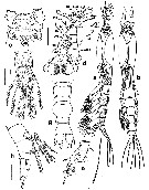
Female (Trieste): a-b, habitus (lateral and dorsal, respectively); c, cephalic region showing preoral ornamentation (ventral); d, frontal area and right A1 with armature (dorsal); e, cephalic region showing preoral ornamentation (lateral); f, urosome and P5 (ventral); g, same (dorsal); h, same (lateral).
Scale bars: 200 µm (a, b); 50 µm (c-h).
Nota:
- Cephalothorax representing 58.7 % of total body length (caudal rami excluded)
- Midventral oral papilla located at 14 % of cephalothorax length.
- Pair of large ocelli present, pigment cups moderately developed, medially separated by less than half of one eye diameter, pigmented only on inner margin; ventral cup slightly larger than lateral cups.
- Cephalic area with strongly produced forehead, ornamented..
- Frontal sensilla absent.
- Cephalic section not constricted, but narrower than medial part of cephalothorax.
- Paired ventral papilla-like processes between A1 bases with few adjacent transverse striae (arrow in Fig.7e)
- Nipple-like processes on preoral surface surrounded by striae arranged in incomplete concentric pattern (Fig. 7c).
- Urosome with 3 somites, together representing 18 % of total body length. Relative lengths of urosomites 34.2 : 42.4 : 23.4 = 100.
- 5th pedigerous somite subrectangular with shallow striation on dorsal surface (Fig. 7g).
- Genital double-somite with globose anterior part; somite with anteroventral protuberance visible in lateral view (Fig. 7 g, h), with shallow ventral suture line and small rounded posterolateral process (arrow in Fig. 7h).
- Caudal ramus subquadrate, armed with 3 subequally long, sparsely setulated caudal setae.
- Ovigerous spines paired, representing 47 % of total body length, reaching well beyond distal end of caudal setae. Spines basally separated, slender, straight at their base and along shaft, without distal expansions and tapering distally, both spines subequally long.
- Anal somite unconstricted, with smooth dorsal and ventral surfaces.
- A1 relatively short, not divergent, representing about 19 % of total body length and 30 % of cephalothorax length. A1 4-segmented, all segments completely separate. Distal segment relatively short, representing 37 % of A1 length.
- P5 basally separate, represented by a single subrectangular lobe armed with 3 subequally long biserially setulated setae on distal position (Fig. 7f).


;)

 Toute utilisation de ce site pour une publication sera mentionnée avec la référence suivante :
