|
|
 |
Fiche d'espèce de Copépode |
|
|
Calanoida ( Ordre ) |
|
|
|
Diaptomoidea ( Superfamille ) |
|
|
|
Pontellidae ( Famille ) |
|
|
|
Calanopia ( Genre ) |
|
|
| |
Calanopia tulina El-Sherbiny & Al-Aidaroos, 2017 (F,M) | |
| | | | | | | Ref.: | | | El-Sherbiny & Al-Aidaroos, 2017 (on line) | 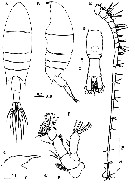 Issued from : M.M. El-Sherbiny & A.M. Aidaroos in Mar. Biodiv., 2017 [Fig. 1]. Female (from central Red Sea): A-B, habitus (dorsal and lateral, respectively); C, forehead with rostrum (lateral); D, urosome (dorsal); E, A1; F, A2. Scale bars in mm. Nota: - Cephalosome and 1st pedigerous somite completely separated, 4th and 5th pedigerous somites completely fused. - Prosome 2.6 times as long as urosome (from the anterior margin of cephalothorax to the tip of posterior wing). - Posterior margins of prosome symmetrical in dorsal view, tapering to pointed process at each lateral corner, slightly inclined ventrally, reaching nearly halfway along genital double-somite. - Rostrum bifid, with broad bases and small medial subterminal notch. - Urosome 2-segmented. - Genital compound somite symmetrical in dorsal view; ventral surface without any process and with smooth evenly rounded operculum located posteroventrally. - Caudal rami asymmetrical, left one slightly longer than right, about 2.5 times as long as wide. - Urosomites 1 and 2 plus left caudal ramus in proportions 47 : 30 : 23 = 100. - A1 18-segmented, reaching slightly beyond posterior border of genital compound somite. - A2 with coxa bearing plumose seta medially. Basis with 2 distomedial subequal setae. Exopod 5-segmented (setal formula: 0, 3, 1, 1,3). Endopod with seta on 1st segment; 2nd segment with 8 setae on proximal lobe and 6 setae and row of spinules on posterior surface of distal lobe.
|
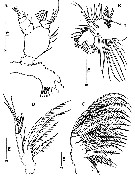 Issued from : M.M. El-Sherbiny & A.M. Aidaroos in Mar. Biodiv., 2017 [Fig. 3]. Female: A, Md; B, Mx1; C, Mx2; D, Mxp. Scale bars in mm. Nota: - Md gnathobase bearing 8 teeth on cutting edge and anteriorly with group of short spinules proximal to 3rd to 7th teeth. Palp basis with 4 unequal setae on medial margin; exopod 5-segmented (setal formula: 1, 1, 1, 1, 3). Endopod 2-segmented, proximal segment with 3 setae at distomedial corner, distal segment crowned with 6 long setae. - Mx1 praecoxal arthrite with 13 setal elements (9 marginal and 4 on posterior surface). Coxal epipodite with 9 setae; coxal and basal endites each with 3 setae; basal exite with 1 long seta. Exopod bearing 9 setae. Endopod incorporated into basis with 2 setae laterally and 9 setae terminally. - Mx2 with 1st praecoxal endites bearing 4 and 3 (2 long and 1 short) setae, respectively; coxal endites with 2 and 3 (2 long and 1 short) setae respectively. Basal endite with 2 long and 1 medium setae. E,dopod 3-segmented, bearing 1, 1 and 4 spinulate setae. Mxp praecoxa and coxa fused; 3 syncoxal endites well developed carrying strong spinulose setae with setal formula: 2, 3 (2 long and 1 short), 2 (1 long and 1 short). Basis relatively long with 2 distal setae. Endopod 4-segmented (setal formula: 2, 1, 1, 3).
|
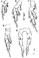 Issued from : M.M. El-Sherbiny & A.M. Aidaroos in Mar. Biodiv., 2017 [Fig. 4]. Female: A, P1; B, P2; C, P3; D, P4; E, P5. Scale bars in mm. Nota: - P5 slightly asymmetrical (right leg slightly shorter than left leg); coxa and intercoxal sclerite fused. Basis with short seta posteriorly. Exopod 2-segmented, 1st exopodal segment of left leg nearly 1.3 times as long as right one, with small laterally serrated spine and midlength and large medially directed serrated terminal spine. 2nd exopodal segment longer than 1st one, bearing 2 serrate lateral spines (distal one longer and nearly about 2/3 the length of distal spiniform process), and ending as a serrated long, distal spiniform process.
|
 Issued from : M.M. El-Sherbiny & A.M. Aidaroos in Mar. Biodiv., 2017 [Table 1]. Spine and setal formula of swimming legs P1 to P4 in female and male.
|
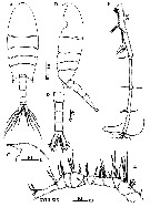 Issued from : M.M. El-Sherbiny & A.M. Aidaroos in Mar. Biodiv., 2017 [Fig. 5]. Male: A-B, habitus (dorsal and lateral, respectively); C, forehead and rostrum (lateral); D, urosome (ventral); E-F, right A1. Scale bars in mm. Nota: Diagnosis Male: - Prosome 1.9 times as long as urosome. - 4th and 5th pedigerous somite fused, produced posterolateraly into asymmetrical pointed corners, the right one wider and slightly longer than the leftone, with distinct notch on its medial margin and reaching end of 1st urosomite. - Rostrum notched as in female. - Urosome 5-segmented, almost symmetrical except for genital somite with genital aperture on left. - 2nd and 3rd urosomites with same length and longer than other urosomites. - Anal somite much shorter than preceding somite. - Caudal rami symmetrical, nearly 4 times longer than wide. Urosomites 1-5 and caudal rami in proportions 18 : 19 : 19 : 15 : 5 : 24, respectively. - Right A1 17-segmented, extending to anterior margin of 2nd urosomite. - Left A1, A2
, mouthparts and P1-P4 as in female.
|
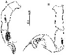 Issued from : M.M. El-Sherbiny & A.M. Aidaroos in Mar. Biodiv., 2017 [Fig. 7]. Male: A, left P5 (posterior view); B, right P5 (posterior view) Scale bar in mm. Nota: - Left P5 shorter than right one; coxa coalesced into intercoxal sclerite; basis 1.9 times longer than coxa carrying plumose seta posteriorly; exopod 2-segmented: 1st segment 0.5 times as long as basis, with pointed spine at distolateral corner, 2nd segment about twice longer than the 1st exopodal one, hirsute on posteromedial surface, armed distally with lateral spine directed medially and 2 relatively long, curved, and medially serrated terminal spines. - Right P5 longer than left; basis slightly shorter than coxa, with plumose seta posteriorly; exopod 2-segmented, forming a stout chela, 1st segment (chela) with very small rounded-tip thumb-like process located nearly halfway, central part of the chela smooth with deep depression laterally and seta near to thumb base, 2nd segment (finger) shorter than 1st, curved at distal 2/3 its length medially with blunt apex and 2 medial setae within depression, and laterally with 2 unequal setae, proximal one long (more than half-length of finger irself) and distal one short, nearly at distal 2/3 of finger.
| | | | | NZ: | 1 | | |
|
Carte de distribution de Calanopia tulina par zones géographiques
|
| | | | Loc: | | | Red Sea (neritic waters off Jeddah)
Type locality: 21°39'34.17'' N, 39°00'54.90''. | | | | N: | 1 | | | | Lg.: | | | 1207) F: 1,88-2,22; M: 2,03; {F: 1,88-2,22; M: 2,03} | | | | Rem.: |
For the authors, C. tulina is most closely related to C. media described from the Suez Canal (Gurney, 1927), but does not fit any existing species-group (see Table in the genus Calanopia), nor does it comprise a new group. The presence of C. tulina during night sampling may refer to its diel vertical migration. | | | Dernière mise à jour : 19/06/2023 | |
|
|
 Toute utilisation de ce site pour une publication sera mentionnée avec la référence suivante : Toute utilisation de ce site pour une publication sera mentionnée avec la référence suivante :
Razouls C., Desreumaux N., Kouwenberg J. et de Bovée F., 2005-2026. - Biodiversité des Copépodes planctoniques marins (morphologie, répartition géographique et données biologiques). Sorbonne Université, CNRS. Disponible sur http://copepodes.obs-banyuls.fr [Accédé le 31 janvier 2026] © copyright 2005-2026 Sorbonne Université, CNRS
|
|
 |
 |


