|
|
 |
Fiche d'espèce de Copépode |
|
|
Calanoida ( Ordre ) |
|
|
|
Diaptomoidea ( Superfamille ) |
|
|
|
Pontellidae ( Famille ) |
|
|
|
Labidocera ( Genre ) |
|
|
| |
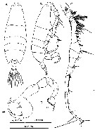
Labidocera churaumi Hirabayashi, 2014 (F,M) | |
| | | | | | | Ref.: | | | Hirabayashi & S. Ohtsuka , 2014 (p.21, Descr. F, M, figs. F,M, Rem.) | 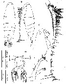 Issued from : T. Hirabayashi & S. Ohtsuka in ZooKeys, 2014, 447. [p.23, Fig. 1]. Female (from Tokashiki Island: Okinawa) A-B, habitus (dorsal and lateral, respectively); C, anterior part of cephalosome (lateral); D, urosome with outline of attached spermatophore and coupler (dorsal); E, left A1; F, A2. Nota: Ratio of prosome to urosome lengths 4 : 1. - Prosome length to width ratio 2.85 : 1. - Cephalic profile rounded in dorsal view, without lateral hooks. - Paired dorsal eyes with cuticular lenses; protuberant ventral eye extending anteroventrally between rostral processes. - Rostrum bifid, directed posteroventrally. - Posterior margins of prosome almost symmetrical in dorsal view, tapering to simple abbreviated, pointed process at each lateral corner. - Urosome 2-segmented, of characteristic shape. - Genital compound somite strongly asymmetrical; anterior left surface with posteriorly-directed rod-like process and posterior right smoothly rounded. - Spermatophore attached dorsally to genital somite. - A1 23-segmented; larger and longer setae on segments 3-6. - A2 biramious: coxa with short plumose distal seta; basis and 1st endopodal segment fused to form elongate allobasis, setation formula 2, 2. Compound distal endopodal segment with 9 and 7 setae on proximal and distal lobes, respectively; exopod 5-segmented, setal formula 0, 0, 2, 2, 3.
|
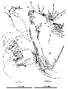 Issued from : T. Hirabayashi & S. Ohtsuka in ZooKeys, 2014, 447. [p.25, Fig. 2]. Female: A, Md; B, Mx1; C, Mx2; D, Mxp. Nota: - Md with wide, heavily chitinized gnathobase; mandibular palp biramous, basis robust, armed with 4 inner setae; endopod 2-segmented, 1st segment armed with 1 short and 3 long setae, 2nd segment with 7 terminal setae; exopod 2-segmented, 1st segment unarmed, 2nd segment with 6 terminal setae; mandibular gnathobase distal edge bearing 8 teeth comprising: from ventral margin 1 apical, 1 subapical, 3 compound medial, and 3 basal; medial teeth with bifurcated cusps; dorsal end of gnathobase with 1 seta. - Mx1 praecoxal arthrite with 15 setal elements, 4 on posterior surface; coxal endite with 2 long and 1 short elements, and 9 setae on epipodite; basis with 3 setae on proximal and distal endites; 1 large seta on basal exite; proximal endopod segment and endopod segment 2 incorporated into basis, proximal endopod segments with 2 setae, endopod segment 2 with 2 setae and distal endopod segment with 5 apical setae; exopod with 10 setae. - Mx2 with 1st praecoxal endite bearing 6 setae, 2nd with 3 setae; coxa with 3 setae each on proximal and distal endites; basis with 3 setae; endopod 3-segmented, setal formula 1, 1, 4. - Mxp with praecoxa and coxa fused, 3 syncoxal endites well developed, setal formula 2, 2, 4; endite setae strong, spinulose; basis fringed with medial row of spiniform processes and 2 distal setae; endopod 4-segmented, setal formula 2, 1, 1, 2.
|
 Issued from : T. Hirabayashi & S. Ohtsuka in ZooKeys, 2014, 447. [p.27, Fig. 3]. Female: A, P1; B, P2; C, P3; D, P4; E, P5. Nota: - P5 biramous, slightly asymmetrical; coxa and intercoxal sclerite fused; basis subrectangular, with posterior seta; endopod rounded distally, about 0.3 times as long as exopod; exopods of both legs 1-segmented, bifurcated tip and with 2 outer spines; outer process on left slightly larger than right and with small spine-like process on proximal part.
|
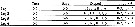 Issued from : T. Hirabayashi & S. Ohtsuka in ZooKeys, 2014, 447. [p.26]. Female and Male: Armature formula of the swimming legs P1-P4 (Arabic numerals : setae; Roman numerals : spines)
|
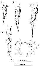
 Issued from : T. Hirabayashi & S. Ohtsuka in ZooKeys, 2014, 447. [p.28, Fig. 4]. Male: A-B, habitus (dorsal and lateral, respectively); C, right A1; D, P5. Nota: - Prosome about 4 times as long as urosome. - Urosome symmetrical with 5 somites. - Anal somite asymmetrical. - Caudal rami asymmetrical. - Right A1 15-segmented, geniculate between segments 11 and 12, reaching middle of 3rd pedigerous somite; segments 11 and 12 with row of teeth. Left A1 as in female. - A2, mouthparts and swimming legs as in female. - P5 asymmetrical. Left P5 short, intercoxal sclerite and left coxa fused; basis cylindrical with seta near base; exopod 2-segmented, 1st segment cylindrical,2nd segment triangular, short, with protruding hairy medial surface and 3 distal and 1 lateral spines, one of them long. Right P5 basis with seta; exopod 2-segmented, forming chela; thumb of chela large, triangular, arising near base of 1st exopodal segment; 1st exopodal segment with 2 small setae, 2nd exopodal segment elongate and curved, with 3 slender marginal setae
| | | | | NZ: | 1 | | |
|
Carte de distribution de Labidocera churaumi par zones géographiques
|
| | | | | | | Loc: | | | NW Pacif. (Tokasshiki Port, Tokashiki Island, Okinawa) | | | | N: | 1 | | | | Lg.: | | | (1210) F: 2,225-2,790; M: 1,0819-2,531; {F: 2,225-2,79; M: 1,081-2,531} | | | | Rem.: | For Hirabayashi & Ohtsuka (2014, p.26) the species is similar to L. madurae Scott, 1909 and L. tasmanica Taw, 1974. These three species constitute a species group within the L. detruncata species complex (see key to Indo-West Pacific species groups in Labidocera detruncata species complex in Labidocera Genus). These three species have a restricted distribution in the subtropical and tropical , coastal waters of the Indo-West Pacific, and are referred by the authors as L. madurae species group (see key of the species in this group). | | | Dernière mise à jour : 19/07/2021 | |
|
|
 Toute utilisation de ce site pour une publication sera mentionnée avec la référence suivante : Toute utilisation de ce site pour une publication sera mentionnée avec la référence suivante :
Razouls C., Desreumaux N., Kouwenberg J. et de Bovée F., 2005-2026. - Biodiversité des Copépodes planctoniques marins (morphologie, répartition géographique et données biologiques). Sorbonne Université, CNRS. Disponible sur http://copepodes.obs-banyuls.fr [Accédé le 22 juin 2026] © copyright 2005-2026 Sorbonne Université, CNRS
|
|
|
|


;)
;)
;)
;)

