|
|
 |
Fiche d'espèce de Copépode |
|
|
Calanoida ( Ordre ) |
|
|
|
Diaptomoidea ( Superfamille ) |
|
|
|
Pseudodiaptomidae ( Famille ) |
|
|
|
Pseudodiaptomus ( Genre ) |
|
|
| |
Pseudodiaptomus japonicus Kikuchi, 1928, 1928 (F,M) | |
| | | | | | | Syn.: | ane Pseudodiaptomus inopinus : Smirnov, 1929 (P.318, fig.F, M); Mashiko & Inoué, 1952 (p.184, figs?F, M); Tanaka, 1966 (p.42, fig.3); Kos, 1976 (Vol. II, figs.F,M, Rem.); Kikuchi & al., 1978 (p.25, figs.7-11); Kos, 1984 (p.225, figs.F, M, Rem.); Harada & al., 1985 (p.51, fig.11, vertical variation abundances); Chihara & Murano, 1997 (p.893, figs.F, M, Rem.); Cordell & coll., 1997 (2012, p.11); Uye & al., 2000 (p.202, fig.7 abundance-tempetature-salinnity diagramm); E.O. Park & al., 2005 (p.370, fig.3: abundance-temperature-salinity); ? Lee & al., 2007; Sakaguchi & Ueda, 2010 (p.62, figs.7A-F, H, I, 8A-F); 2011 (fig.2A, C, E, G, I).
Pseudodiaptomus koreanus Soh & al., 2012 (p.231, figs.2, 3A, 4-8).
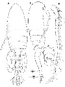
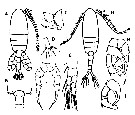
Pseudodiaptomus sp. Eyun & al., 2007 (p.265, molecular biology). | | | | Ref.: | | | Kikuchi, 1928 (p.68, figs.F, M); Ueda & Sakaguchi, 2019 (p.29, Descr. F, M, Rem.) | 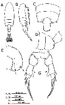 Issued from : Ueda & Sakaguchi in Plankton Benthos Res., 2019, 14 (1). [p.34, Fig.3, A-E, G]; Female (from Lake Suigetsu): A-B, habitus (dorsal and lateral, respectively); C-D, 3rd pediger to genital double-somite (dorsal and lateral, respectively); E, last pediger (posterior); G, P5 (posterior). Nota: - 2nd and 3rd pedigers with row of very fine spinules along posterolateral margin. - Last pediger with weakly protruded posterior projection and 2 groups of spinules rows (Fig.3 E, indicated by arrows) on each side, and devoid of transverse row of fine spinules on lateral surface. - P5 with group of a few spinules, (indicated by arrow) at midlength on medial margin of 1st exopodal segment. Inter-population variation was seen in spinular rows on the female prosome. The last pediger of the Lake Suigetsu specimens has 2 groups of spinules in rows on the distolateral corner and the distomedial surface, respectively, whereas females from the other four localities had 3 groups of spinules as described in P. yamato. Spinules along the posterolateral margin of the 2nd and 3rd pedigers were very fine in the Lake Suigetsu female specimens, whereas in the females from the other localities these were more conspicuous.
|
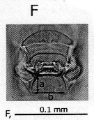 Issued from : Ueda & Sakaguchi in Plankton Benthos Res., 2019, 14 (1). [p.34, Fig.3, F]; Female: photograph of genital operculum. Nota: Genital operculum with short posterior processes, process shorter than distance between tips ('a' < 'b')
|
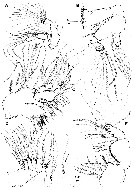
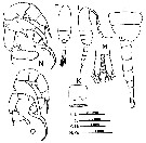 Issued from : Ueda & Sakaguchi in Plankton Benthos Res., 2019, 14 (1). [p.34, Fig.3, H-K, J-]; Male (from Lake Suigetsu): H-I, habitus (dorsal and lateral, respectively); J, last three pedigers and urosome (lateral); K, 2nd urosomite (ventral); M, caudal rami (dorsal); N, P5 with paddle-shaped right terminal segment (posterior); O, P5 with finger-shaped right terminal segment (posterior). Nota: - Prosome with no spinule on each somite or with very fine spinules on ^posterolateral ùmargin of 3rd pediger. - urosome as in P. yamato. - Caudal ramus length 2.7-3.1 times width. - Left P5 with fine spinules on anterior surface of chela arm; 1st exopodal segment with usually concave medial margin.
|
 Issued from : Ueda & Sakaguchi in Plankton Benthos Res., 2019, 14 (1). [p.34, Fig.3, K, L]; Male: K, 2nd urosomite (ventral); L, photograph of ventral spinules in K.
|
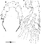
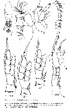
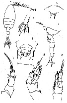 issued from : O. Tanaka in Proceedings Symposium on Crustacea. Symp. Ser. mar. biol. Ass. India, 1966, (2) 1. [p.42, Fig.3]. As Pseudodiaptomus inopinus. Female (from NW copastof Kyushu): a, habitus (dorsal); b, rostrum (frontal view); c, last thoracic segment and urosome (lateral left side); d, genital segment (ventral); e, caudal ramus; f, P1; g, P2; h, P5. Nota: - The urosome segments and furca are in the proportional lengths as 31 : 1 4: 20 : 10 : 25 = 100. - Prosome and urosome are in the proportional lengths as 6 2: 38. - P5 is symmetrical; the 3rd segment has a blunt process on the inner distal margin; the distal segment has b3 spines of which the outer is long and curved.. -A1 22-segmented, reaches the middle of the last thoracic segment.
|
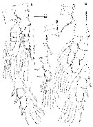
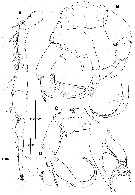
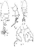
 issued from : S.O. Sakaguchi & H. Ueda in Zootaxa, 2010, 2623. [p.63, Fig.7]. As Pseudodiaptomus inopinus. Female (from Kyushu Island, S Japan): A-B, habitus (dorsal and lateral, respectively); C, 5th pediger (right lateral); D-E, genital double-somite (ventral and right lateral, respectively); F, caudal rami (dorsal); G, caudal rami with thin setae (dorsal); H, P5 (posterior); I, base of 3rd exopodal segment of P5 (anterior). A-F, H, I: the same specimen from the Tsuri-kawa River; G: from Yukiura-River). Nota: 2nd to 4th prosomites laterally with row of fine spinules along posterior margins; 4th and 5th pediger fused with rounded corners, with spiniform process dorsally, small bump terminally (indicated by arrowed a); , several long spinules ventral to bump (arrowed b), and row of spinules on posterolateral corner on each side (arrowed c. Caudal left ramus slightly larger than left. The medial terminal seta especially swollen and as long as ramus, except one specimen from the Yukiura-gawa River with thin, long setae on both rami
|
 issued from : S.O. Sakaguchi & H. Ueda in Zootaxa, 2010, 2623. [p.64, Fig.8]. As Pseudodiaptomus inopinus. Male (from Tsuri-kawa River, Kyushu Island): A-B, habitus (dorsal and lateral, respectively); C, 2nd urosomite (ventral); D, right A1 (segments 16-17th); E, P5 (posterior); F, paddle-shaped 2nd exopod of left P5 (posterior).
|
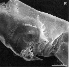 issued from : S.O. Sakaguchi & H. Ueda in Zootaxa, 2010, 2623. [p.57, Fig.3]. As Pseudodiaptomus inopinusSEM photographs of genital flaps of Female (from the Tsuri-kawa River), ventrolateral view.
|
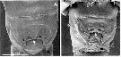 ssued from : H.Y. Soh, S.W. Kwon, W. Lee & Y.H. Yoon in Zootaxa, 2012, 3368. [p.233, Fig.3]. Female: SEM photographs of the genital double-somite from P. koreanus (A) (= P. japonicus) and P. inopinus (B). White arrows indicate process between gonopores; black arrows indicate the posterior process of genital flap.
|
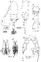
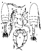
 Issued from : H.Y. Soh, S.W. Kwon, W. Lee & Y.H. Yoon in Zootaxa, 2012, 3368. [p.232, Fig.2]. As Pseudodiaptomus koreanus. Female (from 36°54'43.21''N, 129°22'2248''E): A-B, habitus (dorsal and lateral, respectively); C, genital double-somite (ventral); D, A1. Scale bars in µm. Nota: Prosome/Urosome ca. 1.71. 1st pediger coalesced with cephalosome; 2nd and 3rd pedigers with row of fine spinules along posterolateral margins; 4th and 5th pedigers completely fused with rounded corners, posterolateral corner on each side with row of spinules and with small bump posteromedially Urosome of 4 free somites. Genital double-somite 1.1 times wider than long, greatest width anterior in dorsal view with several spinules on each anterolateral projection; in ventral view posterolateral process of female lateral genital flaps, wide and extend well short of posterior border of genital double-somite (Fig.2C); anal somite shortest. Each somite except anal somite serrated along posterior margin. Caudal rami nearly symmetrical, with 6 setae (outer seta IV basally swollen). A1 symmetrical, 22-segmented.
|
 Issued from : H.Y. Soh, S.W. Kwon, W. Lee & Y.H. Yoon in Zootaxa, 2012, 3368. [p.234, Fig.4]. As Pseudodiaptomus ikoreus. Female: A, A1; B, Md; C, Mx1; D, Mx2; E, Mxp. Scale bars in µm. Nota: A2 coxa with 1 seta; basis and 1st enpopodite segment fused into allobasis; exopod 6-segmented. Md with endopod 2-segmented; exopod 5-segmented. Mx1 praecoxal arthrite with 15 elements; coxa with 4 setae on endite and 9 setae on epipodite; basis with 4 and 5 setae on proximal and distal endite, respectively, with single seta on exite; endopd 3-segmented, with setal formula 4, 4, 7; exopod unisegmented, with 11 setae. Mx2 praecoxa elongated, proximal endite with 4 setae, distal endite with 3 setae; two coxal endites with 3 setae each; basis with sclerotized stout seta in addition to single short and 2 long setae; endopod 4-segmented with 2, 3, 2, 2, respectively. Mxp syncoxa with setal formula 0, 2, 3, 4 on endites; basis with 3 setae, with 1st endopodal segment separated; endopod 6-segmented, with setal formula 2, 3 (including bifurcate setae), 2 (both bifurcate setae), 2, 2+1, 4
|
 Issued from : H.Y. Soh, S.W. Kwon, W. Lee & Y.H. Yoon in Zootaxa, 2012, 3368. [p.235, Fig.5]. As Pseudodiaptomus koreanusFemale: A, P1; B, P2; C, P3. Scale bar in µm.
|
 Issued from : H.Y. Soh, S.W. Kwon, W. Lee & Y.H. Yoon in Zootaxa, 2012, 3368. [p.236, Fig.6]. As Pseudodiaptomus koreanus. Female: A, P4; B, P5. Scale bar in µm. Nota: P5 symmetrical, uniramous; intercoxal scerite completely incorporated into both coxae; basis with small posterior outer seta; exopod 3-segmented, 1st segment furnished with spinules on inner margin, with outer spine and mediodistal blunt projection; 2nd and 3rd segments incompletely fused, 2nd segment with short outer spine and larger serrate inner spines, 3rd segment with long terminal spine and smaller inner spine.
|
 Issued from : H.Y. Soh, S.W. Kwon, W. Lee & Y.H. Yoon in Zootaxa, 2012, 3368. [p.233]. Female: Seta and spine formula of swimming legs P1 to P4. Spines Roman numerals; setae Arabic numerals (note for each segment from outer to inner margins)
|
Issued from : H.Y. Soh, S.W. Kwon, W. Lee & Y.H. Yoon in Zootaxa, 2012, 3368. [p.233, Fig.3]. As Pseudodiaptomus koreanus. Female: SEM photographs of the genital double-somite from P. koreanus (A) and P. inopinus (B). White arrows indicate process between gonopores; black arrows indicate the posterior process of genital flap. Nota: Genital double-somite of P. koreanus, in ventral view, posterolateral process of lateral genital flaps, wide and extend well short of posterior border of genital double-somite; pair of uncovered gonopores and rounded posteriocentral projection between those (Fig.3A, white arrowed) located posteroventrally. P. koreanus differs from P. inopinus in having broad, short posterior processes of the female lateral genital flaps, a rounded posteriocentral projection between genital flaps.
|
 Issued from : H.Y. Soh, S.W. Kwon, W. Lee & Y.H. Yoon in Zootaxa, 2012, 3368. [p.237, Fig.7]. As Pseudodiaptomus koreanus. Male (from 36°54'43.21''N, 129°22'2248''E): A-B, habitus (dorsal and lateral, respectively); C, 2nd urosomite (ventral view). Scale bars in µm. Nota: Cephalosome and 1st pediger, 4th and 5th pedigers completely fused. 1st to 5th pediger with row of posterolateral spinules; Urosome of 5 free somites; genital somite with genital opening on left; 2nd urosomite with ventral transverse row of spinules; genital and anal somites naked, 2nd to 4th somites armed with posterodorsal spinules. ledft A1 same as in female; right A1 21-segmented, geniculated.
|
 Issued from : H.Y. Soh, S.W. Kwon, W. Lee & Y.H. Yoon in Zootaxa, 2012, 3368. [p.239, Fig.8]. As Pseudodiaptomus koreanus. Male: A, right A1; B, P5, paddle-shape; C, P5, finger-shape; D, detail of P5. Scale bars in µm. Nota: P5 asymmetrical; coxa with spinule patch on both surfaces, left basis and endopod completely fused, produced into 2 large medial processes. In finger-shaped morph (Fig.5C) larger medial process of left basoendopodal segment curved, with patch of minute spinules at mid-length; proximal endopodal segment having inner seta proximally widest on central part; distal segment with 5 setae and serrated outer spine covered with hairs; right basis with outer seta and 2 unequal inner knobs; 1st exopodal segment with long stout distal outer spine and medial seta; 2nd segment medially swollen with medial inner seta and spine; 3rd segment elongate, with stout terminal spine, medial seta, and distal seta. In paddle-shape morph (Fig.8B) patch of minute spinules absence at mid-length of medial process of left basoendopodal segment; proximal exopodal segment with inner seta proximally widest on proximal part; distal segment covered with hairs with 3 setae and serrated outer spine; 2 unequal inner lobes absence on right basis; width of 2nd exopodal segment nearly equal.
|
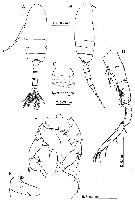
 Issued from : M.S. Kos in Issled. Fauny Morei, 1984 (1985), 30 (38). [Fig.8]. As Pseudodiaptomus inopinus. From Ueda (pers. comm.) Female: 1, habitus (dorsal) with egg-sacs; 2, A1; 3, A2; 4, Mxp; 5, P1; 6, P2; 7, P3; 8, P4; 9, P5.
|
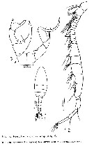 Issued from : M.S. Kos in Issled. Fauny Morei, 1984 (1985), 30 (38). [Fig.9]. As Pseudodiaptomus inopinus. From Ueda (pers. comm.) Male: 1, habitus (dorsal); 2, right A1; 3, P5.
|
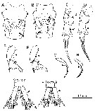
 Issued from : M.S. Kos in Field guide for plankton. Zool Institute USSR Acad., Vol. II, 1976. Female: 1, habitus (dorsal); 2, P5. Male: 3, habitus (dorsal); 4, P5.
|
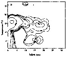
 issued from : S.O. Sakaguchi & H. Ueda in Plankton Benthos Res., 2011, 6 (2). [p.126, Fig.2]. As: Morphologies of Pseudodiaptomus inopinus specimens from the Japan Sea and Pacific coasts of western Japan. Female: A-B, genital double-somite (ventral); C-DP5; Male: E-F, exopod of left P5; G-H, 3rd exopodal segment of right P5; I-J, anal somite and caudal rami. Left and right figures of each pair are of specimens from the Japan Sea and Pacific groups, respectively. Morphologies to be compared are indicated by arrowheads.
|
 issued from : S.O. Sakaguchi & H. Ueda in Plankton Benthos Res., 2011, 6 (2). [p.127, Fig.3]. Comparisons of morphometric characters of Pseudodiaptomus inopinus specimens from the Japan Sea and Pacific coasts of western Japan. A: mean length of left and right posterior processes of the genital operculum B: depth of medial concavity of 1st exopodal segment of the male left P5. C: positions of medial process of 3rd exopodal segment of the male left P5. D: proportional length of the left caudal ramus. Filled and open symbols represent specimens from the Japan Sea and Pacific groups, respectively. Circles and triangles in males indicate specimens with thumb- and paddle-type P5, respectively. Nota: The study revealed that there are two morphological types of P. inopinus in western Japan. These facts indicate that specimens of the two groups belong to different species and P. inopinus in Japan is a species complex. However, it is no possible to decide their taxonomic statuses herein, because it is still uncertain wether one or two types is identical to P. inopinus s. str. or not. Compared with Burckhardt (1913) original figures of P. inopinus s. str. from Taifu Lake near the Yangtze River mouth (China), the Pacific group is more similar to P. inopinus s. str. in having longer posterior processes of the genital operculum. However, the morohology of the male P5 in the original description is somewhat different from that of the Pacific group (the ratios of RPL/RSL measured for the Burckhardt's original figures are 0.62 for the thumb type and 0.58 for the paddle type, which are apparently the same as for the Japan Sea group. Specimen-based morphological comparisons with P. inopinus s. str. from China and genetic comprisons between Japanese and Chinese specimens as well as between the two types in Japan are necessary to make a final decision on their taxonomic status.
|
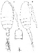
 Issued from : K. Kikuchi in Mem. Coll. Sci. Kyoto Imp. Univ. Ser. B, 1928, 4 (1). From H. Ueda (pers comm.) [p.68, rearranged from Pl.18, figs.9-12, figs.13-17]. Female: A, habitus (dorsal); B-C, posterior region of prosome and genital doubbl-somite; D, caudal ramus; E, P1; F, P5. Male: G, habitus (dorsal); H-I, P5.
|
 Issued from : Y. Kikuchi, R. Yoshida & S. Yamane in Bull. Fac. Educ. Ibaraki Univ. (Nat. Sci.), 1978, 27. [p.25, figs.7-11]. As Pseudodiaptomus inopinus (from Lake Kita-ura on the Pacific coast of middle Honshu, Japan). From H. Ueda (2019, pers.comm.). Female: 7, habitus (dorsal); 8, P5. Male: 9, habitus (dorsal); 10, right P5.
| | | | | NZ: | 2 | | |
|
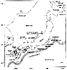
Carte de distribution de Pseudodiaptomus japonicus par zones géographiques
|
| | |  Issued from : Ueda & Sakaguchi in Plankton Benthos Res., 2019, 14 (1). [p.37, Fig.5]. Issued from : Ueda & Sakaguchi in Plankton Benthos Res., 2019, 14 (1). [p.37, Fig.5].
Locations from which Psedodiaptomus yamato and P. japonicus have been recorded
Broken lines represent boundaries betweenhj their ranges.
Gray arrow represents the general path of the Kuroshio Current. |
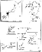 issued from : S.O. Sakaguchi & H. Ueda in Zootaxa, 2010, 2623. [p.55, Fig.1]. issued from : S.O. Sakaguchi & H. Ueda in Zootaxa, 2010, 2623. [p.55, Fig.1].
Map of the Nansei Islands and Kyushu Island. Locations of the rivers where sampling occurred indicated by black spots, among which those with the numbers represent the rivers from which specimens of P. nansei or P. inopinus (= P. japonicus where collected. |
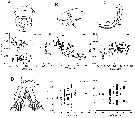
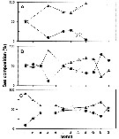 Issued from : E-O. Park, J.R. Cordell & H.Y. Soh in Zool. Stud., 2013, 52 (7). [p.7, Fig.7]. Issued from : E-O. Park, J.R. Cordell & H.Y. Soh in Zool. Stud., 2013, 52 (7). [p.7, Fig.7].
Seasonal variations in the adult sex composition of P. inopinus (= P. japonicus) from Mankyung River estuary (South Korea).
(A): Oligohaline, (B): mesohaline, (C): polyhaline. Circle : female; triangle: male.
Nota: Males were more abundant than females throughout the study period (January to December 2000) and across the entire range of salinity, but the ratio of males to females exceeded 50% under oligohaline conditions in January, mesohaline conditions in late fall and winter, and polyhaline conditions in early winter..
Gravid females appeared in oligohaline conditions in March, July, and September and accounted for 50% of all females present in September. Under mesohaline conditions, they occurred in June to October, and under polyhaline conditions, they occurred only in June and October, and >50% of all females were gravid in October.
These facts indicate that abundance of P. inopinus (= P. japonicus) as P. poplesia may be controlled by Chl a concentrations and salinity conditions. |
 Issued from : E.O. Park, H.-L. Suh & H.Y. Soh in Korean J. Environ. Biol., 2005, 23 (4). [p.370, Fig.3]. As Pseudodiaptomus sp. Issued from : E.O. Park, H.-L. Suh & H.Y. Soh in Korean J. Environ. Biol., 2005, 23 (4). [p.370, Fig.3]. As Pseudodiaptomus sp.
Abundance-temperature- salitity diagram of Pseudodiaptomus sp. in Seomjin River estuary, soyhern Korea.
Nota: this species was later described as P. koreanus (= P. japonicus by Soh & al. (2012). |
 Issued from : J. Cordell & coll., 1997 in PICES Annual meeting (2012). [p.11]. Issued from : J. Cordell & coll., 1997 in PICES Annual meeting (2012). [p.11].
Abundance of Pseudodiaptomus inopinus (probably = P. japonicus) in relation to salinity in seven Pacific northeast estuaries.
Salinity (PSU) in abscissa and abundance (no. per sample) in ordinate. |
| | | | Loc: | | | S & E Korea, Japan (SW Honshu, Hiruma & Kita-ura Lakes, NW Hokkaido), Sakhalin | | | | N: | 8 | | | | Rem.: | Freshwater, bracish.
Agfter Ueda (2019, pers. comm.) P. japonicus
was originally described from brackish lakes of the Japan Sea coast of Japan in 1928, but the species had been considered as a synonym of P. inopinus for a long time.
Sakaguchi & Ueda (2011, p.124) found two forms in the mainland of Japan and Sakaguchi & Ueda (2012, p.177) revived the species name japonicus for one of them because of genetic distance between the two forms and morphological differences from the original description of P. inopinus
Ueda & Sakaguchi (2019) indicated that P. japonicus is widespread in northern East Asia without overlapping the range of the other form, i.E. P. inopinus.
For Sakaguchi & al. (2011, Table 1) P. japonicus (as P. inopinus) is often the most abundant copepod in river estuaries on the Japan Sea coast of northern Kyoshu to northern San-in District, Japan.
P. yamato is distinguishable from wP. japonicus by having long posterior processes of the female genital operculum and lacking medial spinules at midlength of the 1st exopodal segment of the female P5. The males of P. yamato and P. japonicus are not distinguishable at present.
P. inopinus specimens described from South Korea (Lee & al., 2007) probably include P. japonicus, because the specimens were collected from various coasts of South Korea including the east and south coasts, from which P. koreanus (= P. japonicus) was described (Soh & al., 2012). However, their specimens from the west coast are probably not P. japonicus, because Soh & al. (2012) indicated that specimens from the west coast are genetically different from P. koreanus | | | Dernière mise à jour : 15/04/2020 | |
|
|
 Toute utilisation de ce site pour une publication sera mentionnée avec la référence suivante : Toute utilisation de ce site pour une publication sera mentionnée avec la référence suivante :
Razouls C., Desreumaux N., Kouwenberg J. et de Bovée F., 2005-2026. - Biodiversité des Copépodes planctoniques marins (morphologie, répartition géographique et données biologiques). Sorbonne Université, CNRS. Disponible sur http://copepodes.obs-banyuls.fr [Accédé le 17 juin 2026] © copyright 2005-2026 Sorbonne Université, CNRS
|
|
|
|


;)
;)
;)
;)
;)
;)
;)
;)
;)
;)
;)
;)
;)
;)
;)
;)
;)
;)
;)
;)
;)
;)
;)
;)
;)
;)
