|
|
 |
Fiche d'espèce de Copépode |
|
|
Calanoida ( Ordre ) |
|
|
|
Diaptomoidea ( Superfamille ) |
|
|
|
Acartiidae ( Famille ) |
|
|
|
Acartia ( Genre ) |
|
|
|
Odontacartia ( Sous-Genre ) |
|
|
| |
Acartia (Odontacartia) nadiensis Lee S, Soh & Lee W, 2019 (F,M) | |
| | | | | | | Ref.: | | | Lee S. & al., 2019 (p.71 (p.71, Descr. F, M, figs.F, M, phylogeny, , Table 3: comparison between species of the subgenus Odontacartia, species key of the Odontacartia). | 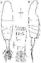 Issued from : S. Lee, H.Y. Soh & W. Lee in ZooKeys, 2019, 893. [p.73, Fig.1]; Female (from Nadi Bay, Fiji): A-B, habitus (dorsal and lateral); C, urosome (ventral. Scale bars in µm. Nota: - Prosome/Urosome length ratio = 3.5 : 2.1. - Prosome 5-segmented. Head and 1st pediger completely separate, 4th and 5th pedigers fused. - Posterior corners of 5th pediger somite rounded, each with 3 spines. - Rostral filaments thick and short. - Urosome 3-segmented. - Genital double-somite slightly swollen anterolaterally, with paired of gonopores ventromedially, each covered with pointed operculum. - 1st and 2nd urosomites each with 4 spines on posterodorsal margin. - Caudal rami bearing short hairs on lateral margin. - Proportional lengths of urosomites and caudal rami 38 : 23 : 17 : 22 = 100.
|
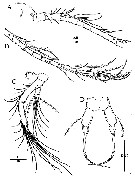 Issued from : S. Lee, H.Y. Soh & W. Lee in ZooKeys, 2019, 893. [p.74, Fig.2]; Female: A, rostrum and A1 (part., 1st to 8th segment); B, A1 (part., 8th to 18th segment); C, A2; D, P5. Scale bars in µm. Nota; - A1 incompletely 18-segmented, 4th to 7th segments partly fused on dorsal surface, 9th to 11th each with 1 row of setules, 12th segment with 3 rows of sdetules, 13th and 17th segments each with 1 row of setules. - A2 coxa with 1 seta; basis and 1st endopod segment fused to form elongated allobasis bearing 8 setae medially and 1 seta terminally along inner margon, and spinular row on distal arca; 2nd endopod segment elonjgated, with 7 setae, row of spinules o,n lateral margin:; 3rd exopod segment short, with 7 setae. Exopod 4-segmented (setation formula 1, 2, 2, 3). - P5 symmetrical, 3-segmented; basis ovate, with outer seta; exopod tapering, thick, bent at midlength, distal portion with minutely serrated terminal spine , base slightly swollen.
|
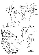 Issued from : S. Lee, H.Y. Soh & W. Lee in ZooKeys, 2019, 893. [p.75, Fig.3]; Female: A, Md; B, Mx1; C, Mx2; D, Mxp. Scale bar in µm. Nota: - Md: Coxa with well developed gnathobase bearing 11 teeth; basis with seta and row of setules on lateral and posterior margins; endopod 2-segmented, 1st segment with 2 setae, 2nd segment with 7 setae: exopod 5-segmented (setation formula 1, 1, 1, 1, 2). - Mx1 precoxa and coxa incompletely fused, precoxal arthrite with 8 setae; coxal endite with 3 setae: 1 short seta and 8 long setae on coxal epipodite; basal endite with 1 seta; basal exite with 1 seta; 1-segmetd exopod with 2 setae laterally and 8 setae terminally; endopod absent. - Mx2 precoxa and coxa incompletely fused; setation formula of endites 4, 2, 2, 3; basal endite with 1 seta and row of spinules on distal margin; endopod 3-segmented (setation formula 2, 2, 3). - Mxp comprisind syncoxa with 6 setae; basis with spiniform seta; endopod 2-segmented, 1st segment with 3 setae, 2nd segment with 2 setae.
|
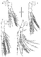 Issued from : S. Lee, H.Y. Soh & W. Lee in ZooKeys, 2019, 893. [p.77, Fig.4]; Female: A, P1; B, P2; C, P3; D, P4. Scale bar in µm.
|
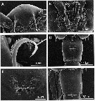 Issued from : S. Lee, H.Y. Soh & W. Lee in ZooKeys, 2019, 893. [p.80, Fig.7]; Female (Scanning electron micrographs): A, rostrum (ventral view); B, P5; C, P5 terminal spine; D, genital double-somite (ventral view); E, genital field; F, genital double-somite (dorsal view).
|
 Issued from : S. Lee, H.Y. Soh & W. Lee in ZooKeys, 2019, 893. [p.76]; Female & male : Spine and setal formulae for P1 to P4. Roman numerals: spines; Arabic numerals: setae.
|
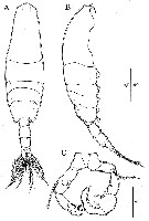 Issued from : S. Lee, H.Y. Soh & W. Lee in ZooKeys, 2019, 893. [p.78, Fig.5]; Male: A-B, habitus (dorsal and lateral); C, P5. Scale bars in µm. Nota: - Body surface armed with some sensilla. - Prosome/Urosome length ratio 3.1 : 2.1. - Prosome 5-segmented. - Rostral filaments thin. - 5th prosomite with 6 spines on posterior margin. - Urosome 5-segmented. - 2nd urosomite with 4 spines on posterodorsal margin and 2 spines on posteroventral margin; pair of sensillae on dorsal surface; 3rd and 4th urosomites each with 4 spines on posterodorsal margin. - Caudal rami bearing short hairs on lateral margin. - Length proportions of urosomites to caudal rami 16 : 31 : 21 : 7 : 12 : 14 = 100. - P5 asymmetrical, intercoxal sclerite distinct. Left leg 4-segmented; basis armed with posterolateral seta and rounded lobe on posterior surface; exopod 2-segmented, 1st segment unarmed, 2nd segment with hairs and 1 spine with teeth on medial margin and 1 small spine distally. Right leg 5-segmented, basis armed with posterolateral seta; exopod 3-segmented, 1st segment with long slender seta, 2nd segment with oblong inner lobe bearing 1 spine on distal margin, 3rd segment with 1 spine on medial margin and 1 spine distally.
|
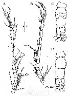 Issued from : S. Lee, H.Y. Soh & W. Lee in ZooKeys, 2019, 893. [p.79, Fig.6]; Male: A, left A1; B, right A1; C-D, urosome (dorsal and ventral, respectively). Scale bars in µm. Nota: - Left A1 22-segmented;. Right A1 18-segmented, with geniculation (14th and 15th segments). - Other mouthparts and P1-P4 as in female.
|
 Issued from : S. Lee, H.Y. Soh & W. Lee in ZooKeys, 2019, 893. [p.81, Fig.8]; Male: (Scanning electron micrographs): B, rostrum (ventral view); G, 5th urosomite and caudal rami (dorsal view); H, same (ventral view).
|
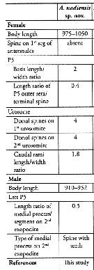 Issued from : S. Lee, H.Y. Soh & W. Lee in ZooKeys, 2019, 893. [p.84, Table 3]. Issued from : S. Lee, H.Y. Soh & W. Lee in ZooKeys, 2019, 893. [p.84, Table 3]. Acartia (Odontacartia) nadiensis: Morphological characters. Compare with other Odontacartia species. Nota: 1 - Absence of spine on 1st to 2nd segments of female A1 .....2. 2 - Spine present on dorsal surface of female genital double-somite ..... 3. 3 - Four strong spines on dorsal surface of female genital double-somite and 2nd urosomite.
|
 Issued from : S. Lee, H.Y. Soh & W. Lee in ZooKeys, 2019, 893. [p.82, Fig.9] Ohylogenetic tree based on mtCOI sequences (58 bp) of Odontacartia species including A. (Acartiura) omorii as outgroup. One-thousand bootstrap replicates were performed by MEGA6 using neighbor joining and minimum evolution methods. Neighbor joining bootstrap values shown above branches; minimum evolution bootstrap values are below branches.
| | | | | NZ: | 1 | | |
|
Carte de distribution de Acartia (Odontacartia) nadiensis par zones géographiques
|
| | | | Loc: | | | SW Pacif. (Nadi Bay, Fiji)
Type locality: 17°45.848' S, 177°22.348' E. | | | | N: | 1 | | | | Lg.: | | | (1243) F: 0,975-1,050; M: 0,910-0,952; {F: 0,975-1,050; M: 0,910-0,952} | | | | Rem.: | For Lee & al. (2019, p.82) the subgenus Odontacartia, including the new species, can be divided into 2 groups based on the presence or no of a spine on the 1st segment of A1. A. nadiensis lacks a spine on this segment, second, the outer seta of the female P5 is much shorter than the terminal spine, and the length ratio of the outer seta/ terminal spine is 0.4. The male P5 is clearly distinguishable from the rest of species based on its length and the type of medial process on the exopodal segment 2 of the left leg.; furthermore, the new species shows other minor differences compared to the other 13 Odontacartia species, such as the number of dorsal spines on the urosomite, the length/width ratio of the female P5 basis, and the length/width ratio of caudal rami. To supplement the morphological evidences, molecular analysis (using partial mtCOI sequences) of six Odontacartia species showed that A. nadiensis differed by 24.1-29.0% from the sequences of congeneric species, which is greater tha the range of interspecific differences reported in previous studies. | | | Dernière mise à jour : 09/03/2020 | |
|
|
 Toute utilisation de ce site pour une publication sera mentionnée avec la référence suivante : Toute utilisation de ce site pour une publication sera mentionnée avec la référence suivante :
Razouls C., Desreumaux N., Kouwenberg J. et de Bovée F., 2005-2025. - Biodiversité des Copépodes planctoniques marins (morphologie, répartition géographique et données biologiques). Sorbonne Université, CNRS. Disponible sur http://copepodes.obs-banyuls.fr [Accédé le 05 janvier 2026] © copyright 2005-2025 Sorbonne Université, CNRS
|
|
 |
 |


