Fiche d'espèce de Copépode
Monstrilloida ( Ordre )
    Monstrillidae ( Famille )
        Monstrilla ( Genre )
Monstrilla chetumalensis  Suarez-Morales & Castellanos-Osorio, 2019   (M)
Ref.:
Suarez-Morales & Castellanos-Osorio, 2019 (p.113, Descr. M, figs. M, , Caribbean Key F, M))
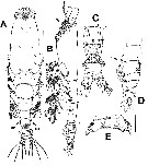
Espèce Monstrilla chetumalensis - Planche 1 de figures morphologiquesIssued from : E. Suarez-Morales & A. Castellanos-Osorio in ZooKeys, 219, 876. [p.115, Fig.2].
Male (from: Chetumal Bay, Mexican Caribbean): A-B, habitus (dorsal); B, habutus (lateral; arrow indicates medial ventral protuberance); C-D, urosome (ventral and lateral; arrows indicate globular prcxesses on 5th pedigerous somite); E, genital complex with lappets (ventral view).
Scale bars: µm (A-B); µm (C-D); µm (E).

Nota: Cephalothorax robust, representing 47.5 % of total body length.
- Succeeding pediger somites 2-4 each with pair of biramous swimming legs; pediger somites 2-4 combined accounting for 31 % of total body length in dorsal view.
- Cephalic region wide, bilaterally protuberant in dorsal view, narrower than cephalothorax; outer margin of cephalic protuberances corrugate.
- Forehead weakly rounded, with coarsely rugose anterior margin and field of transverse striations on dorsal anterior surface; no other cephalic ornamentation discernible on dorsal anterior surface
- Pair of small dorsal pit setae between A1 bases; ventral anterior surface also with 2 pit setae.
- Cephalothorax dorsal surface with scattered dorsal pores.
- Midventral oral papilla moderately protyberant, located at about proximal 1/3 along ventral surface of cephalothorax.
- Pair of relatively small lateral pigment cups moderately developed, separated by length of less than one eye diameter, weakly pigmented; ventral cup slightly larger than lateral cups.
- Urosome consisting of 5th pediger somite, genital somite (carrying genital complex), 2 short, free pstgenital somites divided by incomplete dorsal suture, and short anal somite.
- 5th pediger somite with ventrally producede proximal half; dorsal surface smooth; distal half with pair of small medial rounded processes visible in lateral view ( arrowed in fig.2C, D); posterolateral margins of 5th somite produced, partially overlapping succeeding genital somite both dorsally and laterally (''ple in fig.2A, arrow in fig.2B).
- Genital somite slightly shorter than 5th pediger somite; genital complex of type I (see Suarez-Morales & McKinnon, 2014<), represented by short, robust ventrally expanded shaft; complex with short, widely divergent lappets tapering distally into apical subtriangular opercular process (fig.2C-E.; lappets with rugose anterior surface, branches connectede medially by wide debntate margin.
- Anal somite 1.3 times as long as genital somite.
- Caudal rami subquadrate, approximally 1.1 times as long as wide and about 0.7 times as long as anal somite. Each ramus armed with 4 subequally long caudal setae.


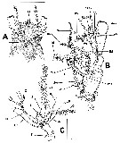
Espèce Monstrilla chetumalensis - Planche 2 de figures morphologiquesIssued from : E. Suarez-Morales & A. Castellanos-Osorio in ZooKeys, 219, 876. [p.117, Fig.3].
Male: A, anterior part of cephalosome (ventral view; arrow indicates nipple-like processes; 1, 2 = sensillae ): B, A1 segments 1-4 (dorsal view) showi,g setal elements (sensu Grygier & Ohtsuka, 1995); C, 5th antennulary segment with setal elements (sensu Huys & al., 2007).
Scale bars: µm (A-B); µm (C).
- Preoral ventral surface with low, wide-based rounded process protruding between A1 bases (arrowed in fig.2B); nipple-like cuticular processes surrounded by striated surface (fig.3A).
- A1 5-segmented, representing 38 % of total body length, and 75 % of cephalothorax length; segments 1-4 separated by complete sutures. Intersegmental division bertween segments 3 and 4 lacking suture, division marked by constriction; segment 4 being longest: geniculation between segments 4 and 5. Armature using terminology of Grygier & Ohtsuka (1995) for female monstrilloid antennular armature of segments 1-4 complemented with nomenclature by Huys & al. (2007) for elements on male 5th antennulary segment.


Espèce Monstrilla chetumalensis - Planche 3 de figures morphologiquesIssued from : E. Suarez-Morales & A. Castellanos-Osorio in ZooKeys, 219, 876. [p.114, Table 1].
Male: Armature of swimming legs P1 to P4 including basis, exopods and endopods.
Roman numerals indicate spiniform elements, Arabic numerals indicate setiform elements, set from inner to outer positions.- Smooth and rectangular intercoxal sclerites.

NZ: 1

Carte de distribution de Monstrilla chetumalensis par zones géographiques
Loc:
NW Atlant. (Chetumal Bay)
Type locality: 18°26'54'' N, 88°04'00'' W.
N: 1
Lg.:
(1246) M: 0,69; [M: 0,69] *
* Total body length from anterior end of cephalothorax to posterior margin of anal somite.
Rem.: For Suarez-Morales & Castellanos-Osorio (2019, p.116)This male of M. chetumalensis most closely resembles the Indian species M. lata Desai & Bal (1962), however, in M. chetumalensis , the lappets are strongly curved and have an inverted U-shape, moreover, (the A1 armature differs strongly between these two species, particularly, in the size and number of setal elements of segments 1-4. In addition, M. kata has 6 caudal setae vs 4 in the new species (chetumalensis. In M. lata the 5th pediger somite has a weak concavity in its proximal half, whereas the same structure is ventrally produced in chetumalensis. The genital complex of chetumalensis shares some features with M. papilliremispapilliremis, the lappets are medially connected by a smooth margin with a medial notch and also have an inverted U-shape, thus diverging from the conditions observede in the genital complex of chetumalensis. Morphological comparison shows that chetumalensis is assignable to Monstrilla, as it clearly diverges from Caromiobenella.
Dernière mise à jour : 19/06/2023

 Toute utilisation de ce site pour une publication sera mentionnée avec la référence suivante :

Razouls C., Desreumaux N., Kouwenberg J. et de Bovée F., 2005-2024. - Biodiversité des Copépodes planctoniques marins (morphologie, répartition géographique et données biologiques). Sorbonne Université, CNRS. Disponible sur http://copepodes.obs-banyuls.fr [Accédé le 21 novembre 2024]

© copyright 2005-2024 Sorbonne Université, CNRS

Webmaster
Mentions légales et informations nominatives
CNRS   Observatoire Océanologique de Banyuls sur Mer - Laboratoire Arago
Sorbonne Université


Version française
English version


Rechercher

Sur le WEB du CNRS


Copépodes planctoniques marins

Copépodes planctoniques marins


Imprimer Contact Accueil Plan du site Accès restreint Retour Une du Labo Imprimer Contact Plan du site Crédits Téléchargez les Plug-Ins