|
|
 |
Fiche d'espèce de Copépode |
|
|
Calanoida ( Ordre ) |
|
|
|
Clausocalanoidea ( Superfamille ) |
|
|
|
Aetideidae ( Famille ) |
|
|
|
Undeuchaeta ( Genre ) |
|
|
| |
Undeuchaeta incisa Esterly, 1911 (F,M) | |
| | | | | | | Syn.: | Undeuchaete superba With, 1915 (p.137, figs.F,M);
Undeuchaeta incisa : Lysholm & Nordgaard, 1921 (p.17);
Mesundeuchaeta asymmetrica Wolfenden, 1911 (p.244, figs.F); De Decker & Mombeck, 1964 (p.13);
Undeuchaeta superba With, 1915 (p.137, figs.F,M);
Pseudochirella superba : Jespersen, 1934 (p.67); 1940 (p.27); Sewell, 1948 (p.500); Vervoort, 1952 g (n°48, p.3, figs.F,M);
U. magna Tanaka, 1957 b (p.203, figs.F); 1969 (p.266, figs.F, Rem.); Paiva, 1963 (p.39, figs.F); Tanaka & Omori, 1970 a (p.144); U. major (part.): Vervoort, 1957 (p.71); 1963 b (p.154);
Pseudochirella incisa : Grice, 1964 (p.262, Figs.F, Rem.); Grice & Hulsemann, 1968 (tab.2); ?
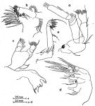
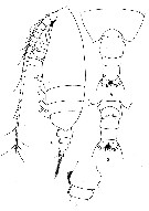
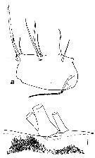
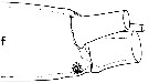
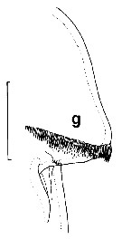
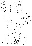
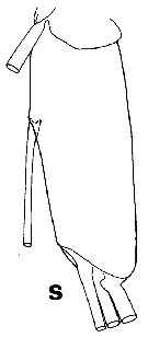
Undeuchaeta sp. Mulyadi, 2004 (p.53, fig.F, Rem.) | | | | Ref.: | | | Esterly, 1911 (p.319, Descr.F, figs.F); Brodsky, 1950 (1967) (p.184, figs.F); Vervoort, 1957 (p.72: Rem.); Park, 1978 (p.183, figs.F,M, Rem.: redescriptions); Bradford & Jillett, 1980 (p.80, figs.F,M, fig.73, distribution chart); Vaupel Klein, 1984 a (p.54, figs.F, Table II: characters, Rem.); He & al., 1992 (p.250); Markhaseva, 1996 (p.302, figs.F,M); Chihara & Murano, 1997 (p.689, Pl.48,49: F,M); Koomen & Vaupel Klein, 1998 (p.401, figs.F); Bradford-Grieve & al., 1999 (p.880, 923, figs.F,M); Boxshall & Halsey, 2004 (p.59: fig.M) | 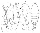 issued from : Tanaka O. in Publ. Seto Mar. Biol. Lab., 1957 b, 6 (2). [Fig.60, p.204]. As Undeuchaeta magna. Female (from Suruga): a, habitus (dorsal aspect); b, head (lateral aspect); c, last thoracic segment and urosome (lateral aspect, left side); d, last thoracic segment and genital segment (lateral aspect, right side); e, genital segment (ventral aspect); f, P1; g, P2. Nota Female: - Cephalothorax about 4.1 times the andomen length (4.89 : 1.18). - Head and 1st pediger segment separate, 4th and 5th pedigers fused. - Forehead with a median crest. - Rostrum 1-pointed, short and broad at the base. - Last thoracic segment slightly asymmetrical; left side triangularly produced, and pointed at the apex.; right side irregularly rounded, with a small prominence on the postero-lateral corner. - Genital segment with a strong protuberance; ventral spine near genital opening smaller than that of U. major- A1 23-segmented, extends to the end of caudal rami. - Abdominal segments and caudal rami in proportional lengths 41 : 21 : 21 : 5 : 12 = 100. - P2 to P4 with with terminal exopodal spine of 40, 32 and 33 teeth respectively. - P4 without spinules on posterior surface of basis.
|
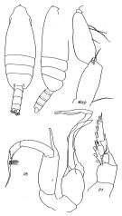
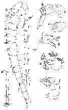
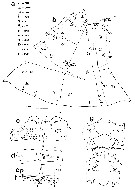
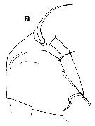
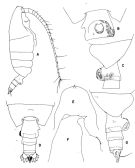 issued from : T. Park in Antarctic Res. Ser. Washington, 1978, 27. [p.184, Fig.54]. Female: A, habitus (right lateral side); B, posterior part of metasome and genital segment (ventral); C, last metasomal and first two urosomal segments (lateral); D, last metasomal and urosome (dorsal); E, forehead (dorsal); F, idem (lateral); G, last metasomal and urosome (lateral).
|
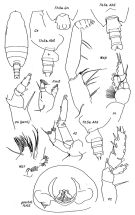
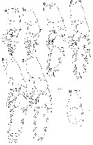
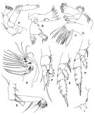 issued from : T. Park in Antarctic Res. Ser. Washington, 1978, 27. [p.185, Fig.55]. Female: A, A2; B, Md; C, Mx1; D, Mx2; E, Mxp; F, P1; G, P2; H, P4; I, medial part of coxa of P4 (anterior). P1-4: legs (anterior).
|
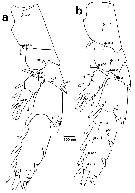
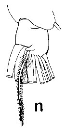
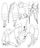 issued from : T. Park in Antarctic Res. Ser. Washington, 1978, 27. [p.186, Fig.56]. Male: A, habitus (dorsal view); B, idem (lateral); C, A2; D, Md; E, P1 (anterior); F, P2 (idem); G, P5 (viewed from left side); H, two exopodal segments of P5, medial); I, P5 (anterior).
|
 issued from : E.L. Markhaseva in Proc. Zool. Inst. RAN, St. Petersburg, 1996, 268. [p.303, Fig.243]. Female (from NW Pacif.: Marian Trench). Ce: forehead (lateral); P.md: mandibular palp; Th5 & GN: distal part of metasome and genital segment (ventral); Th5 & Abd: Th5 and urosome (dorsal, right side and left side lateral, respectively); P4 (part.): fourth leg (coxopod, basipod and endopod).
|
 issued from : E.L. Markhaseva in Proc. Zool. Inst. RAN, St. Petersburg, 1996, 268. [p.304, Fig.244]. Male (from S Australia).
|
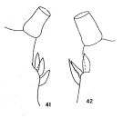 issued from : G.D. Grice in Crustaceana, 1964, 6 (4). [p.260, Figs.41-42]. As Pseudochirella incisa. Female: 41, basal segment of P4 (Pacific specimen); 42, idem (Atlantic specimen).
|
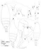 issued from : J.M. Bradford & J.B. Jillett in Mem. N.Z. oceanogr. Inst., 86, 1980. [p.81, Fig.56]. Female: A, habitus (dorsal); B, idem (lateral right side); C, left side of posterior of matasome; D-h, inner edge of basipod 1 of P4 (different specimens frm different stations. Male: I, habitus (dorsal); J, forehead (lateral left side); K, P5.
|
 issued from : O. Tanaka in Publ. Seto Mar. Lab., 1969, XVII (4). [Fig.7]. As Undeuchaeta magna. Female (from Pacific): a, A2; b, Mx1; c, Md; d, Mx2; e, Mxp. Nota: A2 with exopod 1.8 times as long as the endopod; endopod with 7 setae on the outer lobe and 8 setae on the inner lobe. Mx1: outer lobe with 9 setae; exopod with 11 setae; endopod with 13 setae; 2nd basal segment with 4 long and 2 small setae; 3rd inner lobe with 4 setae; 2rd inner lobe with 3 setae and the 1st inner lobe with 14 setae. Mxp: segments of the endopod short and robust, 1st basal segment half as long as the 2nd one.
|
 issued from : R.N. Wolfenden in Die Marinen Copepoden der Deutschen Südpolar-Expedition 1901-1903, 1911. [p.245, Fig.28]. As Mesundeuchaeta asymmetrica. Female: basipod of P4.
|
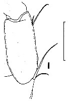
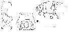
 issued from : R.N. Wolfenden in Die Marinen Copepoden der Deutschen Südpolar-Expedition 1901-1903, 1911. [Pl.XXIX, Figs.4-7]. As Mesundeuchaeta asymmetrica. Female: 4, habitus (lateral); 5, posterior part cephalothorax and urosome (dorsal); 6, urosome (ventral); 7, genitla segment (lateral, anotther specimen).
|
 issued from : J.C. von Vaupel Klein in Crustaceana, Supplt 9, Studies on Copepoda, III, 1984. [p.75, Fig.13, a, i]. Female: a, condition of the single seta (arrowed) of group I on basipodite 1 of Mxp (right appendage in medial aspect); i, patch of spinules adjacent to the insertions of the setae of group III on basipodite 1 of Mxp (detail of left appendage in lateral view).
|
 issued from : J.C. von Vaupel Klein in Crustaceana, Supplt 9, Studies on Copepoda, III, 1984. [p.74, Fig.12, f]. Female: f, spinules on the distal margin of 5th endite of Mx2 (right appendage, antero-lateral view). Nota: with 10 spinules in a small patch.
|
 issued from : J.C. von Vaupel Klein in Crustaceana, Supplt 9, Studies on Copepoda, III, 1984. [p.76, Fig.14, d, g]. Female: d, accessory seta of endopodite 4th segment of Mxp (right one in medial aspect) (note: seta large and stout, and armed with a least two rows of denticles, lateralrow not visible in the figure); g, arrangement of spinules on the lateral tubercle of endopod of P1 (left appendage,anterior face) (note: well delimited basal row with an adjoining field of distally decreasing spinules). Scale bar 0.1mm for d; 0.067 mm for g.
|
 issued from : J.C. von Vaupel Klein in Crustaceana, Supplt 9, Studies on Copepoda, III, 1984. [p.63, Fig.5, l]. Female: l, setal armature of endopodite 1 of A2 (note: at least one of which long). Scale bar 0.2 mm.
|
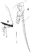
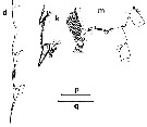 issued from : J.C. von Vaupel Klein in Crustaceana, Supplt 9, Studies on Copepoda, III, 1984. [p.84, Fig.18, d, k, m]. Female: d, number and structure of spines along the lateral margin of exopodal segments 1, 2 and 3 of P1 (left appendage, posterior view) (note: 2 subequal spines and both bipectinate, the one on exopodal segment 1 missing); k, condition of the lateral margin of exopodal segment 2 of P2 (left appendage, posterior view) (note: a hairy margin, the same conditions as found in legs 3 and 4); m, endopodal segment 2 of P3 (left leg, anterior aspect) (note: a row of 7 thin, needle-shaped spinules inserting antero-laterally, adjacent to the hinge-join endopodal segments 2/3). Scale bar: q = 0.2 mm for d; 0.1 mm for m; p = 0.2 mm for k.
|
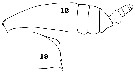 issued from : C.O. Esterly in Univ. Calif. Publs Zool., 1911, 6 (14). [Pl.27, Figs.12, 19]. Female (from San Diego Region): 12, habitus (lateral); 19, forehead (lateral). Nota: A1 extend a little beyond the end of the body. A2 with endopod half the exopod length. Prosome (4-segmented) a little over 4 times as long as urosome. The last thoracic segment is rounded on the right side and produced on the left into a process which is notched at the end. Urosome 4-segmented. Genital segment is as long as the 2nd and 3rd together, the 2nd about half as long as the 3rd and the anal half as long as the 2nd, caudal rami about as long as the anal segment.Exopod of P1 indistinctly 3-segmented (suture between the 1st and 2nd segments indicated by a line); there are 2 outer marginal spines; the spines of the outer margin of the exopod of P1 are much longer than in the P2 and P3.
|
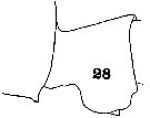 issued from : C.O. Esterly in Univ. Calif. Publs Zool., 1911, 6 (14). [Pl.28, Fig.28]; Female: part of last thoracic segment and genital segment (lateral, left side).
|
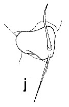
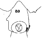 issued from : C.O. Esterly in Univ. Calif. Publs Zool., 1911, 6 (14). [Pl.29, Fig.59]; Female: part of last thoracic segment and genital segment (ventral). Nota: Genital segment markedly protuberant ventrally; there is a lamellar process at the right orifice, and on the right of the segment about the middle there is a wing-like extension.
|
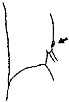
 issued from : J.C. von Vaupel Klein in Crustaceana, Supplt 9, Studies on Copepoda, III, 1984. [p.82, Fig.17, b, g, i]. As Euchirella messinensis messinensis. Female: b, arrangement of hair-sensilla on the posterior face of the proximal segments of right P4; g, genital somite (ventral; showing asymmetrical presence of 2 spiniform processes: arrows); i, terminal part of basipodal segment 2 and endopod of right Mxp (medial view).
|
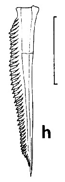 issued from : J.C. von Vaupel Klein in Crustaceana, Supplt 9, Studies on Copepoda, III, 1984. [p.80, Fig.16, h]. Female: h, number of teeth on the serrate lateral mrgin of the terminal spines of exopodal segmnt 3 of P2-P4 (medial plumosy omitted). Scale bar 0.2 mm. Nota: Spine of P2 with 40 teeth.
|
 issued from : J.C. von Vaupel Klein in Crustaceana, Supplt 9, Studies on Copepoda, III, 1984. [p.69, Fig.9, e]. Female: e, complement of terminal setae on the 2nd inner lobe of basipodal segment 1 of right Mx1 (details of right appendages in anterior view; to the posterior side 2 relatively stout setae are always present, which are combined bipectinate and spinulose; anteriorly with 2 well developed, spinulose setae and a fifth, smaller spinulose seta). Scale bar: 0.2 mm.
|
 issued from : J.C. von Vaupel Klein in Crustaceana, Supplt 9, Studies on Copepoda, III, 1984. [p.76, Fig.14, g]. Female: g, arrangement of spinules on the lateral tubercle of endopod of left P1 (anterior face). Scale bar: 0.067 mm. Nota: Note a well-delimited basal row with an adjoining field of distally decreasing spinules.
|
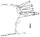
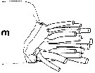
 issued from : P. Koomen & J. C. von Vaupel Klein in J. Mar. Systems, 1998, 15. [p.405, Fig.3]. In situ mapping of the integumental organs of the female (from 04°03'N, 123°26'E): a, a', a\", left A1 (in lateral view, with segment numbers specified); b, left A2 (lateral view); c, proximal part of the lrft mandibular gnathobasis (ventral view; hatched area marks insertion of palpus mandibularis); d, left palpus mandibularis (postero-medial view); e, right Mx1 (anterior view); f, right Mx2 (antero-lateral aspect). Sutures drawn in dotted lines are situated on the reverse side of the appendage. Pictogramm coding see fig.1)
|
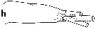
 issued from : P. Koomen & J. C. von Vaupel Klein in J. Mar. Systems, 1998, 15. [p.406, Fig.4]. In situ mapping of the integumental organs of the female: a, right Mxp (medial aspect); b, left caudal ramus (dorsal view); c, right caudal ramus (ventral aspect); d, internal face of the upper lip (top = distal; bottom = proximal). Pictogramm coding see fig.1).
|
 issued from : P. Koomen & J. C. von Vaupel Klein in J. Mar. Systems, 1998, 15. [p.407, Fig.3]. In situ mapping of the integumental organs of the female: a, left P1 (anterior view); b, right P1 (posterior aspect); c, left P2 (posterior); d, right P2 (posterior); e, left P3 (anterior); f, right P3 (posterior face); g, ventral aspect of anteriormost sternal keel without any perforations (top = rostral; bottom = caudal edge). Pictogram coding see fig.1)
|
 issued from : P. Koomen & J. C. von Vaupel Klein in J. Mar. Systems, 1998, 15. [p.408, Fig.6]. In situ mapping of the integumental organs of the female: a, left P4 (anterior); b, right P4 (posterior). Pictogram coding see fig.1.
|
 issued from : P. Koomen & J. C. von Vaupel Klein in J. Mar. Systems, 1998, 15. [p.403, Fig.1]. In situ mapping of the integumental organs of the female: a, pictogram coding; b, integument of cephalon, cut in two with three additional incisions in the left part to make a flat spread possible with line ''m'' representing the dorsal midline (dorsal aspect); c-f, urosomal somites 1+2 (genital double-somite), 3, 4 and 5, respectively (cut in several parts with ''m'' representing the dorsal midline); g, ventral aspect of 'oral field' and adjacent regions. Structures drawn in dotted lines are seen through the (hyaline) integument of parts in the foreground. Integumental organs within the oral cavity (hidden by the flattened upper lip) have not been depicted (see fig. 4d). Abbreviations used for integumental organs and pictogram coding: SLIT = regular slit-shaped glandular pore. CONC = (partly) concealed slit-shaped glandular pore. CLO = large closing-flap glandular pore. TUB = tubular glandular pore. PIT = pit-sensillum. PEG = peg-sensillum. HAIR = hair-sensillum. SPIN = spine-sensillum and/or spinular pore. CIRC = circular pore. URN = urn-shaped glandular pore. BUT = button-shaped pore. UNC = integumental organ of uncertain nature.
|
 issued from : P. Koomen & J. C. von Vaupel Klein in J. Mar. Systems, 1998, 15. [p.404, Fig.2]. In situ mapping of the integumental organs of the female of the thoracic somites spread out flatly: a-c, thoracic somites 1 -3; d, thoracic somites 4+5 together. Line 'm' represents the dorsal midline. Pictogram coding see fig.1a).
|
 Undeuchaeta incisa Undeuchaeta incisa female: 1 - Cephalon with crest. 2 - Posterior corners of last thoracic segment not equally prolonged, one of them shorter and rounded (lateral view).
|
 Undeuchaeta incisa Undeuchaeta incisa male: 1 - Cephalon with crest. 2 - Exopodal segment 2 of left P5 twice, or more longer than wide.
|
 issued from : O. Tanaka in Publ. Seto Mar. Biol. Lab., 1957 b, 6 (2). [Fig.59, p.202]. Female A1: Proportional lengths of segments.
|
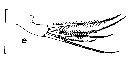
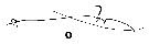 issued from : J.C. von Vaupel Klein in Crustaceana, Supplt 9, Studies on Copepoda, III, 1984. [p.66, Fig.7, o]. Undeuchaeta incisa: Structural feature of exopodite 1 and exopodite 2 of A2 (lateral aspects of left appendages and medial views of right antennal parts; compare with Fig.7n which represents left A2 medial face drawn from a lateral view). o, medial.
|
 issued from : J.C. von Vaupel Klein in Crustaceana, Supplt 9, Studies on Copepoda, III, 1984. [p.66, Fig.7, s]. Undeuchaeta incisa: Structural feature of the exopodite 2 of A2 (7th segment), right appendage in medial aspect showing 4th, appendicular seta inserting halfway the segment.
|
 issued from : J.C. von Vaupel Klein in Crustaceana, Supplt 9, Studies on Copepoda, III, 1984. [p.68, Fig.8, a]. Undeuchaeta incisa: Structure and setal armature of basipode 2 of Md (right appendage in anterior view). 3-setae condition.
|
 issued from : J.C. von Vaupel Klein in Crustaceana, Supplt 9, Studies on Copepoda, III, 1984. [p.68, Fig.8, J]. Undeuchaeta incisa: Structure and setal complement of endopod 1 of Md (right, anterior). 2 large setae.
|
 issued from : J.C. von Vaupel Klein in Crustaceana, Supplt 9, Studies on Copepoda, III, 1984. [p.68, Fig.8, n]. Undeuchaeta incisa: Accessory setae on endopodite 2 of Md (left, posterior). A single seta.
|
 Issued from : J.C. von Vaupel Klein in Crustaceana, Supplt 9, Studies on Copepoda, III, 1984. [p.70, Fig.10, J]. Undeuchaeta incisa: Setal armature on proximo-distal corner of basis (Ba2), adjacent to the endopod (Ri) of Mx1 (left appendage in postrior view; secondary structures on large setae are not shown, and neither are the hairs on the basis proper). 5 large and stout but unequal detae, 4 in an anterior row, the 5th inserting more posteriorly.
|
 Issued from : J.C. von Vaupel Klein in Crustaceana, Supplt 9, Studies on Copepoda, III, 1984. [p.72, Fig.11, h]. Undeuchaeta incisa. Structure of Mx1, terminal configuration of setae plus the 'blunt tooth (= tubular pore) on the endite of basis (Ba2) (plumosity and/or spinules on setae, as well as hairs on lobe, omitted, right appendage in anterior view) (See Euchirella curticauda, Fig.11, a; Chirundinella magna Fig.11, g). Contrary to E. curticauda (see Fig.11, a) or other euchirellinid forms, three well developed (though not exactly equal) setae, presumably corresponding to (p, c, d, as in Euchirella) in .
|
 Issued from : J.C. von Vaupel Klein in Crustaceana, Supplt 9, Studies on Copepoda, III, 1984. [p.70, Fig.11, m]. Undeuchaeta incisa: Setal armature of the endopod of Mx1 (detail of left appendage in posterior aspect; setules/spinules omitted). Crowded complement in the other euchirenillid genera studied by the author: 15 setae in U. incisa (compare with E. formosa Fig.11, i; and E. truncata Fig.11, J).
|
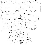
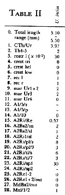 Issued from : J.C. von Vaupel Klein in Crustaceana, (Supplement) 9, 1984. [p.93, Table II ]. Undeuchaeta incisa Female: Datamatrix stating observed states of characters from Table I (p.87-90) presently examined; nos. refer to the input nos. used in Table I (see to the family Aetideidae).
|
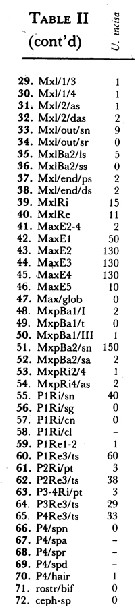 Issued from : J.C. von Vaupel Klein in Crustaceana, (Supplement) 9, 1984. [p.94, Table II (cont' d) ]. Undeuchaeta incisa Female: Datamatrix stating observed states of characters from Table I (p.87-90) presently examined; nos. refer to the input nos. used in Table I (see to the family Aetideidae).
|
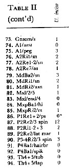 Issued from : J.C. von Vaupel Klein in Crustaceana, (Supplement) 9, 1984. [p.95, Table II (cont' d) ]. Undeuchaeta incisa Female: Datamatrix stating observed states of characters from Table I (p.87-90) presently examined; nos. refer to the input nos. used in Table I (see to the family Aetideidae).
| | | | | Ref. compl.: | | | Sewell, 1948 (p.563, 564, Rem.); Guangshan & Honglin, 1984 (p.118, tab., Rem.: p.165);Heinrich, 1990 (p.17); Shih & Young, 1995 (p.67); Errhif & al., 1997 (p.423); Razouls & al., 2000 (p.343, Appendix): Hsiao & al., 2004 (p.325, tab.1); Ikeda & al., 2006 (p.1791, Table 2); Park & Ferrari, 2009 (p.143, Table 4, Appendix 1, biogeography); in CalCOFI regional list (MDO, Nov. 2013; M. Ohman, comm. pers.) | | | | NZ: | 15 | | |
|
Carte de distribution de Undeuchaeta incisa par zones géographiques
|
| | | | | | | | | | | | 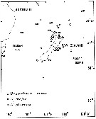 Issued from : J.M. Bradford & J.B. Jillett in New Zealand Ocean. Inst. Memoir, 86, 1980. [p.90-91, Fig.69]. Issued from : J.M. Bradford & J.B. Jillett in New Zealand Ocean. Inst. Memoir, 86, 1980. [p.90-91, Fig.69].
Distribution of several species of Undeuchaeta in the Tasman Sea and around New Zealand. |
| | | | Loc: | | | Antarct. (S & SE Pacif.), sub-Antarct. (SE Pacif.), South Africa (E), off Tristan da Cunha (N & E), Atlant. (sub-tropical & temperate), Cape Verde Is., Davis Strait, Strait of Denmark, Iceland, Faroe Is., SW Ireland, Indian, S Indian (subtropical convergence), ? Flores Sea, Celebes Sea, China Seas (East China Sea, South China Sea), Japan, Okhotsk Sea, Pacif. (N - S), California, W Pacif. (equatorial), off S Australia, New Zealand, Galapagos, Pacif. (SE tropical), off Juan Fernandez Is., Chile.
Type locality: NE Pacific (off California). | | | | N: | 20 | | | | Lg.: | | | (7) F: 6,42; M: 5,35; (10) F: 6-5,5; (20) F: 6,66-6; M: 5,58; (37) F: 6,6-5,7; M: 5,58-4,08; (56) F: 6,07; (105) F: 6,03; (112) F: 5,9-5,5; (143) F: 6,1; (201) F: 6,6-5,7; M: 5,2; (1257) F: 5,1-5,5; {F: 5,10-6,66; M: 4,08-5,58} | | | | Rem.: | méso-bathypélagique.
Sampling depth (Antarct., sub-Antarct.): 0-1000+ m.
Espèce qui devrait être considérée comme le type du Genre.
Voir aussi les remarques en anglais | | | Dernière mise à jour : 22/02/2021 | |
|
|
 Toute utilisation de ce site pour une publication sera mentionnée avec la référence suivante : Toute utilisation de ce site pour une publication sera mentionnée avec la référence suivante :
Razouls C., Desreumaux N., Kouwenberg J. et de Bovée F., 2005-2025. - Biodiversité des Copépodes planctoniques marins (morphologie, répartition géographique et données biologiques). Sorbonne Université, CNRS. Disponible sur http://copepodes.obs-banyuls.fr [Accédé le 30 novembre 2025] © copyright 2005-2025 Sorbonne Université, CNRS
|
|
|
|


;)
;)
;)
;)
;)
;)
;)
;)
;)
;)
;)
;)
;)
;)
;)
;)
;)
;)
;)
;)
;)
;)
;)
;)
;)
;)
;)
;)
;)
;)
;)
;)
;)
;)
;)
;)

;)
