|
|
 |
Fiche d'espèce de Copépode |
|
|
Calanoida ( Ordre ) |
|
|
|
Diaptomoidea ( Superfamille ) |
|
|
|
Candaciidae ( Famille ) |
|
|
|
Candacia ( Genre ) |
|
|
| |
Candacia bradyi A. Scott, 1902 (F,M) | |
| | | | | | | Syn.: | no Candacia curva (F) Mori, 1932 (p.175, figs.F);
no C. bradyi : Carl, 1907 (p.9, part.? figs.F,M, Rem.);
no C. bradyi : A. Scott, 1909 (p.156, figs.M); 1912 (p.49); Sewell, 1912 (p.366, figs.F); 1914 a (p.229); Tanaka,1935 (p.212); Mori, 1937 (1964) (p.80, figs.F); Krishnaswamy, 1953 (p.130); Grice, 1963 (p.174, figs.F,M); Tanaka, 1964 c (p.244); Chen & Zhang, 1965 (p.88, figs.F,M); ? Suarez-Morales & Gasca, 1998 a (p108); ? Hernandez-Trujillo & Esqueda-Escarcega, 2002 (in Appendix) | | | | Ref.: | | | A. Scott, 1902 (p.406, Descr.M, figs.M); Thompson & Scott, 1903 (p.235, 250); Pesta, 1912 a (p.49, fig.F,M); 1941 (p.163, figs.F,M); Sewell, 1932 (p.335); Grice, 1963 (p.174); ? Kasturirangan, 1963 (p.44, 47, figs.F,M); Saraswathy, 1966 (1967) (p.81); Lawson, 1973 a (p.487, figs.F, M, Rem.); 1977 (p.71, 75, tab.2,3,4, fig.3,5); Greenwood, 1978 (p.7, figs.F,M); Zheng Zhong & al., 1984 (1989) (p.250, figs.F,M); Mulyadi, 1997 a (p.80, Redescr.F,M, figs.F,M); Bradford-Grieve, 1999 b (p.163, figs.F,M, figs.182, 193); Al-Yamani & Prusova, 2003 (p.75, figs.F,M); Conway & al., 2003 (p.105, figs.F,M, Rem.); Mulyadi, 2004 (p.80, figs.F,M, Rem.); Phukham, 2008 (p.61, figs.F,M); Al-Yamani & al., 2011 (p.57, figs.F,M)
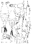
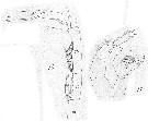
| 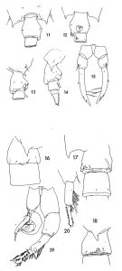 issued from : T.J. Lawson in Proc. Biol. Soc. Washington, 1973, 86 (11). [Figs.11-15, p.487; Figs.16-20, p.489]. Female: 11-13, urosome segments 1 and 2 (dorsal, ventarl, right side respectively); 14, urosome (right side); 15, P5 (posterior). Male: 16, urosome segments 1 and 2 (right side); 17, urosome segments 1 and 2 (ventral and slightly from the right side); 18, urosome segment 1 (right side); 19, P5 (anterior); 20, left P5, segments 3 and 4 (left side).
|
 issued from : J.G. Greenwood in Proc. R. Soc. Qd, 1978, 89. [p.8, Fig.3]. Female (from Moreton Bay, E Australia): a, urosome (lateral left side); b, idem (ventral); c, P5. Male: d, posterior part of prosome and urospme (dorsal); e, genital and 2nd urosomal segments (dorsal); f, Mx1; g, P5. Nota: Basipodal segment 2 of female P5 with single seta on each leg; this not figured by Sewell (1912) probably because broken during dissection. Female characteristics (from Bradford-Grieve, 1999 b, p.163): - Posterior metasome corners each terminate in a short spine. - Genital segment symmetrical in dorsal view and globular in shape, with a well-marked genital swelling. - Urosomal segment 2 is produced ventrally in midline into a short spine which is half the length of genital segment. - Caudal rami about twice as long as broad, slightly asymmetrical, that on right being slightly broader than on left. - A1 23-segmented. Mx2 basipod 2 with proximal spine longer and much stouter than distal spine. - P5 segment 3 slightly curved inwards with 2 setae on inner margin, with 3 outer edge spines on distal half of segment; these external spines are blunt and pigmented on left side and sharp and devoid of pigment on the right. Male characteristics (from Bradford-Grieve, 1999 b, p.165) : - Posterior metasome symmetrical, tip of right process does not reach beyond midpoint of genital segment. - Genital segment produced into a toothed tubercle on right side. - Urosomal segment 2 with a patch of small spines near posterior end. - Left A1 23-segmented and extends to posterior border of metasome. - Mx2 segment 2 with 3 short spines. - Left P5 segment 3 is produced at outer distal angle into a short stout pigmented tooth-like process which is divided into 3 blunt points, segment 4 is elongate and narrow with 3 terminal small spines.
|
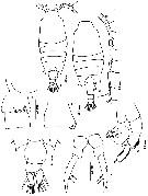
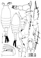 issued from : Mulyadi in Treubia, 1997, 31 (2). [p.81, Fig.4]. Female (from Ambon Bay): A, habitus (dorsal); B, 5th metasomal somite and urosome (lateral right side); C, Mx2; D, P1; E, 3rd exopodal segment of P3; F, P5. Male: G, habitus (dorsal); H, 5th metasomal somite and urosome (lateral right side); I, angle of the 5th metasomal somite and genital somite (lateral right side); J, 15th to 19th segments of right A1; K, P5 (posterior view). Nota: This species is distinguished from other species in the genus by the ventral spine-like process on urosomal segment 2 in female, and the toothed tubercle on the right side of urosomal segment 1 and the structure of P5 male.
|
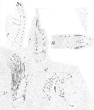
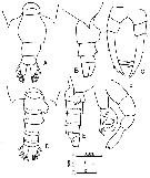 issued from : F.Y. Al-Yamani & I. Prusova in Common Copepods Northwestern Arabian Gulf : Identification Guide. Kuwait Institute for Scientific Research, 2003. [p.74, Fig.26]. Female: A, urosome (dorsal); B, idem (lateral right side); C, P5. Male: D, urosome (dorsal); E, idem (lateral right side); F, P5.
|
 issued from : N. Phukham in Species diversity of calanoid copepods in Thai waters, Andaman Sea (Master of Science, Univ. Bangkok). 2008. [p.144, Fig.18]. Female (from W Malay Peninsula): a, habitus (dorsal); b, urosome (dorsal); c, P5. Male: d, habitus (dorsal); e-f, genital segment; g, P5. Body length after the drawings: F: = 1.589 mm; M = 1.455 mm.
|
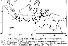
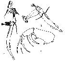 issued from : J. Wickstead in J. Anim. Ecol., 1959, 28. [p.70, Fig.1]. a-b, examples of characteristic attitudes of Candacia bradyi (from Singapore region) affixed to a chaetognath; c, Mx2 of male. Nota: Males and females Candacia bradyi were affixed to a Sagitta enflata, Sagitta pulchra, Sagitta planctonis, to a ctenophore and juvenile Eucalanus (= Subeucalanus) subcrassus, Paracalanus aculeatus, and to an actinotrocha larva of Phoronis. Therefore 73 % were affixed to a chaetognath and 62 % were affixed to a Sagitta enflata. There were no examples of more than 1 copepod being affixed to a simple chaetognath in these samples. The analysis of two samples shows that, in the first haul about 1.1 % of the chaetognaths were attacked by this species copepod, and in the second haul 0.35 %.
|
 issued from : Mulyadi in Published by Res. Center Biol., Indonesia Inst. Sci. Bogor, 2004. [p.81, Fig.45]. Female (from Ambon Bay): A, habitus (dorsal); B, 5th metasomal somite with P5 and urosome (lateral right side); C, Mx2; D, P1; E, 3rd exopodal segment of P3; F, P5. Male: G, habitus (dorsal); H, 5th metasomal somite and urosome (lateral right side); I, angle of the 5th metasomal somite and genital somite (lateral right side); J, 15th to 19th segments of right A1; K, P5 (posterior view). Nota: This species is distinguished from other species in the genus by the ventral spine-like process on urosomal segment 2 in female, and the toothed tubercle on the right side of urosomal segment 1 and the structure of P5 male.
|
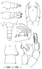
 Issued from : J. Carl in Rev. Suisse Zool., 1907, 15. [Pl. I, figs.8, 9, 10, 12, 13]. Candacia Bradyi Female (from Baie d'Amboine = Ambon Bay, Indonesia): 8, P5; 9, exopodite of P3 (segments 2 and 3); 10, Md gnathobase.
|
 Issued from : J. Carl in Rev. Suisse Zool., 1907, 15. [Pl. I, figs.11, 14]. Candacia Bradyi Male (from Ambon Bay): 11, P5; 14, A1 (segments 16-20).
| | | | | Ref. compl.: | | | Carl, 1907 (p.17); Sewell, 1948 (p.323, 418, 426, 433, 437); Chiba & al., 1957 (p.310); 1957 a (p.11); Yamazi, 1958 (p.151, Rem.); Wickstead, 1959 (p.69, fig. 1, carnivorous behaviour); 1962 (p.546, food & feeding); Ganapati & Shanthakumari, 1962 (p.9, 15); Delalo, 1968 (p.138); Itoh, 1970 a (p.8: tab.2); Brodsky, 1972 (p.256); Patel, 1975 (p.659); Madhu Pratap & al., 1977 (p.138, Table 3: abundance vs. stations); Carter, 1977 (1978) (p.36); Chen Q-c, 1980 (p.794); Guangshan & Honglin, 1984 (p.118, tab.); Madhupratap & Haridas, 1986 (p.105, tab.1); Sarkar & al., 1986 (p.178); Othman & al., 1990 (p.561, 564, Table 1); Dai & al., 1991 (p.47); Shih & Young, 1995 (p.69); Sharaf & Al-Ghais, 1997 (tab.1); Achuthankutty & al., 1998 (p.1, Table 2, fig.6: salinity range, seasonal abundance vs monsoon); Hwang & al., 1998 (tab.II); Wong & al, 1998 (tab.2); Hsieh & Chiu, 1998 (tab.2); El-Serehy, 1999 (p.172, Table 1, occurrence); Hwang & al., 2003 (p.193, tab.2); Razai & al., 2004 (p.490, tab.2); Osore & al., 2004 (p.195); Lan & al., 2004 (p.332, tab.1); Hwang & al., 2006 (p.943, tabl. I); Rakhesh & al., 2006 (p.93, Table 2, spatial distribution); Dur & al., 2007 (p.197, Table IV); Fernandes, 2008 (p.465, Tabl.2); Tseng L.-C. & al., 2008 (p.153, Table 2, occurrence vs geographic distribution); W.-B. Chang & al., 2010 (p.735, Table 2, abundance); Shanthi & Ramanibai, 2011 (p.132, Table 1); Maiphae & Sa-ardrit, 2011 (p.641, Table 2); Guo & al., 2011 (p.567, table 2, indicator); Kâ & Hwang, 2011 (p.155, Table 3: occurrence %); Tseng L.-C. & al., 2011 (p.47, Table 2, occurrences vs mesh sizes); Tseng & al., 2012 (p.621, Table 3: abundance); Tseng & al., 2013 (p.507, seasonal abundance); Jagadeesan & al., 2013 (p.27, Table 3, seasonal abundance); Rakhesh & al., 2013 (p.7, Table 1, abundance vs stations); Hwang & al., 2014 (p.43, Appendix A: seasonal abundance); Nakajima & al., 2015 (p.19, Table 3: abundance); Zakaria & al., 2016 (p.1, Table 1, Rem.) | | | | NZ: | 8 + 1 douteuse | | |
|
Carte de distribution de Candacia bradyi par zones géographiques
|
| | | | | | | | | | | |  issued from : T.J. Lawson in Marine Biology, 1977, 43. [Fig.5, p.81]. issued from : T.J. Lawson in Marine Biology, 1977, 43. [Fig.5, p.81].
Distribution map for the Indian Ocean. |
 issued from : Mulyadi in Treubia, 1997, 31 (2). [p.109, Fig.16]. issued from : Mulyadi in Treubia, 1997, 31 (2). [p.109, Fig.16].
Distribution of Candaciidae in Indonesian waters. 2: C. bradyi. |
 issued from : C.T. Achuthankutty, N. Ramaiah & G. Padmavati in Pelagic biogeography ICoPB II. Proc. 2nd Intern. Conf. Final report of SCOR/IOC working group 93, 9-14 July 1995. Workshop Report No. 142, Unesco, 1998. [p.8, Fig.6]. issued from : C.T. Achuthankutty, N. Ramaiah & G. Padmavati in Pelagic biogeography ICoPB II. Proc. 2nd Intern. Conf. Final report of SCOR/IOC working group 93, 9-14 July 1995. Workshop Report No. 142, Unesco, 1998. [p.8, Fig.6].
Salinity ranges for C. bradyi in coastal and estuarine waters of Goa (India).
Shaded area indicates the range of higher abundance. |
| | | | Loc: | | | South Africa (E, off Durban, Natal), E Medit. (W Egyptian coast), G. of Suez, Red Sea, G. of Aden, Arabian Gulf, UAE coast, Arabian Sea, Kenya, Madagascar (Nosy Bé), Indian, Karavatti & Agatti Is., India (Goa, Lawson's Bay, G. of Mannar, Hooghly estuary, Godavari region), Bay of Bengal, W Malay Peninsula (Andaman Sea), Straits of Malacca, Singapore region, Java (off Labuan, off Jakarta), Flores Sea, Manado Bay (N Celebes), Baie d'Amboine (= Ambon Bay), Thailand, Java (off Labuan, Jakarta-Seibu Islands), Flores Sea, Tioman Is., N Celebes, Viet-Nam (Cauda Bay), Gulf of Tonkin, Hong Kong, China Seas (Yellow Sea, East China Sea, Taiwan Strait, South China Sea), Japan, Izu region, (Tanbe Bay in Yamazi, 1958), Taiwan, NE Taiwan, off SW Taiwan, S Korea (in Kim & al.,1993), ? Pacif. E (off Mexico), Pacif. (W equatorial), Australia (G. of Carpentaria, Moreton Bay) | | | | N: | 66 ( E Medit.: 1, Red Sea: 1; Indian: 29; Indo-Malaysia: 8; W Pacif.: 27) | | | | Lg.: | | | (343) F: 2,1-1,4; M: 1,46-1,08; ? (334) F: 1,8; M: 1,9; (530) F: 2; M: 1,8; (778) F: 1,93-1,85; M: 1,8-1,74; (991) F: 1,4-2,1; M: 1,4-1,8; (1085) F: 1,86-1,94; M: 1,74-1,88; (1122) F: 1,85; M: 1,75; {F: 1,40-2,10; M: 1,08-1,90}.
The mean female size is 1.839 mm (n = 11; SD = 0.2384), and the mean male size is 1.668 (n = 11; SD = 0.2508. . The size ratio (male : female) is 0.92 (n = 7; SD = 0.0988). | | | | Rem.: | Lawson (1973 a) montre que C. tuberculata n'est pas synonyme de cette espèce doù certaines confusions probables dans l'Indien.
Localisations Pacifique incertaines. Lan al. (2004) signalent les deux espèces au NW de Taiwan.
Voir aussi les remarques en anglais | | | Dernière mise à jour : 03/12/2020 | |
|
|
 Toute utilisation de ce site pour une publication sera mentionnée avec la référence suivante : Toute utilisation de ce site pour une publication sera mentionnée avec la référence suivante :
Razouls C., Desreumaux N., Kouwenberg J. et de Bovée F., 2005-2025. - Biodiversité des Copépodes planctoniques marins (morphologie, répartition géographique et données biologiques). Sorbonne Université, CNRS. Disponible sur http://copepodes.obs-banyuls.fr [Accédé le 04 décembre 2025] © copyright 2005-2025 Sorbonne Université, CNRS
|
|
|
|


;)
;)
;)
;)
;)
;)
;)
;)
;)

;)
;)
