|
|
 |
Fiche d'espèce de Copépode |
|
|
Calanoida ( Ordre ) |
|
|
|
Diaptomoidea ( Superfamille ) |
|
|
|
Acartiidae ( Famille ) |
|
|
|
Acartia ( Genre ) |
|
|
|
Acanthacartia ( Sous-Genre ) |
|
|
| |
Acartia (Acanthacartia) fossae Gurney, 1927 (F,M) | |
| | | | | | | Syn.: | ? Acartia hamata Mori, 1937 (1964) (p.104, figs.F); 1942 (p., Figs.M); Kos, 1972 (Vol. I, figs. F, M, Rem.); Peterson, 1975 (p.28, Rem., abundance); Ueda & al., 1983 (p.165, table 1, 2, 4, swarms); Park & Choi, 1997 (Appendix); Kazmi, 2004 (p.228, Rem.: p.232); Jungbluth & Lenz, 2013 (p.630, Table I);
A. ransoni : Vaissière, 1954 (p.358) | | | | Ref.: | | | Gurney, 1927 (p.156, figs.F,M, Rem.); Grice, 1964 (p.262, figs.F,M, Rem.); Lakkis, 1984 (p.298, figs.F,M, Rem.); Nishida, 1985 (p.136, figs.F,M, Rem.); Dussart, 1989 (p.14, 116: figs.F,M); Chihara & Murano, 1997 (p.670, Pl.18,19: F,M); Bradford-Grieve, 1999 (N°181, p.5, figs.F,M); Barthélémy, 1999 (p.858, 864, 869: Rem., figs.F); 1999 a (p.9, Fig.24, A-F); Conway & al., 2003 (p.102, figs.F,M, Rem.); Vives & Shmeleva, 2007 (p.431, figs.F,M, Rem.); Al-Yamani & al., 2011 (p.82, figs.F,M) | 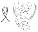 issued from : G.D. Grice in Crustaceana, 1964, 6 (4). [p.260, Figs.38-40]. Female: 38, P5. Male: 39, P5; 40, idem (other side).
|
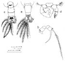 issued from : S. Nishida in Publ. Seto Mar. Biol. Lab., 1985, 30 (1/3). [p.135, Fig.6, a-d]. Female (from Kabira Bay, Ishigaki Is.): a, 5th thoracic segment and urosome (dorsal); b, P5. Male: c, 5th thoracic segment and urosome (dorsal); d, P5.
|
 issued from : T. Mori in The pelagic Copepoda from the neighbouring waters of Japan, 1937 (2nd edit., 1964). [Pl.51, Figs.1-5]. ? As Acartia hamata; (Cf. Acartia bispinosa) Female: 1, forehead (ventral); 2, anal segment and caudal rami (dorsal); 3, P5; 4-5, habitus (lateral and dorsal, respectively).
|
 issued from : R.-M. Bathélémy in J. Mar. Biol. Ass. U.K., 1999, 79. [p.862, Fig.5, A-C]. Scanning electon miccrograph. Female (from Tahiti): A, genital double-somite (ventral), note the fragmented genital area with lateral genital stuctures (arrows); B, idem (left lateral); C, detail of the left genital structure; note the anterior thickening (arrow) prolonged by a small cuticular flap similar to an operculum (*). Scale bars: 0.050 mm (A, B); 0.010 mm (C).
|
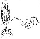 issued from : T. Mori in Palao trop. biol. Stn Stud., 1942, 2 (3). [Pl.X, Figs.1, 2]. As Acartia hamata. Male (from Palao Island): 1, habitus (dorsal); 2, P5 (posterior view).
|
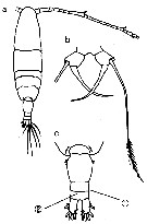 Issued from : M. Chihara & M. Murano in An Illustrated Guide to Marine Plankton in Japan, 1997. [p.676, Pl. 18, fig.7 a-c]. Female: a, habitus (dorsal); b, P5; c, last thoracic segment and urosome (dorsal). Nota: numbers show characteristics of this species to compare with A. tumida, A. steueri.
|
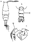 Issued from : M. Chihara & M. Murano in An Illustrated Guide to Marine Plankton in Japan, 1997. [p.677, Pl. 19, fig.7 a-d]. Male: a, habitus (dorsal); b, last thoracic segment and urosome (dorsal) c, P5. Nota: numbers show characteristics of this species to compare with A. steueri, A. tumida.
|
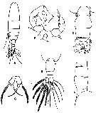 Issued from : R. Gurney in Trans. zool. Soc. London, 1927, 22. [p.157, Fig.22]. Female (Suez Canal): A, habitus (dorsal); B, abdomen (dorsal); C, P5. Male: D, abdomen (dorsal); E, abdomen (lateral); F, P5. Nota female: - Body slender. - last thoracic somite rounnded, with a row of 4 to 5 small teeth (as in A. clausi). - Proportion of thorax to abdomen (included furca) 84 : 24. - Abdomen 3-segmented. - Genital somite (= double-somite) longer than somites 2 and 3 combined, and as wide as long. This double-somite has a few small lateral hairs, but there are no dorsal spines on this or succeeding somites. Rostral filaments present, very slender. - A1 reaches to end of 2nd abdominal somite. - P5P5 basal segment short and broad; length 16, breadth 12. Terminal spine rather stout and curved, smooth. Nota male: - Abdomen and thorax 58 : 110. - last thoracic somite rounded and with teeth as in female. - Abdomen 5-segmented - 1st abdominal somite with lateral hairs. - Abdominal somites 2, 3, 4 with posterior row of very minute denticles, dorsally. - Furcal rami [= caudal rami] short and broad, with inner hairs. - A1 reaches nearly to end of thorax. - Right P5: exopod with a process on posterior side bearing a slender seta; 2nd exopodal segment produced into a large inner lobe. 2nd exopodal segment of left leg with a stout terminal spine and a curved anterior seta. For Gurney, this species very closely resembles A. bifilosa, from which it is separated only by some minute details.
| | | | | Ref. compl.: | | | Sewell, 1948 (p.441); Lakkis, 1976 (p.84); Kovalev & Shmeleva, 1982 (p.85); Kimmerer & McKinnon, 1985 (p.149); M. Lefèvre, 1986 (p.33, 40); Yen & al., 1992 (p.495, tab.1, mechanorecptors); 25); Ueda, 1985 (p.1McKinnon & Ayukai, 1996 (p.595); Go & al., 1997 (tab.1, comme Acarita: lapsus calami); Mauchline, 1998 (tab.8, 65, 78); Lenz & al., 2000 (p.338); Belmonte & Potenza, 2001 (p.173); El-Serehy & al., 2001 (p.116, Table 1: abundance vs transect in Suez Canal); Zenetos & al., 2005 (p.63, Rem.: p.82, casual occurrence); McKinnon & al., 2008 (p.843: Tab.1); Zenetos & al., 2010 (p.397); Saitoh & al., 2011 (p.85, Table 4, 5); El-Serehy & al., 2013 (p.2099, Rem.: p.2101); Mojib & al., 2014 (p.2740, blue-pigment); Ohtsuka & Nishida, 2017 (p.565, Table 22.1); Abo-Taleb & Gharib, 2018 (p.139, Table 5, occurrence %). | | | | NZ: | 6 + 1 douteuse | | |
|
Carte de distribution de Acartia (Acanthacartia) fossae par zones géographiques
|
| | | | | | | | | | | |  Carte de 1996 Carte de 1996 | |
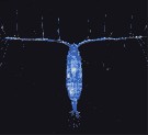 Issued from : N. Mojib & al. in Molecular Ecology, 2014, 23. [p.2742, Fig.1 (part.) Issued from : N. Mojib & al. in Molecular Ecology, 2014, 23. [p.2742, Fig.1 (part.)
Photomicrograph of the blue-pigmented copepod Acartia fossae from the Red Sea: 22.4575°N, 39.0305°E in May 2012, to analyse carotenoid-protein complexes responsible for blue coloration. |
 issued from : J. Yen, P.H. Lenz, D.V. Gassie & D.K. Hartline in J. Plankton Res., 1992, 14 (4). [p.502, Fig. 4]. issued from : J. Yen, P.H. Lenz, D.V. Gassie & D.K. Hartline in J. Plankton Res., 1992, 14 (4). [p.502, Fig. 4].
Neural activity from the A1 of Acartia fossae from Kaneohe Bay, Hawaii.
Left side: spontaneous background activity. Right side: responses to a variety of mechanical stimuli (tap). All recordings are 40 ms long.
Compare with Labidocera madurae, Euchirella curticauda, Undinula vulgaris, Pleuromamma xiphias. |
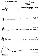 issued from : J. Yen, P.H. Lenz, D.V. Gassie & D.K. Hartline in J. Plankton Res., 1992, 14 (4). [p.504, Fig. 5B]. issued from : J. Yen, P.H. Lenz, D.V. Gassie & D.K. Hartline in J. Plankton Res., 1992, 14 (4). [p.504, Fig. 5B].
Neural activity recorded from the A1 of Acartia fossae (from Kaneohe Bay, Hawaii) in response to an increase in the amplitude of the stimulus displacement from 0.14 to 2.2 µm.
Form of the stimulus (output of displacement gauge) is depicted as the bottom displacement trace
Compare with Labidocera madurae. |
 issued from : J. Yen, P.H. Lenz, D.V. Gassie & D.K. Hartline in J. Plankton Res., 1992, 14 (4). [p.508, Fig. 10]. issued from : J. Yen, P.H. Lenz, D.V. Gassie & D.K. Hartline in J. Plankton Res., 1992, 14 (4). [p.508, Fig. 10].
Neural activity recorded from the A1 of Acartia fossae (from Kaneohe Bay, Hawaii) in response to a single-cycle, 1.76 µm displacement of the entire A1.
(A) Initial distal bend followed by a proximal bend of all setae. (B) Proximal followed by distal bend of all setae.
The lower trace depicts the stimulus. |
| | | | Loc: | | | Medit. (Lebanon), Suez Canal, Bitter Lakes, El Kantara, Lake Timsah Hurghada, Port Taufik Red Sea, Arabian Gulf (Kuwait), Indian (Madagascar, Nosy Bé), Arabian Sea, China Seas (East China Sea), Ishigaki Is., Okinawa (Kabira Bay, Lagoon), ? Japan (Tanabe Bay ! (inYamazi, 1958), Palau Is., SE & W Australia (Melbourne, Exmouth Gulf), Pacif. (Hawaii, Kanneohe Bay, Society Is., Tuamotu, Moorea Is.: lagoon), Okinawa, S Korea. | | | | N: | 36 | | | | Lg.: | | | (91) F: ±1,06; (178) F: 1,03; M: 1-0,91; (325) F: 1,4-1,03; M: 1,3-0,91; (866) F: 1,1-1,3; M: 1,2; (991) F: 1,03-1,4; M: 0,91-1,3; (1085) F: 0,9-1,2; M: 0,9-1,1; {F: 0,90-1,40; M: 0,91-1,30} | | | | Rem.: | La présence de cette espèce en Méditerranée E (côtes libanaises) pourrait être due à une migration lessepsienne.
Voir aussi les remarques en anglais | | | Dernière mise à jour : 18/11/2022 | |
|
|
 Toute utilisation de ce site pour une publication sera mentionnée avec la référence suivante : Toute utilisation de ce site pour une publication sera mentionnée avec la référence suivante :
Razouls C., Desreumaux N., Kouwenberg J. et de Bovée F., 2005-2025. - Biodiversité des Copépodes planctoniques marins (morphologie, répartition géographique et données biologiques). Sorbonne Université, CNRS. Disponible sur http://copepodes.obs-banyuls.fr [Accédé le 02 avril 2025] © copyright 2005-2025 Sorbonne Université, CNRS
|
|
 |
 |


