|
|
 |
Fiche d'espèce de Copépode |
|
|
Calanoida ( Ordre ) |
|
|
|
Clausocalanoidea ( Superfamille ) |
|
|
|
Euchaetidae ( Famille ) |
|
|
|
Euchaeta ( Genre ) |
|
|
| |
Euchaeta acuta Giesbrecht, 1892 (F,M) | |
| | | | | | | Syn.: | Euchäta acuta Giesbrecht, 1892 (p.246, 262, 772, figs.F,M);
Euchaete acuta : With, 1915 (p.187, figs.F);
Paraeuchaeta acuta : Bradford al., 1983 (p.25, figs.F,M); Siokou-Frangou, 1999 (p.476); Brugnano & al., 2010 (fig.8); Brugnano & al., 2012 (p.207, Table 3); Minutoli & Guglielmo, 2012 (p.91, carbon requirement vs vertical distribution); in CalCOFI regional list (MDO, Nov. 2013; M. Ohman, comm. pers.);
Pareuchaeta acuta : Heinrich, 1995 (tab.1); Hafferssas & Seridji, 2010 (p.353, Table 3); Uysal & Shmeleva, 2012 (p.909, Table I);
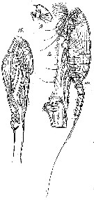
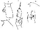
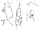
Euchaeta acuta s;l. : Brinton & al., 1986 (p.228, Table 1) | | | | Ref.: | | | Giesbrecht & Schmeil, 1898 (p.38, Rem. F,M); I.C. Thompson, 1903 a (p.18); Thompson & Scott, 1903 (p.234, 244); Esterly, 1905 (p.157, figs.M); Farran, 1908 b (p.40); A. Scott, 1909 (p.65, figs.F); Wolfenden, 1911 (p.299); Pesta, 1920 (p.511); Sars, 1925 (p.108, figs.F,M); Farran, 1926 (p.256); 1929 (p.208, 237); Rose, 1933 a (p.114, figs.F,M); Lysholm & al., 1945 (p.22); Brodsky, 1950 (1967) (p.199, figs.F,M); Vervoort, 1957 (p.84, 85, Rem.); Paiva, 1963 (p.42); Gaudy, 1963 (p.23); Mazza, 1964 b (p.273, figs.F,M); Crisafi, 1965 a (p.66, fig.F); Owre & Foyo, 1967 (p.52, figs.F,M); Mazza, 1967 (p.152, 161, 390, figs.F,M, juv.); Vidal, 1968 (p.30, figs.F); Bradford, 1972 (p.40, figs.F); Tanaka, 1973 (p.130, figs.F,M); Park, 1978 (p.208, figs.F,M); Björnberg & al., 1981 (p.633, 635, figs.F,M); Park, 1995 (p.23, Redescr.F,M, figs.F,M); Mauchline, 1999 (n°182, p.8, figs.F,M); Lapernat, 1999 (p.17, 55); Bradford-Grieve & al., 1999 (p.880, 925, figs.F,M); Braga & al., 1999 (p.84, 89, Rem.: Biol. mol., Table 1, figs.6, 7, 8); Avancini & al., 2006 (p.84, Pl. 53, figs.F,M, Rem.); Vives & Shmeleva, 2007 (p.646, figs.F,M, Rem.) | 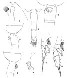 issued from : T. Park in Antarctic Res. Ser. Washington, 1978, 27. [p.211, Fig.69]. Female: A, forehead (lateral); B, last metasomal and genital segments (left side); C, idem (right side); D, genital segment (ventral); E, urosome (dorsal); F, outer lobe of Mx1; G, P1; H, P2; P1-2: legs (anterior). Nota : Proportional lengths of prosome and urosome 67 : 33. Frontal eminence of forehead pronounced, bearing auprafrontal sensilla ; rostrum large, pointing obliquely forward at angle about 70° with reference to body. Posterolateral corner of metasome not prolonged posteriorly; its posteror margin broadly rounded in either dorsal and lateral view. Dorsally or ventrally, genital segment asymmetrical with conspicuous conical protuberance anteriorly on its left side and more convex margin on its right side. Genital field, when viewed ventrally, also asymmetrical with dissimilar genital flanges. Genital prominence arising from anterior half of genital segment, in lateral view, pointing anteroventrally, with genital field facing in the same direction. Right genital flange produced at middle into pointed process; posterior half of left genital flange enlarged into large lobe. Appendicular caudal setae straight, much thicker and longer than terminal setae. A1 extending beyond distal end of metasome by its last 2 segments. Mx1 : outer coxal lobe with 5 setae of about equal length. Mx2 : 1 of 6 apical setae bearing long, widely separated spines in addition to short spinules found throughout its entire length Mxp : long endopodal setae terminating in spinule. P1 : 1st and 2nd exopodal segments fused without visible trace of segmentation ; resulting compound segment with single external spine reaching distal end of following segment. P2 : external spine of 2 nd exopodal sgment reaching middle of 1st external spine od 3rd. In 3rd exopodal segment, 1st and 3rd external spines about equally small ; 2 nd large, reaching close to distal end of segment ; incision posterior to 2nd spine deep.
|
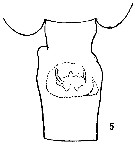
 issued from : T. Park in Antarctic Res. Ser. Washington, 1978, 27. [p.212, Fig.70]. Male: A, forehead (lateral); B, last metasomal and genital segments (lateral); C, P5 (anterior); D, middle part of exopod of left P5 (medial); E, idem (anterior)
|
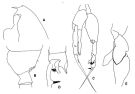
 issued from : T. Park in Bull. Scripps Inst. Oceanogr. Univ. California, San Diego, 1995, 29. [p.122, Fig.12]. Female: a, forehead (left side); b, urosome (left); c, d, e, f, genital somite (left, dorsal, right, ventral, respectively); g, outer lobe of Mx1; h, endopod of Mx2; i, Mxp (coxa separated, posterior, the rest anterior); j, P1 (anterior); k, P2 (anterior). Male: l, forehead (left); m, last pedigerous and genital somites (left); n, P5 (anterior); o, p, exopod of left 5th leg (anterior, medial, respectively).
|
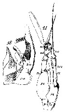
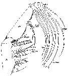
 issued from : J.M. Bradford, L. Haakonssen & J.B. Jillett in Mem. N.Z. Oceanogr. Inst., 1983, 90. [p.26, Fig.9]. As Paraeuchaeta acuta. Female: A, habitus (lateral right side); B-D, genital segment (dorsal, right lateral, left lateral, respectively); E, exopod of P1; F, exopod segment 3 of P2. Nota: - P2 exopod: Aa ± AB; Bb ≤ 1/3 BC; Cc = CD. (see code of lengths outer spines in the Genus' figure of Paraeuchaeta, or in Paraeuchaeta sp. A).
Male: G, habitus (lateral left side); H, P5; I, terminal part of left P5 exopod.
|
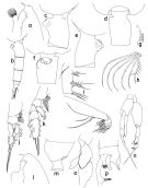
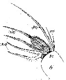
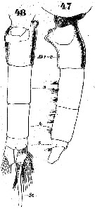
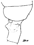
 Issued from : G.O. Sars in Résult. Camp. Scient. Prince Albert I, 69, pls.1-127 (1924). [Pl.XXX, figs.12-15]. Female: 12, habitus (lateral left side); 13, forehead (lateral); 14, genital segment (lateral left side). Male: 15, P5.
|
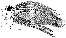
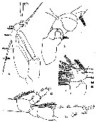
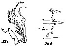
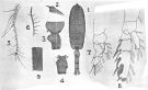 issued from : A. Scott in Siboga-Expedition, 1909, XIX a. [Plate XX, Figs.1-9]. Female (from Indonesia-Malaysia) : 1, habitus (dorsal); 2, forehead (lateral); 3, last thoracic and genital segments (left side); 4, genital segment (dorsal); 5, A1; 6, Mxp (end hair); 7, P1; 8, P2; 9, part of terminal spine of exopodite of P3.
|
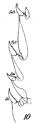
 Issued from : W. Giesbrecht in Systematik und Faunistik der Pelagischen Copepoden des Golfes von Neapel und der angrenzenden Meeres-Abschnitte. Fauna Flora Golf. Neapel, 1892. Atlas von 54 Tafeln. [Taf.16, Figs.18, 21]. As Euchäta acuta. Male: 18, exopodal segments 2 and 3 of P5; 21, 21, P5 (anterior view), with spermatophore. Pd = right leg; Ps = left leg.
|
 Issued from : W. Giesbrecht in Systematik und Faunistik der Pelagischen Copepoden des Golfes von Neapel und der angrenzenden Meeres-Abschnitte. - Fauna Flora Golf. Neapel, 1892. Atlas von 54 Tafeln. [Taf.16, Fig.10]. As Euchäta acuta. Male: 10, outer margin of exopodal segments 2 and 3 of P2.
|
 Issued from : W. Giesbrecht in Systematik und Faunistik der Pelagischen Copepoden des Golfes von Neapel und der angrenzenden Meeres-Abschnitte. - Fauna Flora Golf. Neapel, 1892. Atlas von 54 Tafeln. [Taf.16, Fig.27]. As Euchäta acuta. Male: 27, Mx1.
|
 Issued from : W. Giesbrecht in Systematik und Faunistik der Pelagischen Copepoden des Golfes von Neapel und der angrenzenden Meeres-Abschnitte. - Fauna Flora Golf. Neapel, 1892. Atlas von 54 Tafeln. [Taf.16, Fig.6]. As Euchäta acutaFemale: 6, Mx1 (anterior view).
|
 Issued from : W. Giesbrecht in Systematik und Faunistik der Pelagischen Copepoden des Golfes von Neapel und der angrenzenden Meeres-Abschnitte. - Fauna Flora Golf. Neapel, 1892. Atlas von 54 Tafeln. [Taf.16, Fig.39]. As Euchäta acuta. Female: 39, Mxp ( posterior view).
|
 Issued from : W. Giesbrecht in Systematik und Faunistik der Pelagischen Copepoden des Golfes von Neapel und der angrenzenden Meeres-Abschnitte. - Fauna Flora Golf. Neapel, 1892. Atlas von 54 Tafeln. [Taf.16, Fig.14]. As Euchäta acuta. Female: 14, exopodal segments 2 and 3 of P2. (posterior view).
|
 Issued from : W. Giesbrecht in Systematik und Faunistik der Pelagischen Copepoden des Golfes von Neapel und der angrenzenden Meeres-Abschnitte. - Fauna Flora Golf. Neapel, 1892. Atlas von 54 Tafeln. [Taf.37, Figs.47, 48]. As Euchäta acuta. Female: 47-48, urosome (lateral and ventral, respectively).
|
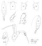
 issued from : C.O. Esterly in Univ. Calif. Publs Zool., 1905, 2 (4). [p.158, Fig.23]; Male (from San Diego Region): a, P5 (Ri.dx = endopodite of right P5; re1, 2, sn = 1st and 2nd segments of exopodite of left P5; proc = process; sph = spermatophore); b, 2nd and 3rd segments of exopod of left P5; c, P2; d, Mx1 (Le.1 = 1st lobe of outer margin; ri = endopodite; re = exopodite; b1 and b2 = basipodites 1 and 2).
|
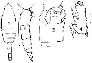 issued from : J.M. Bradford in Mem. N. Z. Oceonogr. Inst., 1972, 54. [p.42, Fig.9, (1-4)]. Female (from Kaikoura, New Zealand): 1, habitus (dorsal); 2, urosome (lateral, right side); 3, genital segment (ventral); 4, exopod segment 3 of P2. Scale bars: 1 mm (1, 2); 0.1 mm (3, 4). Nota: A1 does not reach the hind border of the cephalothorax.
|
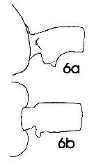 issued from : J. Mauchline in ICES Ident. Leafl. Plankton, 1999, N°182. [p.3, Fig.2: 6a-6b]. Female (Northeast Atlantic): 6a, genital double-somite (left side); 6b, same (dorsal).
|
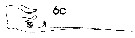 issued from : J. Mauchline in ICES Ident. Leafl. Plankton, 1999, N°182. [p.4, Fig.3: 6c]. Male (Northeast Atlantic): 6c, terminal two segments, exopodal segments of left P5.
|
 issued from : C. With in The Danish Ingolf-Expedition, 1915, III (4). [Pl. VI, Fig.12, a]. As Euchaete acuta. Female (from 47°-51°N, 8-11°43'W): a, last thoracic segment and genital segment (left lateral).
|
 issued from : C. With in The Danish Ingolf-Expedition, 1915, III (4). [Pl. VI, Fig.12, b-c]. As Euchaete acuta. Female: b, labrum (oral view); c, lamina labialis and serrula 6-dentata.
|
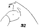 Issued from : W. Giesbrecht in Systematik und Faunistik der Pelagischen Copepoden des Golfes von Neapel und der angrenzenden Meeres-Abschnitte. - Fauna Flora Golf. Neapel, 1892, 19 , Atlas von 54 Tafeln. [Taf.37, Fig.52]. As Euchäta acuta. Female: 52, forehead (lateral). R = rostrum.
|
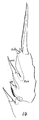
 issued from : O. Tanaka in I O B C Handbook, 1973, IV. Cochin, India [p.131, Fig.1]. Female (from Arabian Sea): A, genital segment (lateral); B, forehead (lateral); C, P2. Nota: Abdominal segments and caudal rami in proportional lengths 35 : 25 : 20 : 6: 14 = 100. A1 23-segmented, extends to the end of the last thoracic segment. Exopod of A2 7-segmented, endopod 2-segmented. Endopod with 6 setae on the outer lobe, and 7 setae on the inner lobe. Md: cutting blade with 6 teeth and 1 inner marginal seta; 2nd basal segment with single seta; exopod with 6 setae, endopod with 1 seta on the 1st segment and 9 setae on the distal segment. Mx1: 6 setae on the outer lobe, 10 on the 1st inner lobe, 1 on the 2nd inner lobe, the 3rd inner lobe is absent. Male: D, forehead (lateral); E, distal segment of left P5. Nota: Abdominal segments and caudal rami in the proportional lengths 19 : 26 : 22 : 18 : 5 : 10 = 100. Caudal rami 1.4 times as long as it is wide at the proximal part. Right A1 exceeds slightly the end of the thoracic segment. mouth parts reduced much. Mx1 agrees with that figured by Giesbrecht in which the outer lobe is firnished with 4 setae, however, it is replaced by 2 setae in the present specimen.
|
 issued from : O. Tanaka in I O B C Handbook, 1973, IV. Cochin, India [p.148, Fig.9: 5]. Female: 5, genital segment (dorsal).
|
 Euchaeta acuta Euchaeta acuta Female: 1 - See Key to acuta species Group. 2 - Dorsally, genital somite asymmetrical. 3 - Dorsally, genital somite without notch on right side (Fig.12-d).
|
 issued from : O. Tanaka in I O B C Handbook, 1973, IV. Cochin, India [p.131]. Proportional lengths of A1 segments from female and male.
|
 issued from : O. Tanaka in I O B C Handbook, 1973, IV. Cochin, India [p.131, Fig.1]. Female (from Indian Ocean): A, genital segment (lateral); B, forehead (lateral); C, P2. Nota: Abdominal segments and caudal rami in the proportional lengths 35 : 25 : 20 : 6 : 14 = 100. Male: D, forehead (lateral); E, distal segment of left P5. Nota: Abdominal segments and caudal rami in the proportional lengths 19 : 26 : 22 : 18 : 5 : 10 = 100.
| | | | | Ref. compl.: | | | Cleve, 1904 a (p.190); Pearson, 1906 (p.17); Wilson, 1942 a (p.185); Massuti Alzamora, 1942 (p.110); Sewell, 1948 (p.345, 487); Wilson, 1950 (p.211); Fagetti, 1962 (p.21); Gaudy, 1962 (p.93, 99, Rem.: p.106); Duran, 1963 (p.16); Giron-Reguer, 1963 (p.30); V.N. Greze, 1963 a (tabl.2); Grice, 1963 a (p.495); Gaudy, 1963 (p.23, Rem.); Ahlstrom & Thrailkill, 1963 (p.57, Table 5, abundance); Björnberg, 1963 (p.39, Rem.); Bary, 1963 a (p.1519, Table 1); 1964 (p.183, T-S diagram-occurrences); Unterüberbacher, 1964 (p.23); De Decker & Mombeck, 1964 (p.12); Mazza, 1964 (p.293, weight); Shmeleva, 1965 b (p.1350, lengths-volume -weight relation); Mullin, 1966 (p.546, Table I, III, diet); Pavlova, 1966 (p.43); Mazza, 1966 (p.70); 1967 (p.367); Ehrhardt, 1967 (p.738, geographic distribution, Rem.); Fleminger, 1967 a (tabl.1); De Decker, 1968 (p.45); Vinogradov, 1968 (1970) (p.268); Morris, 1970 (p.2300); Deevey, 1971 (p.224); Gamulin, 1971 (p.382, tab.3); Binet & al., 1972 (p.55); Bainbridge, 1972 (p.61, Appendix Table III: occurrence); Apostolopoulou, 1972 (p.327, 349); Roe, 1972 (p.277, tabl.1, tabl.2); 1972 b (p.526); Björnberg, 1973 (p.329, 386); Nival & al., 1974 (p.231, respiration & excretion); Vives & al., 1975 (p.42, tab.II, III); Deevey & Brooks, 1977 (p.256, tab.2, Station "S"); Carter, 1977 (1978) (p.35); Dessier, 1979 (p.205); Vaissière & Séguin, 1980 (p.23, tab.1); Vives, 1982 (p.291); Kovalev & Shmeleva, 1982 (p.83); Scotto di Carlo & Ianora, 1983 (p.150); Gaudy & Boucker, 1983 (p.37, Table 1, Rem.: metabolism); Tremblay & Anderson, 1984 (p.5, Rem.); Brenning, 1984 (p.3, T-S diagram, Rem.); Guangshan & Honglin, 1984 (p.118, tab. as acutus); De Decker, 1984 (p.316, 349: carte); Roe, 1984 (p.357); Scotto di Carlo & al., 1984 (p.1041); Regner, 1985 (p.11, Rem.: p.30); Brenning, 1985 a (p.28, Table 2); Madhupratap & Haridas, 1986 (p.105, tab.1); Chen Y.-Q., 1986 (p.205, Table 1: abundance %, Table 2: vertical distribution); Rudyakov, 1986 (tab.2); Jimenez-Perez & Lara-Lara, 1988; Lozano Soldevilla & al., 1988 (p.58); Herman, 1989 (p.247); Cervantes-Duarte & Hernandez-Trujillo, 1989 (tab.3); Suarez & al., 1990 (tab.2); Scotto di Carlo & al., 1991 (p.271); Hattori, 1991 (tab.1, Appendix); Suarez & Gasca, 1991 (tab.2); Suarez, 1992 (App.1); Mauchline, 1992 a (p.2); 1994 a (p.561); Hays & al., 1994 (tab.1); Kouwenberg, 1994 (tab.1); Hajderi, 1995 (p.542); Tranter, 1996 (p.596, 598); Hure & Krsinic, 1998 (p.48, 101, 112); Mauchline, 1992 a (p.3); Suarez-Morales & Gasca, 1998 a (p.109); Mauchline, 1998 (tab.42); Lapernat, 2000 (tabl. 3, 4); Razouls & al., 2000 (p.343, Appendix); Seridji & Hafferssas, 2000 (tab.1); Lopez-Salgado & al., 2000 (tab.1); Madhupratap & al., 2001 (p. 1345, vertical distribution vs. O2, figs.4, 5: clusters, p.1353); d'Elbée, 2001(tabl. 1); Lapernat & Razouls, 2001 (tab.1); Holmes, 2001 (p.51); Fransz & Gonzalez, 2001 (p.255, tab.1); Beaugrand & al., 2002 (p.1692); Beaugrand & al., 2002 (p.179, figs.5, 6); Vukanic, 2003 (139, tab.1); CPR, 2004 (p.54, fig.153); Lo & al., 2004 (p.89, tab.1); Berasategui & al., 2005 (p.313, fig.2); Isari & al., 2006 (p.241, tab.II); Lavaniegos & Jiménez-Pérez, 2006 (Tab.2, 3, Rem.); Valdés & al., 2007 (p.103: tab.1); Ayon & al., 2008 (p.238, Table 4: Peruvian samples); Morales-Ramirez & Suarez-Morales, 2008 (p.519); Raybaud & al., 2008 (p.1765, Table A1); Licandro & Icardi, 2009 (p.17, Table 4); Schnack-Schiel & al., 2010 (p.2064, Table 2: E Atlantic subtropical/tropical); Dias & al., 2010 (p.230, Table 1); Mazzocchi & Di Capua, 2010 (p.425); Medellin-Mora & Navas S., 2010 (p.265, Tab. 2); Tutasi & al., 2011 (p.791, Table 2, abundance distribution vs La Niña event); Miloslavic & al., 2012 (p.165, Table 2, transect distribution); Lidvanov & al., 2013 (p.290, Table 2, % composition); Zakaria & al., 2016 (p.1, Table 1); Benedetti & al., 2016 (p.159, Table I, fig.1, functional characters); El Arraj & al., 2017 (p.272, table 2); Benedetti & al., 2018 (p.1, Fig.2: ecological functional group); Belmonte, 2018 (p.273, Table I: Italian zones); Hure M. & al., 2018 (p.1, Rem.: p.12); Acha & al., 2020 (p.1, Table 3: occurrence % vs ecoregions, Table 5: indicator ecoregions). | | | | NZ: | 20 | | |
|
Carte de distribution de Euchaeta acuta par zones géographiques
|
| | | | | | | | | | | | | | | | | | 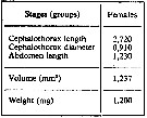 issued from : A.A. Shmeleva in Bull. Inst. Oceanogr., Monaco, 1965, 65 (n°1351). [Table 6: 20 ]. Euchaeta acuta (from South Adriatic). issued from : A.A. Shmeleva in Bull. Inst. Oceanogr., Monaco, 1965, 65 (n°1351). [Table 6: 20 ]. Euchaeta acuta (from South Adriatic).
Dimensions, volume and Weight wet. Means for 50-60 specimens. Volume and weight calculated by geometrical method. Assumed that the specific gravity of the Copepod body is equal to 1, then the volume will correspond to the weight. |
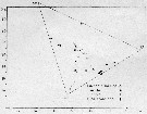 issued from : U. Brenning in Wiss. Z. Wilhelm-Pieck-Univ. Rostock - 33. Jahrgang 1981. Mat.-nat. wiss. Reihe, 6. [p.2, Fig.1]. issued from : U. Brenning in Wiss. Z. Wilhelm-Pieck-Univ. Rostock - 33. Jahrgang 1981. Mat.-nat. wiss. Reihe, 6. [p.2, Fig.1].
T-S Diagram for Euchaeta marina, E. acuta, E. hebes (= Paraeuchaeta hebes), E. paraconcinna from Cap Blanc (Mauritania).
SO: Southern Surface Water (S °/oo: 34,50; T°C: 29,0); ND: Northern Water of the Surface Layer (S °/oo: 37,5; T°C: 21,0); SD: Southern Deep Water of the surface layer (S °/oo: 35,33; T°C: 13,4). See commentary in Temora stylifera and Brenning (1985 a, p.6). |
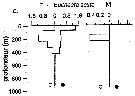 Issued from : P.-E. Lapernat in DEA Océanogr. Biol., Univ. P. & M. Curie, Paris VI. July 5, 2000. [Fig.9 c ]. Issued from : P.-E. Lapernat in DEA Océanogr. Biol., Univ. P. & M. Curie, Paris VI. July 5, 2000. [Fig.9 c ].
Verical distribution of Euchaeta acuta at an eutrophic site (off Mauritanian coast: 20°32 'N, 18°36' W) in females (F) and males (M) (ind. per m3) in the day (white circle) and night (black circle).
Nota: Sampling in the water column 0-1000 m, one during the day and another during the night with BIONESS multiple-net: 0-75; 75-150; 150-250; 250-350; 350-450; 450-550; 550-700; 700-850; 850-965 m. In May-June 1992. |
| | | | Loc: | | | sub-Antarct. (S Tasmania), South Africa (E & W), Namibia, Tristan da Cunha Is., Congo, off Lagos, Ivorian shelf, off Ascension Is., off E St. Paul Is., Cape Verde Is., off Mauritania, Morocco-Mauritania, Canary Is., off Madeira, off W Cabo Finisterre, Bay of Biscay, Azores, Argentina, Brazil, off Macaé, off Amazon, Caribbean Sea, Caribbean Colombia, G. of Mexico, Florida, off Bermuda: Station "S" (32°10'N, 64°30'W), S Cape Hatteras, Cabot Strait, Flemish Cap, off Rockall Is., W & SW Ireland, North Sea, Ibero-moroccan Bay, Mediterranean Sea (Alboran Sea, Marseille (rare), Ligurian Sea, Tyrrhenian Sea, Strait of Messina, off Malta, Adriatic Sea, Ionian Sea, Aegean Sea, W Egyptian coast, Lebanon Basin, Marmara Sea), Natal, Arabian Sea, S Indian Ocean, off S Madagascar, off SE La Réunion Is., W Australia, Indonesia-Malaysia, Philippines, China Seas (East China Sea), Japan, off Sanriku, off Hokkaido SE, Okhotsk Sea, Pacif. (W equatorial), New Caledonia, New Zealand (Kaikoura), off Hawaii, California, W Baja California, Gulf of California, W Mexico, Central America, Costa Rica (Dome), W Costa Rica, off Galapagos, Galapagos-Ecuador, off Peru, E Easter Is., E Marquesas Is., Fidji Is., Chile, off Valparaiso | | | | N: | 151 | | | | Lg.: | | | (1) F: 4,3; (3) F: 4,28-3,6; M: 4,08-3,36; (5) F: 4,2; (7) F: 4,02; (9) F: 4,1- 3,4; M: 3,3-3,2; (20) F: 4,04-3,68; M: 3,92-3,28; (24) F: 4,4; (38) F: 4,15-4,1; M: 3,72-3,6; (47) F: 4,1; M; 3,8-3,55; (73) F: 4,05; (98) F: 3,59; M: 3,20; (116) F: 4,2; (142) F: 4; M: 4-3,5; (187) F: 4-3,88; (199) F: 4,1-3,57; M: 3,8-3,42; (207) F: 4,08-4; M: 3,8-3,5; (237) F: 4,5-4,0; M: 4,0; (244) F: 4; (340) M: 3,45; 3,4; (393) F: 4,413-4,055; M: 4,13-3,753; (432) F: 4,7-3,9; (449) F: 4,1; M: 4,8-3,5; (920) F: 3,92; (1111) F: 3,5-4,41; M: 3,5-4,03; {F: 3,40-4,70; M: 3,20-4,80}
The mean female size is 4.041 mm (n = 33; SD = 0.2945), and the mean male size is 3.677 mm (n : 26; SD = 0.3617). The size ratio (male : female) is 0.92 (n = 8; SD = 0.0305). In samples, the sex ratio (female : male) is temporary 1.57. | | | | Rem.: | épi-mésopélagique, 2000-3000 m (off Malte).
Sampling depth (Antarct., sub-Antarct.) : 0-1000 m.
Certaines confusions sont possibles entre cette espèce et E. paracuta.
Voir aussi les remarques en anglais | | | Dernière mise à jour : 21/10/2022 | |
|
|
 Toute utilisation de ce site pour une publication sera mentionnée avec la référence suivante : Toute utilisation de ce site pour une publication sera mentionnée avec la référence suivante :
Razouls C., Desreumaux N., Kouwenberg J. et de Bovée F., 2005-2026. - Biodiversité des Copépodes planctoniques marins (morphologie, répartition géographique et données biologiques). Sorbonne Université, CNRS. Disponible sur http://copepodes.obs-banyuls.fr [Accédé le 22 mai 2026] © copyright 2005-2026 Sorbonne Université, CNRS
|
|
|
|


;)
;)
;)
;)
;)
;)
;)
;)
;)
;)
;)
;)
;)
;)
;)
;)
;)
;)
;)
;)
;)
;)
;)

;)
;)
;)
