|
|
 |
Fiche d'espèce de Copépode |
|
|
Calanoida ( Ordre ) |
|
|
|
Clausocalanoidea ( Superfamille ) |
|
|
|
Euchaetidae ( Famille ) |
|
|
|
Euchaeta ( Genre ) |
|
|
| |
Euchaeta spinosa Giesbrecht, 1892 (F,M) | |
| | | | | | | Syn.: | Euchäta spinosa Giesbrecht, 1892 (p.246, 263, 772, figs.F);
Paraeuchaeta spinosa Bradford & al., 1983 (p.54, figs.F,M, Rem.); Chihara & Murano, 1997 (p.802, Pl.106,110: F,M);
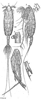
Pareuchaeta spinosa : Raybaud & al., 2008 (p.1765, Table A1); | | | | Ref.: | | | Giesbrecht & Schmeil, 1898 (p.39, Rem. F); I.C. Thompson, 1903 a (p.19); Thompson & Scott, 1903 (p.234, 244); Esterly, 1905 (p.159, figs.F); A. Scott, 1909 (p.75, figs.M); Sharpe, 1910 (p.410, figs.); Wolfenden, 1911 (p.298); Pesta, 1920 (p.513); Sars, 1925 (p.104, figs.F,M); Farran, 1926 (p.256); Rose, 1929 (p.25); Sewell, 1929 (p.149); Wilson, 1932 a (p.62, figs.F); Rose, 1933 a (p.115, figs.F,M); Lysholm & al., 1945 (p.22); Sewell, 1947 (p.111, 112, 117); C.B. Wilson, 1950 (p.217, figs.F,M); Brodsky, 1950 (1967) (p.200, figs.F,M); Tanaka, 1958 (p.332, figs.F); Grice, 1962 (p.203, figs.F); Vervoort, 1963 b (p.162, Rem.); Mazza, 1964 b (p.278, figs.F,M, juv.); Crisafi, 1965 a (p.66, fig.F); Owre & Foyo, 1967 (p.55, figs.F,M, Rem.); Mazza, 1967 (p.155, 161, figs.F,M, juv.); Tanaka & Omori, 1968 (p.224); Park, 1968 (p.551, figs.M, Rem.F,M); Razouls, 1972 (p.94, Annexe: p.50); Tanaka, 1973 (p.138: Rem.); Chen & Shen, 1974 (p.127, figs.F); Park, 1975 c (p.8, figs.F,M); 1978 (p.213, figs.F, M); Björnberg & al., 1981 (p.633, figs.F,M); Gardner & Szabo, 1982 (p.266, figs.F,M); Park, 1995 (p.27, 95, Redescr.F,M, figs.F,M); Mauchline, 1999 (n°182, p.8, figs.F,M); Bradford-Grieve & al., 1999 (p.880, 924, figs.F,M); Vives & Shmeleva, 2007 (p.654, figs.F,M, Rem.) | 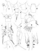 issued from : T. Park in Bull. Scripps Inst. Oceanogr. Univ. California, San Diego, 1995, 29. [p.128, Fig.18]. Female: a, forehead (left side); b, urosome (left); c, d, e, f : genital somote (left, dorsal, right, ventral, respectively); g, Mx1 (first inner lobe separated), posterior; h, P1 (anterior); i, P2 (anterior). Nota: Laterally, dorsal margin of forehead nearly straight; frontal eminence pronounced but not conspicuously projecting forward; rostrum very elongate, only slightly curved backward, pointing in an anteroventral direction. Outer lobe of Mx1 with 9 setae, of which 2 proximal setae are smaller than 6 long distal setae and 1 seta between the proximal and distal groups of setae is very small; 1st endopodal segment of Mx1 often appearing 2-segmented, 3 proximal setae belonging to 1st segment and the 4th seta to 2nd. One endopodal seta of Mx2 armed with long spinules in addition to rows of very short spinules. Basis of Mxp with a row of only small spinules along proximal part of medial margin. Dorsally, genital somite nearly symmetrical with marked swelling at middle; ventrally, strongly asymmetrical with genital field projecting far beyond left side of somite; laterally, both genital flanges appearing similar in shape, but left one a litthe larger than and slightly posterior in position to right. Appendicular and marginal setae of caudal ramus as in E. marina. A1 extending beyond distal end of caudal ramus by its last 2 segments. In morphological features, A1, A2 and Md similar to those of E. marina female. Male: j, forehead (left); k, last pedigerous and genital somites (left); l, P5 (anterior); m, exopod of left 5 th leg (anterior, tilted counterclockwise); idem (medial)
|
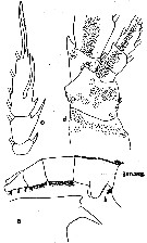
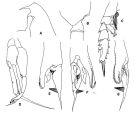 issued from : T. Park in Antarctic Res. Ser. Washington, 1978, 27. [p.216, Fig.72]. Male: A, forehead (lateral); B, last metasomal and genital segments (lateral); C, P2 (anterior); D, P5 (anterior); E, F, G, distal part of exopod of left P5 (lateral, medial and anterior). Nota from Bradford & al. (p.56): - P2 exopod male: Aa = 1/2 AB; Bb = 1/4 BC; Cc = 1/4 CD; Dd = 1/6 CD. (see code of lengths outer spines in the Genus' figure, or in Paraeuchaeta sp. A).
|
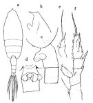 issued from : O. Tanaka in Publs Seto Mar. Biol. Lab., 1958, VI (3). [p.332, Fig.64]. Female: a, habitus (dorsal); b, forehead (lateral left side); c, last thoracic segment and urosome segments 1 & 2 (lateral left side); d, genital somite (ventral); e, exopod of P1; f, P2. Nota: The urosome segments and furca are in the proportional lengths as 42:23:16:8:11 = 100. A1 extends to the middle of the distal margin of the 3rd urosome segment.
|
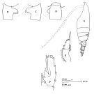 issued from : J.M. Bradford, L. Haakonssen & J.B. Jillett in Mem. N.Z. Oceanogr. Inst., 1983, 90. [p.56, Fig.30]. As Paraeuchaeta spinosa. Female: A, habitus (lateral left side); B-D, genital segment (dorsal, left, right, respecively); E, exopod of P1; F, exopod segment 3 of P2. Nota: - P1 exopod female: Bb = 1/2 BC; Cc = BC. - P2 exopod female: Aa > Ab; Bb = 1/4 BC; Cc = CD + 1/2 Dd; Dd = 1/10 CD. (see code of lengths outer spines in the Genus' figure, or in Paraeuchaeta sp. A).
|
 Issued from : G.O. Sars in Résult. Camp. Scient. Prince Albert I, 69, pls.1-127 (1924). [Pl.XXX, figs.1-6]. Female: 1, habitus (dorsal); 2, idem (lateral left side); 3, forehead (lateral); 4, genital segment (lateral left side). Male: 5, P5; 6, distal end of left P5 (enlarged).
|
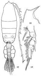 issued from : Q.-c Chen & C.-j. Shen in Studia Marina Sinica, 1974, 9. [p.129, Figs.17-19]. Female: 17, habitus (dorsal); 18, forehead (lateral); 19, P2.
|
 issued from : C.O. Esterly in Univ. Calif. Publs Zool., 1905, 2 (4). [p.160, Fig.24]; Female (from San Diego Region): a, forehead (lateral); b, urosome (lateral); c, exopod of P2; d, basipodites, endopodite and proximal segments of exopod of P4.
|
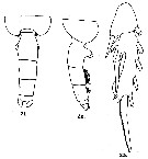 issued from : G.D. Grice in Fish. Bull. Fish and Wildl. Ser., 1962, 61. [p.202, Pl.14, Figs.21-23]. Female (from 00°00', 149°38'W): 21, posterior part of thorax and urosome (dorsal); 22, same (lateral, right side); 23, 2nd and 3rd segments of exopod of P2. Nota: The ventral projections of the genital segment are visible in dorsal view. The middle spine on the outer margin of the 3rd exopodal segment of P2 is greatly elongate, and the spine on the 2nd exopodal segment surpasses the tip of the first spine on the 3rd segment.
|
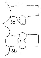 issued from : J. Mauchline in ICES Ident. Leafl. Plankton, 1999, N°182. [p.3, Fig.2: 3a-3b]. Female (Northeast Atlantic): 3a, genital double-somite (left sde); 3b, same (ventral).
|
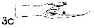 issued from : J. Mauchline in ICES Ident. Leafl. Plankton, 1999, N°182. [p.4, Fig.3: 3c]. Male (Northeast Atlantic): 3c, terminal two segments, exopodal segments of left P5.
|
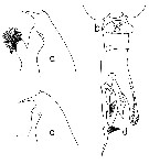 issued from : T. Park in Smiths. Contr. Zool., 1975, 196. [p.8, Fig.5]. Female (G. of Mexico): a, forehead (lateral); b, genital segment (ventral). Male: c, forehead (lateral); d, exopod of left P5 (medial).
|
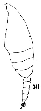 issued from : H.B. Owre & M. Foyo in Fauna Caribaea, 1967, 1, Crustacea, 1: Copepoda. [p.56, Fig.341]. Male (from Florida Current): 341, habitus (lateral).
|
 issued from : H.B. Owre & M. Foyo in Fauna Caribaea, 1967, 1, Crustacea, 1: Copepoda. [p.56, Figs.343-344]. Female: 343, Mx2; 344, Basis of P4.
|
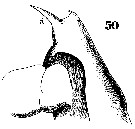 Issued from : W. Giesbrecht in Systematik und Faunistik der Pelagischen Copepoden des Golfes von Neapel und der angrenzenden Meeres-Abschnitte. Fauna Flora Golf. Neapel, 1892, 19 , Atlas von 54 Tafeln. [Taf.37, Fig.50]. As Euchäta spinosa. Female: 50, forehead (lateral). R = rostrum.
|
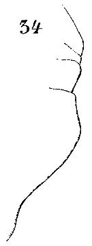
 Issued from : W. Giesbrecht in Systematik und Faunistik der Pelagischen Copepoden des Golfes von Neapel und der angrenzenden Meeres-Abschnitte. - Fauna Flora Golf. Neapel, 1892, 19 , Atlas von 54 Tafeln. [Taf.37, Figs.34, 35]. As Euchäta spinosa. Female: 34, urosome (lateral); 35, same (ventral).
|
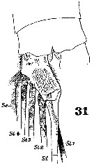 Issued from : W. Giesbrecht in Systematik und Faunistik der Pelagischen Copepoden des Golfes von Neapel und der angrenzenden Meeres-Abschnitte. - Fauna Flora Golf. Neapel, 1892, 19 , Atlas von 54 Tafeln. [Taf.37, Fig.31]. As Euchäta spinosa. Female: 31, anal segment and caudal ramus (ventral).
|
 Issued from : W. Giesbrecht in Systematik und Faunistik der Pelagischen Copepoden des Golfes von Neapel und der angrenzenden Meeres-Abschnitte. - Fauna Flora Golf. Neapel, 1892, 19 , Atlas von 54 Tafeln. [Taf.16, Fig.47]. As Euchäta spinosa. Female: 47, distal segments of A1.
|
 Issued from : W. Giesbrecht in Systematik und Faunistik der Pelagischen Copepoden des Golfes von Neapel und der angrenzenden Meeres-Abschnitte. - Fauna Flora Golf. Neapel, 1892, 19 , Atlas von 54 Tafeln. [Taf.16, Fig.34]. As Euchäta spinosa. Female: 34, outer margin of Mx2.
|
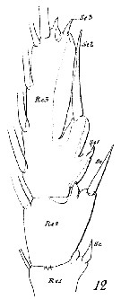 Issued from : W. Giesbrecht in Systematik und Faunistik der Pelagischen Copepoden des Golfes von Neapel und der angrenzenden Meeres-Abschnitte. - Fauna Flora Golf. Neapel, 1892, 19 , Atlas von 54 Tafeln. [Taf.16, Fig.12]. As Euchäta spinosa. Female: 12, exopod of P2 (posterior view).
|
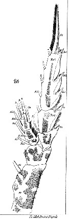 Issued from : W. Giesbrecht in Systematik und Faunistik der Pelagischen Copepoden des Golfes von Neapel und der angrenzenden Meeres-Abschnitte. - Fauna Flora Golf. Neapel, 1892, 19 , Atlas von 54 Tafeln. [Taf.16, Fig.26]. As Euchäta spinosa. Female: 26, P3 (anterior view).
|
 Euchaeta spinosa Euchaeta spinosa. 1 - Basis of female Mxp with short spinules only along proximal half of medial margin. Exopod of male left P5 withoit poorly sclerotized lobes (Fig.18-l); serrated lamella of male left P5 exopod far short of reaching tuft of stiff hairs (Fig.18-m). 2 - 1st endopodal segment (often appearing 2-segmented) of female Mx1 with 3 proximal setae belonging to 1st segment and the 4th seta to 2nd (Fig.18-g). Left exopod of male P5 much shorter than right exopod (Fig.18-l).
| | | | | Ref. compl.: | | | Cleve, 1904 a (p.190); Wilson, 1942 a (p.185); Massuti Alzamora, 1942 (p.110); Sewell, 1948 (p.330, 501, 508, 521, 530, 546, 556); Fagetti, 1962 (p.22); V.N. Greze, 1963 a (tabl.2); De Decker & Mombeck, 1964 (p.12); Shmeleva, 1965 b (p.1350, lengths-volume -weight relation); Mazza, 1966 (p.70); 1967 (p.367); Fleminger, 1967 a (tabl.1); Grice & Hulsemann, 1968 (tab.2); Vinogradov, 1968 (1970) (p.268); Morris, 1970 (p.2300); Shih & al., 1971 (p.147); Dowidar & El-Maghraby, 1970 (p.268); Roe, 1972 (p.277, tabl.1, tabl.2); Deevey & Brooks, 1977 (p.256, tab.2, Station "S"); Carter, 1977 (1978) (p.35); Vaissière & Séguin, 1980 (p.23, tab.1); Kovalev & Shmeleva, 1982 (p.83); Vives, 1982 (p.291); Gaudy & Boucker, 1983 (p.37, Table 1, Rem.: metabomism); ; Scotto di Carlo & al., 1984 (p.1044); Guangshan & Honglin, 1984 (p.118, tab.); Sazhina, 1985 a (p.491, tab.3); Longhurst, 1985 (tab.2); Regner, 1985 (p.11, Rem.: p.31); Petipa & Borichenko, 1985 (tab.1,2); Brinton & al., 1986 (p.228, Table 1); Chen Y.-Q., 1986 (p.205, Table 1: abundance %, Table 2: vertical distribution); Madhupratap & Haridas, 1986 (p.105, tab.1); Lozano Soldevilla & al., 1988 (p.58); Suarez & al., 1990 (tab.2); Scotto di Carlo & al., 1991 (p.271); Suarez & Gasca, 1991 (tab.2); Suarez, 1992 (App.1); Seguin & al., 1993 (p.23); Shih & Young, 1995 (p.70); Hure & Krsinic, 1998 (p.101); Gilabert & Moreno, 1998 (tab.1, 2); Suarez-Morales & Gasca, 1998 a (p.109); Mauchline, 1998 (tab.42); Lopez-Salgado & al., 2000 (tab.1); Pakhomov & al., 2000 (p.1663, Table 2, transect Cape Town-SANAE antarctic base); Holmes, 2001 (p.52); Beaugrand & al., 2002 (p.179, figs.5, 6); Vukanic, 2003 (139, tab.1); Hsieh & al., 2004 (p.397, tab.1); Park, W & al., 2004 (p.464, tab.1); Hwang & al., 2007 (p.24); Lan Y.C. & al., 2008 (p.61, Table 1, % vs stations); Gaard & al., 2008 (p.59, Table 1, N Mid-Atlantic Ridge); Galbraith, 2009 (pers. comm.); Licandro & Icardi, 2009 (p.17, Table 4); Mazzocchi & Di Capua, 2010 (p.425); in CalCOFI regional list (MDO, Nov. 2013; M. Ohman, comm. pers.); Mazzocchi & al., 2014 (p.64, Table 4, abundance); Zaafa & al., 2014 (p.67, Table I, occurrence); Benedetti & al., 2016 (p.159, Table I, fig.1, functional characters); Marques-Rojas & Zoppi de Roa, 2017 (p.495, Table 1); El Arraj & al., 2017 (p.272, table 2, spatial distribution); Belmonte, 2018 (p.273, Table I: Italian zones); Chaouadi & Hafferssas, 2018 (p.913, Table II: occurrence). | | | | NZ: | 19 | | |
|
Carte de distribution de Euchaeta spinosa par zones géographiques
|
| | | | | | | | | | | | 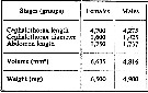 issued from : A.A. Shmeleva in Bull. Inst. Oceanogr., Monaco, 1965, 65 (n°1351). [Table 6: 20 ]. Euchaeta spinosa (from South Adriatic). issued from : A.A. Shmeleva in Bull. Inst. Oceanogr., Monaco, 1965, 65 (n°1351). [Table 6: 20 ]. Euchaeta spinosa (from South Adriatic).
Dimensions, volume and Weight wet. Means for 50-60 specimens. Volume and weight calculated by geometrical method. Assumed that the specific gravity of the Copepod body is equal to 1, then the volume will correspond to the weight. |
| | | | Loc: | | | South Africa (E), off St. Helena Is., off Ascension Is., off E St. Paul Is., G. of Guinea, off Morocco-Mauritania, Canary Is., off Madeira, Bay of Biscay, Azores, off W Ireland, off Uruguay, S Brazil, off Amazon, G. of Mexico, Bahia de Mochima (Venezuela), Caribbean, Florida, off Bermuda: Station "S" (32°10'N, 64°30'W), off Woods Hole, off S Iceland, off W Tangier, Medit. (Alboran Sea, Habibas Is., Banyuls, G. of Lion, Ligurian Sea, Tyrrhenian Sea, Strait of Messina, Adriatic Sea, Ionian Sea, Lebanon Bassin, Alexandria), G. of Aden, Arabian Sea, Natal, off Sri Lanka, Indian, Nicobar Is., Philippines, Viet-Nam (Cauda Bay), China Seas (East China Sea, South China Sea), Taiwan (S, W), Japan (Suruga Bay), Bering Sea, Aleutian Is., G. of Alaska (Icy Strait), British Columbia, California, Gulf of California, Pacif. (equatorial, N central subtropical), Hawaii, off W Guatemala, America (W central), G. of Panama, Galapagos, N Easter Is., Pacif. (W equatorial), off Juan Fernandez Is., off Chile | | | | N: | 91 | | | | Lg.: | | | (1) F: 6,4; (3) F: 7-6,2; M: 6,6-5,8; (11) F: 6,7; (19) F: 6,83-5,83; M: 6,17-5,75; (20) F: 6,91-6,35; M: 6,25; (22) F: 6,4-6; M: 6,3; (38) F: 6,9; (45) F: 6,2-6; (46) F: 6,2; (47) F: 6,2; (63) F: 7,21-6,39; M: 6,9-6,08; (72) F: 7,2-6,24; M: 6,52-5,97; (99) F: 7,2; (101) F: 6,94; (142) F: 6 or +; (143) M: 5,22; (199) F: 6,88-6,24; M: 6,4-5,76; (207) F: 6,7-6,38; M: 6,25-6,15; (393) F: 6,638-5,658; (394) F: 6,87-6,83; (394) F: 6,83-6,87; (449) F: 6; (864) F: 5,18-5,5; {F: 5,18-7,21; M: 5,22-6,90}
The mean female size is 6.467 mm (n = 34; SD = 0.5008), and the mean male size is 6.141 mm (n = 15; SD = 0.4091). The size ratio (male : female) is 0.940 (n = 6; SD = 0.0105), or ± 94 %. | | | | Rem.: | épi-bathypélagique.
Voir aussi les remarques en anglais | | | Dernière mise à jour : 28/10/2022 | |
|
|
 Toute utilisation de ce site pour une publication sera mentionnée avec la référence suivante : Toute utilisation de ce site pour une publication sera mentionnée avec la référence suivante :
Razouls C., Desreumaux N., Kouwenberg J. et de Bovée F., 2005-2026. - Biodiversité des Copépodes planctoniques marins (morphologie, répartition géographique et données biologiques). Sorbonne Université, CNRS. Disponible sur http://copepodes.obs-banyuls.fr [Accédé le 17 juin 2026] © copyright 2005-2026 Sorbonne Université, CNRS
|
|
|
|


;)
;)
;)
;)
;)
;)
;)
;)
;)
;)
;)
;)
;)
;)
;)
;)
;)
;)

;)
