|
|
 |
Fiche d'espèce de Copépode |
|
|
Calanoida ( Ordre ) |
|
|
|
Clausocalanoidea ( Superfamille ) |
|
|
|
Euchaetidae ( Famille ) |
|
|
|
Paraeuchaeta ( Genre ) |
|
|
| |
Paraeuchaeta comosa Tanaka, 1958 (F,M) | |
| | | | | | | Syn.: | Pareuchaeta comosa (F) Tanaka, 1958 (p.363, Descr.F, figs.F, no M);
Pareuchaeta dubia (F) : Tanaka & Omori, 1967 (p.245); 1968 (p.234: F);
Pareuchaeta hanseni (M) Tanaka, 1958 (p.362, figs.M, no F);
Euchaeta regalis (M) Grice & Hulsemann, 1968 (p.329, figs.M, no F);
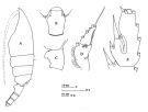
Euchaeta comosa : Park, 1975 c (p.12, figs.F,M, Rem.); 1978 (p.268, figs.F,M, Rem.) | | | | Ref.: | | | Bradford & al., 1983 (p.20, 33, figs.F,M, Rem.); Park, 1994 (p.322, 325, biogéo.); Park, 1995 (p.56, Rem.F,M, figs.F,M); Chihara & Murano, 1997 (p.799, Pl.108,112: F,M); Bradford-Grieve & al., 1999 (p.880, 926, figs.F) | 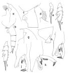 issued from : T. Park in Bull. Scripps Inst. Oceanogr. Univ. California, San Diego, 1995, 29. [p.160, Fig.50]. Female: a, forehead (left side); b, urosome (left); c, genital somite (left); d, outer lobe of Mx1; e, exopod of P1 (anterior); f, exopod of P2 (anterior). Nota: Laterally, rostrum elongate, pointing bliquely forward at an angle of about 10° with respect to anterodorsal margin of forehead, and only slightly curved downward. Dorsal margin of genital somite smoothly curved. Genital flange with nearly straight or slightly bulging ventral margin and produced posteriorly into a large lobe. Posterior edge of genital field also produced distally into a relatively large lobe followed by posterior margin of genital prominence, which is bulging into characteristic curve and longer than posterior ventral wall of somite by about 1/4 its length. A low ridge is present close to anterodorsal margin on left side of genital somite. Cephalosomal appendages similar to those of P. malayensis except that outer lobe of Mx1 with 6 long setae and 1 short seta proximally. In P1 exopod, outer spine of 2nd segment short of reaching base of following outer spine. Male: g, forehead (left); h, last pedigerous and genital somites (left); i, exopod of P2 (anterior); j, distal exopodal segments of left 5th leg (lateral, tilted clockwise); k, idem (showing variation in teeth of serrated lamella); l, idem (lateral, also showing variation in teeth of serrated lamella).
|
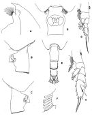 issued from : T. Park in Antarctic Res. Ser. Washington, 1978, 27. [p.270, Fig.111]. As Euchaeta comosa. Female: A, forehead (lateral); B, C, D, distal end of metasome and genital segment (left side, right side, and ventral, respectively); E, posterior part of metasome and urosome (dorsal); F, outer lobe of Mx1; G, P1; H, P2. P1-2: legs (anterior).
|
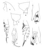 issued from : T. Park in Antarctic Res. Ser. Washington, 1978, 27. [p.271, Fig.112]. As Euchaeta comosa. Male: A, forehead (lateral); B, C, last metasomal and genital segments (left side and right side, respectively); D, P1; E, P2; F, P5; G, H, distal part of exopod of left P5 (anterolateral and medial, respectively). P1-5: legs (anterior). -P1 exopod: Aa minute; Bb = 1/4 BC; Cc < BC.
- P2 exopod: Aa = 1/3 AB; Bb = 1/2 BC; Cc = 1/3 CD. (see code of lengths outer spine in the Genus' figure of Paraeuchaeta, or in Paraeuchaeta sp. A).
|
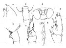 Issued from : O. Tanaka in Publs Seto mar. Biol. Lab., 1958, VI (3). [p.364, Fig.79 a-g]. As Pareuchaeta comosa. Female: a, forehead (lateral); b, last thoracic segment and genital segment (lateral left side); c, idem (dorsal); d, genital complex (ventral); e, P1; f, exopod of P2; g, outer margin of 3rd segment of exopod of P3. Nota: The proportional lengths of the first 3 urosomal segments are 19:9:8.
|
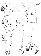
 issued from : J.M. Bradford, L. Haakonssen & J.B. Jillett in Mem. N.Z. Oceanogr. Inst., 1983, 90. [p.34, Fig.14]. Female: A, habitus (lateral left side); B, genital segment (dorsal); C, idem (lateral left side); D, exopod of P1; E, exopod segment 3 of P2. Nota: - P1 exopod: Aa minute; Bb = 3/4 BC; Cc > CD. - P2 exopod: Aa ≥ AB; Bb = 1/2 BC; Cc = 4/5 CD. (see code of lengths outer spine in the Genus' figure of Paraeuchaeta, or in Paraeuchaeta sp. A). Remarks: The south-west Pacific female differs from the original description by having a deeper incision at the base of the 2nd spine of P2 segment 3.
|
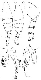 issued from : G.D. Grice & K. Hulsemann in Pacif. Sci., 1968, 22 (3). [p.330, Figs.41-48]. As Euchaeta regalis. Male (from SE Pacific): 41-42, habitus (dorsal and lateral, respectively); 43, forehead (lateral); 44, posterior end of thorax and urosome (left lateral view); 45, posterior end of thorax and 1st and 2nd urosome segments (right lateral view); 46, P5 (distal end of right exopod missing); 47, P5 (another view); 48, distal end of exopod of left P5.
|
 issued from : T. Park in Smiths. Contr. Zool., 1975, 196. [p.12, Fig.10]. As Euchaeta comosa. Female (G. of Mexico): a, forehead (lateral); b, genital segment (lateral); c, 3rd exopodal segment of P2 (anterior). Male: d, forehead (lateral); e, 3rd exopodal segment of P2 (anterior); f, exopod of left P5 (anterior). Nota: A1 reaching about distal end of 2nd urosomal segment. Posterolateral corners asymmetrical, each with a low toothlike process dorsally. Exopod of P1 3-segmented, with external spines of reduced size. Endopod of P2 1-segmented. For Park, the male described is believed to belong to E. comosa because of its close similarity ton the female in size, distribution, and, particularly, form of the P2 (the exceptionally deep incision present in the 3rd exopodal segment of the P2 in both the female and male of this species is not found in any other euchaetid species observed in the present study. The male referred to P. hansenni by Tanaka (1958) and that described as belonging to E. regalis by Grice & Hulsemann (1968) seem to be identical with the male of P. comosa described here. All specimens were found in tows down to depths exceeding 1000 m and constitute the first record of the species in rge Atlantic Ocean.
|
 Paraeuchaeta comosa Paraeuchaeta comosa Female: 1 - See key to species Groups and independent species of Paraeuchaeta (p.30): malayensis species Group. 2 - Outer spine of 2nd exopodal segment (or the 2nd of the first 2 exopodal segments forming a proximal, compound segment) of P1 normally developed (Fig.50-e). 3 - Outer lobe of Mx1 with 6 long setae in addition to 1 short seta proximally (Fig.50-d). 4 - Genital somite without a conical process on left side close to anterior base of genital prominence (Fig.50-c). 5 - Dorsally and ventrally, genital somite symmetrical. 6 - Laterally, genital flange with normal posterior lobe (Fig.50-c). 7 - Laterally, genital somite with more or less smooth dorsal margin (Fig.50-c). 8 - Laterally, genital prominence with conspicuously bulging posterior margin (Fig.50-c). 9 - Laterally, genital prominence with smoothly convex posterior margin (Fig.50-c).
| | | | | Ref. compl.: | | | Suarez-Morales & Gasca, 1998 a (p.109, as cosmosa); Razouls & al., 2000 (p.343, Appendix); Park & Ferrari, 2009 (p.143, Table 5, Appendix 1, biogeography); | | | | NZ: | 11 | | |
|
Carte de distribution de Paraeuchaeta comosa par zones géographiques
|
| | | | | | | | | | | | 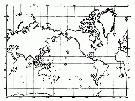 issued from : T. Park in Hydrobiologia 292/293, 1994. [Fig. 1, p.325].
Geographical distribution of Paraeuchaeta comosa (lozange). issued from : T. Park in Hydrobiologia 292/293, 1994. [Fig. 1, p.325].
Geographical distribution of Paraeuchaeta comosa (lozange). |
| | | | Loc: | | | S Atlant., Argentina, Caribbean Sea, G. of Mexico, Indian, China Seas (South China Sea, East China Sea), Indonesia-Malaysia, Japan (Izu), Pacif. (central N-S), Juan Fernandez Is., Tasman Sea, sub-Antarct. (SE Pacif., S Australia) | | | | N: | 8 | | | | Lg.: | | | (3) F: 10-7,8; M: 8,8-7,5; (19) M: 8,08-7,75; (20) F: 9,66-8,66; M: 9,08-8,41; (63) F: 9,88-8,24; (70) F: 9,08-9; (99) F: 9,3-8,8; {F: 7,80-10,00; M: 7,50-8,80} | | | | Rem.: | bathypélagique.
Sampling depth (sub-Antarct.) : 0-1000 m.
Voir aussi les remarques en anglais | | | Dernière mise à jour : 26/02/2015 | |
|
|
 Toute utilisation de ce site pour une publication sera mentionnée avec la référence suivante : Toute utilisation de ce site pour une publication sera mentionnée avec la référence suivante :
Razouls C., Desreumaux N., Kouwenberg J. et de Bovée F., 2005-2026. - Biodiversité des Copépodes planctoniques marins (morphologie, répartition géographique et données biologiques). Sorbonne Université, CNRS. Disponible sur http://copepodes.obs-banyuls.fr [Accédé le 17 juin 2026] © copyright 2005-2026 Sorbonne Université, CNRS
|
|
|
|


;)
;)
;)
;)
;)
;)
;)

;)
