|
|
 |
Fiche d'espèce de Copépode |
|
|
Calanoida ( Ordre ) |
|
|
|
Clausocalanoidea ( Superfamille ) |
|
|
|
Aetideidae ( Famille ) |
|
|
|
Aetideopsis ( Genre ) |
|
|
| |
Aetideopsis armata (Boeck, 1872) (F,M) | |
| | | | | | | Syn.: | Euchaeta armata Boeck, 1872;
Chiridius armatus : Sars, 1901 a (1903) (p.27, figs.F,M); 1925 (p.45); Farran, 1903 (p.15, figs.F); Vanhöffen, 1907 (p.519, figs.F,M); Damas & Koefoed, 1907 (p.398, tab.II, III); Farran, 1908 b (p.30, Rem.); With, 1915 (p.77, figs.F,M); Sars, 1925 (p.45); Farran, 1926 (p.247); Wilson, 1932 a (p.47, figs.F,M); Rose, 1933 a (p.94, figs.F,M); Jespersen, 1934 (p.55, fig.12); 1939 (p.46, Rem., Table 29); 1940 (p.18); Lysholm & al., 1945 (p.11); Sewell, 1948 (p.384, 499); C.B. Wilson, 1950 (p.189, fig.M); Gundersen, 1953 (p.1, 26, seasonal abundance); Matthews, 1964 (p.6, figs.N, juv., F,M); Elofsson, 1966 (p.92, fig.: eye); Pavlova, 1966 (p.43); Matthews, 1967 (p.159, Table 1, Rem.);Vives, 1967 (p.551, fig.M, tab.III); Vinogradov, 1968 (1970) (p.195); Tanaka & Omori, 1970 (p.114, figs.F); Björnberg, 1972 (p.25, Rem.N); Björnberg, 1973 (p.323, 385); Matthews & Sands, 1973 (p.19, Table 4); Alvarez & Matthews, 1975 (p.67, feeding behaviour, assimilation); Skjoldal & Bamstedt, 1977 (p.197, adenosides); Vives, 1978 a (p.265); Dessier, 1979 (p.204); Pipe & Coombs, 1980 (p.223, figs. 1, 2, table 1, vertical distribution); Bamstedt & Skjoldal, 1980 (p.304, weight-RNA); Kovalev & Schmeleva, 1982 (p.83); Bamstedt, 1983 (p.291, RNA variation); Norrbin & Bamstedt, 1984 (p;47, Table 2, 3: calorific value); Bamstedt, 1985 (p.607, excretion rate); Bamstedt, 1988 (p.15, protein content); Pancucci-Papadopoulou & al., 1990 (p.199); Fransz & al., 1991 (p.9); Krause & al., 1995 (p.81, Fig.17, abundance, Rem.: p.132); Hanssen, 1997 (tab.3.1); Falkenhaug & al., 1997 (p.449, spatio-temporal pattern); Hure & Krsinic, 1998 (p.46, 101); Mauchline, 1998 (tab.26, 30, 58); Halvorsen & Tande, 1999 (p.279, tab.2, 3, Rem.: p.282); d'Elbée, 2001 (tabl.1); Sameoto & al., 2002 (p.12); Schøyen & Kaartvedt, 2004 5p;159, verticql distribution, feeding); Veistheim & al., 2005 (p.382, tab.1, fig.1); Gaard & al., 2008 (p.59, Table 1, N Mid-Atlantic Ridge);
Pseudaetideus armatus (Boeck,1872): Wolfenden, 1904 (p.115); Pearson, 1906 (p.11); Pesta, 1920 (p.509); Brodsky, 1950 (1967) (p.157, figs.F,M); Vervoort, 1952 c (n°44, p.3, figs.F,M); Østvedt, 1955 (p.14: Table 3, p.60); Vervoort, 1963 b (p.125, Rem.); Grice & Hulsemann, 1965 (p.223); Mazza, 1965 a (p.310, figs. juv.F,M, F, Rem.); 1966 (p.70); 1967 (p.119, 139, figs.juv., F,M); 1968 (p.531, figs.); Grice, 1969 a (p.454); Gamulin, 1971 (p.382, tab.3); Roe, 1972 (p.277, tabl.1, tabl.2); Scotto di Carlo & Ianora, 1983 (p.150); Scotto di Carlo & al., 1984 (p.1044); Scotto di Carlo & al., 1991 (p.270); Mumm, 1993 (tab.1);
Pseudoaetideus armatus : Park, 1970 (p.476); Kovalev & Schmeleva, 1982 (p.83) | | | | Ref.: | | | Bradford, 1969 (p.94, Rem.); 1969 b (p.502); Park, 1975 b (p.276, figs.F,M); Bradford & Jillett, 1980 (p.19); Markhaseva, 1996 (p.27, figs.F,M); Yamaguchi & al., 2005 (p.835, Tabl. VIII); Vives & Shmeleva, 2007 (p.537, figs.F,M, Rem.) | 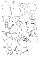 issued from : E.L. Markhaseva in Trudy Zool. Inst. RAN, St. Petersburg, 1996, 268. [p.29, Fig.14]. Female (from SE Pacif.). Different specimens. Th5 (a) & Abd (a) from 29°50'S, 93°34'W), other figures from 36°05'S, 134°05'W.
|
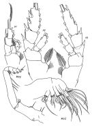 issued from : E.L. Markhaseva in Trudy Zool. Inst. RAN, St. Petersburg, 1996, 268. [p.30, Fig.15]. Female (from 36°05'S, 134°05'W).
|
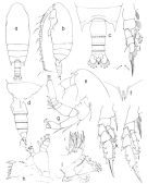 issued from : T. Park in Bull. Mar. Sci., 1975, 25 (2). [p.277, Fig.3] Female (from Gulf of Mexico): a, b, habitus (dorsal, lateral, respectively); c, d, posterior part of metasome and urosome (dorsal, lateral, respectively); e, forehead, (lateral); f, rostrum (anterior); g, A2; h, Md; i, Mx1; j, P1; k, P2; l, P3. P1-3: legs (anterior).
|
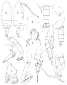 issued from : T. Park in Bull. Mar. Sci., 1975, 25 (2). [p.278, Fig.4] Male (from Gulf of Mexico): a, b, habitus (dorsal, lateral, respectively); c, forehead (lateral); d, e, posterior part of metasome and urosome (dorsal, lateral); f, rostrum (anterior); g, A2; h, mandibular palp of Md; i, Mx1; j, Mxp; k, P1; l, P2; m, P5 (posterior); P1-2: legs (anterior; P5: fifth pair of legs.
|
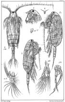 issued from : G.O. Sars in An Account of the Crustacea of Norway. Vol. IV. Copepoda Calanoida. Published by the Bergen Museum, 1903. [Pl. XV]. As Chiridius armatus. Female & Male. Nota: R = rostrum; M = Md; m = Mx1; mp1 = Mx2. Nota Female: Front narrowly rounded and armed below with a very small bifid rostrum. Eye unusually large and distinctly bilobate, dark red in colour. Exopod of A2 about 1/3 longer than endopod. P1 with a well-marked setiform spine outside the 1st segment of the exopod. P2 with endopod distinctly biarticulate. Nota Male: A1 (23-segmented) considerably shorter than in female, not nearly as long as the prosome, and somewhat incrassated at the base. mandibular palps much more strongly built than in female, with endopod well developed and carrying at the tip very coarse diverging setae. Right P5 stronger than the left, both with a very small uniarticulate appendage inside the 2nd segment, representing a rudiment of endopod
|
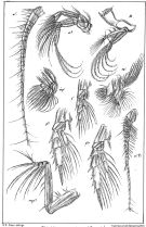 issued from : G.O. Sars in An Account of the Crustacea of Norway. Vol. IV. Copepoda Calanoida. Published by the Bergen Museum, 1903. [Pl. XVI]. Female & Male. M = Md; m = Mx1; mp1 = Mx2; mp2 = Mxp.
|
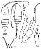 Issued from : K.A. Brodskii in Calanoida of the Far Eastern Seas and Polar Basin of the USSR. Opred. Fauna SSSR, 1950, 35 (Israel Program for Scientific Translations, Jerusalem, 1967) [p.156, Fig.70]. As Pseudaetideus armatus. Female (from Arctic): habitus (dorsal and lateral left side); R, rostrum. Nota: Cephalothorax 2.5 times length of abdomen. Eye very large. Spikes of terminal thoracic segment slightly asymmetrical, right one larger than left, both parallel, reaching to middle of genital segment. Male: S5, P5. Nota: A1 23-segmented.
|
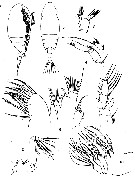 issued from : G.P. Farran in Qcient. Invest. Fish. Brch, Ireland, 1901 (2), Appendix 7, 1903. [Plate XVI, Figs.1-2, 4-5, 8-12]. As Chiridius armatus. Female (from off Cleggan, Irelandl): 1-2, habitus (lateral and dorsal, respectively); 4, A2; 5, Md (mandibular palp); 8, Mx1 (lower lobes omitted); 9, Mxp; 10, Mx2; 11, P2 (anterior).
|
 issued from : G.P. Farran in Qcient. Invest. Fish. Brch, Ireland, 1901 (2), Appendix 7, 1903. [Plate XVI, Fig.3]. As Chiridius armatus. Female: 3, A1.
|
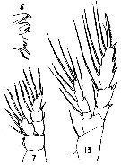 issued from : G.P. Farran in Qcient. Invest. Fish. Brch, Ireland, 1901 (2), Appendix 7, 1903. [Plate XVI, Figs.6, 7, 13]. As Chiridius armatus. Female: 6, Md (cutting edge); 7, P1 (anterior); 13, P4 (posterior).
|
 issued from : G.P. Farran in Qcient. Invest. Fish. Brch, Ireland, 1901 (2), Appendix 7, 1903. [Plate XVI, Fig.12]. As Chiridius armatus. Female: 12, P3 (anterior).
|
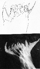 issued from : G.D. Grice & T.J. Lawson in Crustaceana, 1971, 21 (1). [Pl.1]. Teeth of the mandible. Upper figure drawn with the aid of a camera lucida; lower figure an electron-micrograph of the appendage fixed in formalin, freeze dried and gold-coated. Nota: In the upper figure gross structure is readily discernible; however, the type configuration and shape of the teeth which arise from gnathobase are difficult to interpret. The Scanning electron microscope (SEM) permits to examine appendage at a higher level of resolution and with a much greater depth of field. It also suggests a difference in structure or texture between the teeth proper and the remainder of the mandibular blade (gnathobase).
|
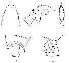 issued from : C. With in The Danish Ingolf-Expedition, 1915, III (4). [p.77, Text-Fig.17]. As Chiridius armatus. Female (NE Atlantic): a, head (dorsal); b, forehead (lateral); c, last thoracic segments and genital segment (dorsal); d, lateral corner and genital segment (left side); e, parasite attached to the right Mx2.
|
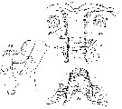 issued from : C. With in The Danish Ingolf-Expedition, 1915, III (4). [Pl.II, Fig.3, b-d]. As Chiridius armatus. Female: b, labrum (oral view); g2-g4 the 2nd and 4th groups of the lateral longitudinal series . Sa the 4th central circular spot. c, lamina labialis and serrulae 6-teeth. d, area labialis and postlabialis. Nota: The anterior surface of the labrum has, in addition to the usual marginal row of setae which are fairly slender in the middle and more like granules laterally, in the middle a transverse row of laterally shorter setae, and in front on each side a group of short setae, and in front on each side a group of short setae. The oral surface of the labrum is characteristic, in front of the chitinous transverse bar behind the median central spot Nr.3, a transverse row of short setae is found, around and behind the median spot 4 (S4) short setae are placed in transverse rows; the skin is everywhere, especially posteriorly, minutely granular; the lateral longitudinal series consists as usual of 5 groups, the two first groups are placed somewhat longitudinally, are distinctly longer than wide, and consist of short spinules, most like granules; the groups 3-5, in contrast, less well separated, and consist of an inner row of longer and an irregular, outer group of scatterad shorter hairs The laminan labialis is in the shape of the dentations almost smooth; the arrangement of the groups of hairs in front and behind the lamina labialis corresponds to a series of bristles on the labial lobes (see 3d).
|
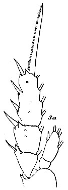 issued from : C. With in The Danish Ingolf-Expedition, 1915, III (4). [Pl.II, Fig.3, a]. As Chiridius armatus. Female: 3a, P2 (anterior view). Nota: 1 glandular pore (formed by a chitinous ring, in the middle of which a generally longitudinal split is seen) on the outer surface of exopod 1, 2 on exopodite 2 and 3 on exopod 3.
|
 issued from : R. Elofsson in Sarsia, 1966, 25. [p.92, Fig62]. . (from Bergen, Norway): Semi-diagrammatic drawing from sections showing a sagittal view of the nauplius eye with nerves, the X-organ and the organ of Gicklhorn. 1 = Gicklhorn's organ; 2 = dorsal part of the dorsal lateral nerve; 3 = ventral part of the same nerve; 4 = X-organ; 5 = Ventral medial nerve. Nota: The naupliar eye has 3 cups, one directed ventrally, and two laterally. The lateral cups are much larger than the ventral one and the sensory cells have undergone radical histological changes. Moreover, there is a new element besides the pigment, taperal and sensory cells, viz. 2 lens cells in each cup. The whole eye lies at some distance from the brain, not far from the anterior surface of the animal.
|
 Aetideopsis armata Aetideopsis armata female: 1 - Endopod of P1 with external lobe. 2 - Posterior corners of last thoracic segment not wing-like, not divergent (dorsal view). Genital segment (dorsal view) barrel-like without lateral swellings. 3 - Crest absent. 1st and 2nd external spines of exopodal segments 1 and 2 of P1 reaching base of next spine. 4 - Endopod of P2 2-segmented (though separation between segments may be incomplete). Points of last thoracic segment reaching the midlength of genital segment. 5 - Anterior part of cephalon (lateral view) fairly smoothly transforming into rostrum, not prominent over base of rostrum. 6 - Rostrum very small, rami rather close-spaced, nearly parallel.
|
 Aetideopsis armata Aetideopsis armata male: 1 - Points of last thoracic segment not reaching posterior border of urosomal segment 1.
|
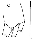 Issued from : E.L. Markhaseva & J. Renz in Crustaceana, 2015, 88 (9). [p.1043, Fig.6, C]. Female: Mx2 (praecoxal endite). Scale bar: 0.1 mm.
| | | | | Ref. compl.: | | | Brenning, 1983 (p.2, Rem.); 1985 a (p.28, Table 2); Roe, 1984 (p.356); Heinrich, 1990 (p.16); Kosobokova & al., 1998 (tab.2); Holmes, 2001 (p.44); Lo & al., 2004 (p.89, tab.1); Kosobokova & al., 2007 (p.929: Tab.7); Darnis & al., 2008 (p.994, Table 1); Mazzocchi & Di Capua, 2010 (p.423); Dvoretsky & Dvoretsky, 2010 (p.991, Table 2); Lidvanov & al., 2013 (p.290, Table 2, % composition); Benedetti & al., 2016 (p.159, Table I, fig.1, functional characters); El-Arraj & al., 2017 (p.272, table 2, distribution vs. seasonal variability); Belmonte, 2018 (p.273, Table I: Italian zones) | | | | NZ: | 14 | | |
|
Carte de distribution de Aetideopsis armata par zones géographiques
|
| | | | | | | | | | | | | | | 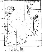 Issued from : M. Krause, J.W. Dippner & J. Beil in Prog. Oceanog., 1995, 35. [p.104, Fig.17]. Issued from : M. Krause, J.W. Dippner & J. Beil in Prog. Oceanog., 1995, 35. [p.104, Fig.17].
Horizontal distribution pattern of Chiridius armatus (= Aetideopsis armata) (individuals per m2) in the winter North Sea.
Collected by WP2-net. Numbers (ind/m3) depth-integrated, extrapoled to the bottom (but at a maximum to a depth of 500 m) and expressed as ind per m2 of water surface. |
| | | | Loc: | | | Atlant. (NW Africa), G. of Guinea, Senegal, off Morocco-Mauritania, Maroccan coast, Caribbean Sea, Sargasso Sea, Bermuda, Azores, off Woods Hole, off Nova Scotia, Bay of Baffin, Davis Strait, SE Greenland, Iceland, Norway Sea, W Norway (Malangen fjord, Korsfjorden, Raunefjorden (all the year), Wyville Thomson Ridge, Kosterfjorden, Nordvestbanken, N Scotland, Nansen Basin, SE Beaufort Sea, Barents Sea, Laptev Sea, North Sea, W Ireland, Bay of Biscay, off W Cape Finisterre, Ibero-moroccan Bay, Medit. (Alboran Sea, Bay of d'Algiers, W Medit., Ligurian Sea, Tyrrhenian Sea, Adriatic Sea, Ionian Sea, Aegean Sea, Lebanon Basin), NW Indian, Philippines, China Seas (East China Sea),Taiwan (N: Mienhua Canyon), Japan, Galapagos (in C.B. Wilson, 1950; Heinrich, 1990), Pacif. (SE tropical), Chile
Type locality: Norway (western coast) | | | | N: | 49 | | | | Lg.: | | | (7) F: 4,43-3,6; M: 3,66; (14) F: 3,6-3,2; (22) F: 4,2-3,3; M: 4-2,9; (37) F: 4,5-3,28; M: 4-3,28; (38) F: 3,6-3,2; (45) F: 4; M: 4; (65) F: 4; M: ± 4; (89) F: 4; (113) F: 4-3,33; (197) F: 3,48-3,28; M: 3,4-3,28; (199) F: 3,32-3,04; (207) F: 3,2-2,8; (260) F: 3,2-2,8; (449) F: 4-3,3; M: 4-3,5; (1109) F: 3,5; {F: 2,80-4,50; M: 2,90-4,00} | | | | Rem.: | épi- à bathypélagique.
-ops ou -opsis est un suffixe (etymologiquement qui a trait à l'oeil). Il peut s'agir de l'organe ou de l'apparence. Ce nom étant féminin l'adjectif s'accorde, donc on doit écrire armata. Cf. Code international de nomemclature zoologique. | | | Dernière mise à jour : 20/10/2022 | |
|
|
 Toute utilisation de ce site pour une publication sera mentionnée avec la référence suivante : Toute utilisation de ce site pour une publication sera mentionnée avec la référence suivante :
Razouls C., Desreumaux N., Kouwenberg J. et de Bovée F., 2005-2026. - Biodiversité des Copépodes planctoniques marins (morphologie, répartition géographique et données biologiques). Sorbonne Université, CNRS. Disponible sur http://copepodes.obs-banyuls.fr [Accédé le 11 février 2026] © copyright 2005-2026 Sorbonne Université, CNRS
|
|
 |
 |


