|
|
 |
Fiche d'espèce de Copépode |
|
|
Calanoida ( Ordre ) |
|
|
|
Clausocalanoidea ( Superfamille ) |
|
|
|
Euchaetidae ( Famille ) |
|
|
|
Paraeuchaeta ( Genre ) |
|
|
| |
Paraeuchaeta weberi A. Scott, 1909 (F,M) | |
| | | | | | | Syn.: | Euchaeta weberi : Vervoort, 1957 (p.78); Grice & Hulsemann, 1967 (p.16); 1968 (tab.2, p.332, Descr.M, figs.M); Park, 1978 (p.224, figs.F,M, Rem.); Björnberg, 1973 (p.386);
Pareuchaeta weberi : Yamanaka, 1976 (p.171, 172, 180); Zvereva, 1976 a (p.70, figs.F); Heinrich, 1990 (p.17). | | | | Ref.: | | | A. Scott, 1909 (p.74, Descr.F, figs.F); Sewell, 1929 (p.133, 169, figs.F, Juv.F,M); Sewell, 1947 (p.123, figs.F,M); Bradford & al., 1983 (p.23); Park, 1994 (p.320); 1995 (p.76, Rem.F,M, figs.F,M). | 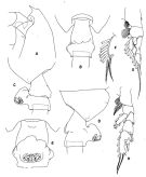 issued from : T. Park in Antarctic Res. Ser. Washington, 1978, 27. [p.225, Fig.80]. As Euchaeta weberi. Female: A, forehead (lateral); B, C,D,E, distal end of metasome and genital segment (dorsal, left side, right side, and ventral, respectively); F, outer lobe of Mx1; G, P1; H, P2. P1-2: legs (anterior).
|
 issued from : T. Park in Antarctic Res. Ser. Washington, 1978, 27. [p.226, Fig.81]. As Euchaeta weberi. Male: A, forehead (lateral); B, last metasomal and genital segments (left side); C, P1; D, P2; E, P5; F, G,H, distal end of exopod of left P5 (anterior, medial, , and anteromedial, respectively). P1-5: legs (anterior). Nota: The male is very close from P. tonsa, P. pseudotonsa, P. tuberculata and the differences by which it can be identified are found only in the rostrum and the left P5 exopod.
|
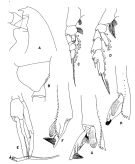
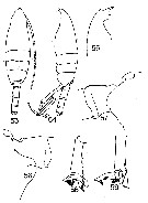
 issued from : T. Park in Bull. Scripps Inst. Oceanogr. Univ. California, San Diego, 1995, 29. [p.183, Fig.73]. Female: a, forehead (left side); b, urosome (left); c, d, genital somite (left, right, respectively); e, exopod of P1 (anterior); f, exopod of P2 (anterior). Male: g, forehead (left); h, exopod of left 5th leg (anterior); i, distal exopodal segments of left 5th leg (anterior, tulted counterclockwise); j, idem (anterior); k, idem (medial).
|
 issued from : R.B.S. Sewell in The John Murray Expedition, 1933-34, Scientific Reports, VIII (1), 1947. [p.124, Fig.28, A]. Female (from Arabian Sea): A, Mx1. Nota: In 1929 (p.170) the 1st inner lobe (or masticatory) of Mx carried 13 setae, and thet the combined 1st and 2nd segments of the endopod carried 7 setae and the 3rd segment 3; in the present specimen, the 1st inner lobe bears 12 setae, the 2nd and 3rd lobes 1 each; the 2nd basal segment has 4 setae; the 1st segment of the endopod bears 6 setae, and the combined 2nd and 3rd carries 3; the exopod bears 11 setae and the outer lobe 9, of which the posterior two are smaller than the others.
|
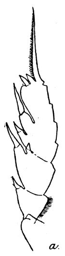 issued from : R.B.S. Sewell in Mem. Indian Mus., 1929, X. [p.171, Fig.63, a]. Female (from Laccadive Sea): a, P2.
|
 issued from : R.B.S. Sewell in Mem. Indian Mus., 1929, X. [p.10, Fig.1]. Female: basal segment 1 of A2. Nota: The 1st basal segment bears a row of long hairs on the posterior and inner aspect. Endopod with 6 and 8 (? 9) setae on the outer and inner lobes respectively of the distal segment.
|
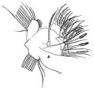
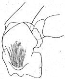
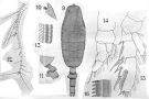 issued from : A. Scott in Siboga-Expedition, 1909, XIX a. [Plate XV, Figs.9-16]. Female (from Indoseia-Malaysia): 9, habitus (dorsal); 10, forehead (lateral); 11, last thoracic and genital segments (left side); 12, A1; 13, Mxp (part of one of the distal hairs); 14, P1; 15, P2; 16, P3 (part of terminal spine of exopodite). Nota: A1 extend to about the middle of the genital segment. This species is easily recognised by the peculiar form of the genital segment.
|
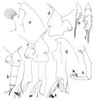
 issued from : G.D. Grice & K. Hulsemann in Pacif. Sci., 1968, 22 (3). [p.330, Figs.53-59]. As Euchaeta weberi. Male (from SE Pacific): 53-54, habitus (dorsal and lateral, respectively); 55, forehead (lateral); 56, posterior end of thorax and 1st urosome segment (left lateral view); 57, idem (right lateral view); 58, distal end of exopod of left P5; 59, idem (another view).
|
 Paraeuchaeta weberi Paraeuchaeta weberi Female: 1 - See key to species Groups and independent species of Paraeuchaeta (p.30). 2 - Laterally, genital field facing ventrad, without large tubercular outgrowth behind genital orifice (Fig.73-c). 3 - Genital somite with large tibercular outgrowths on dorsal side (Fig.73-c).
| | | | | Ref. compl.: | | | Sewell, 1948 (p.329, 530, 532, 534, 539, 540, 541, 552); Bainbridge, 1972 (p.61, Appendix Table III: occurrence); Björnberg, 1973 (p.329); C.E. Morales & al., 2010 (p.158, Table 1); Hidalgo & al., 2010 (p.2089, Table 2); Hidalgo & al., 2012 (p.134, Table 2) | | | | NZ: | 7 | | |
|
Carte de distribution de Paraeuchaeta weberi par zones géographiques
|
| | | | | | | | | | Loc: | | | G. of Guinea (off Lagos in Bainbridge, 1972), Mozambique Channel, Gulf of Aden, Arabian Sea, off SW Sri Lanka, Indian, Indonesia-Malaysia, China Seas (East China Sea), W Pacif. (equatorial), SE Pacif. (tropical), off Juan Fernandez Is., Chile (N, off Santiago, Concepcion).
Type locality: Malay Archipelago. | | | | N: | 15 | | | | Lg.: | | | (3) F: 8-6,7; M: 6,8-6; (5) F: 8,5; (20) F: 7,66-7,16; M: 6,58-6,25; (29) F: 7,562; (100) M: 6,41; {F: 6,70-8,50; M: 6,00-6,41}
The mean female size is 7.597 mm (n = 6; SD = 0.6291), and the mean male size is 6.408 mm (n = 5; SD = 0.3061). The size ratio (male : female) is approximatively ± 86 % | | | | Rem.: | méso-bathypélagique.
W. Zhang confirme la présence de cette espèce dans les mers de Chine (comm. pers., 2006)
Voir aussi les remarques en anglais | | | Dernière mise à jour : 12/08/2016 | |
|
|
 Toute utilisation de ce site pour une publication sera mentionnée avec la référence suivante : Toute utilisation de ce site pour une publication sera mentionnée avec la référence suivante :
Razouls C., Desreumaux N., Kouwenberg J. et de Bovée F., 2005-2026. - Biodiversité des Copépodes planctoniques marins (morphologie, répartition géographique et données biologiques). Sorbonne Université, CNRS. Disponible sur http://copepodes.obs-banyuls.fr [Accédé le 14 juin 2026] © copyright 2005-2026 Sorbonne Université, CNRS
|
|
|
|


;)
;)
;)
;)
;)
;)
;)
;)

