|
|
 |
Fiche d'espèce de Copépode |
|
|
Calanoida ( Ordre ) |
|
|
|
Clausocalanoidea ( Superfamille ) |
|
|
|
Aetideidae ( Famille ) |
|
|
|
Aetideopsis ( Genre ) |
|
|
| |
Aetideopsis multiserrata (Wolfenden, 1904) (F,M) | |
| | | | | | | Syn.: | Faroella multiserrata Wolfenden, 1904 (p.117, Descr.F, figs.F); Pearson, 1906 (p.13, Rem.); Farran, 1908 b (p.29, Rem.);
Chiridius nasutus With, 1915 (p.81, figs.F); Sewell, 1948, (p.384);
? Aetideopsis rostrata : A. Scott, 1909 (p.40);
Chiridius multiserratus : Farran, 1926 (p.248, Rem.);
Aetideopsis pacifica Esterly, 1911 (p.315, figs.F); Brodsky, 1950 (1967) (p.149, figs.F); in CalCOFI regional list (MDO, Nov. 2013; M. Ohman, pers. comm.);
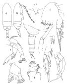
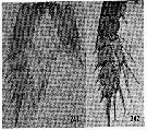
no Aetideopsis multiserrata : Brodsky, 1950 (1967) (p.147, figs.F); Vidal, 12971; Markhaseva, 1984 (p.512) | | | | Ref.: | | | Sars, 1907 a (p.6); 1925 (p.43, figs.F); Rose, 1933 a (p.91, figs.F); Jespersen, 1934 (p.53); Lysholm & al., 1945 (p.11, Rem.); Sewell, 1948 (p.499, 545, 550); Davis, 1949 (p.25, Rem.F); Vervoort, 1951 (p.80, Rem.); 1952 a (n°42, p.3, figs.F); 1957 (p.53, Rem.); Tanaka, 1957 a (p.37, figs.F,M); Vervoort, 1963 b (p.123, Rem.); Owre & Foyo, 1967 (p.43, figs.F,M); Tanaka & Omori, 1970 (p.111); Vidal, 1971 a (p.16, 112, figs.F); Park, 1975 b (p.274, figs.F,M); 1978 (p.111, figs.F); Bradford & Jillett, 1980 (p.19); Markhaseva, 1996 (p.37, figs.F,M, Rem.); Chihara & Murano, 1997 (p.681, Pl.30,31: F,M); Vives & Shmeleva, 2007 (p.538, figs.F,M, Rem.) | 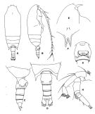 issued from : T. Park in Antarctic Res. Ser. Washington, 1978, 27. [p.112, Fig.6]. Female: A, habitus (dorsal view); B, idem (right lateral view); C, posterior part of prosome and urosome (right lateral view); D, idem (dorsal view); E, forehead (lateral view); F, rostrum (anterior); G, genital segment (ventral view); H, A2.
|
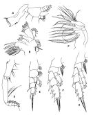 issued from : T. Park in Antarctic Res. Ser. Washington, 1978, 27. [p.113, Fig.7]. Female:A, Md; B, Mx1; C, Mx2; D, Mxp; E, P1 (anterior view); F, P2 (anterior); G, P4.
|
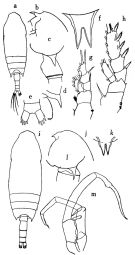
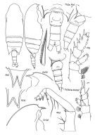 issued from : E.L. Markhaseva in Trudy Zool. Inst. RAN, St. Petersburg, 1996, 268. [p.38, Fig.22]. Female; From different specimens. R (a) and R (b): rostrum (anterior) (a) from 45°11'N, 152°28'E; (b): 48°33'N, 18°52'W; other figures: 43°52'N, 149°49'E.
|
 issued from : E.L. Markhaseva in Trudy Zool. Inst. RAN, St. Petersburg, 1996, 268. [p.39, Fig.23]. Male (from 43°52'N, 149°49'E). R: rostrum (anterior); Gn: genital segment. Male described here differs from Tanaka (1957) and Park (1975) in some details: structure of rostrum (lateral view), length of points of last thoracic segment and details of setation of oral parts.
|
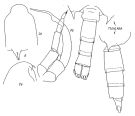
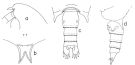 issued from : T. Park in Bull. Mar. Sci., 1975, 25 (2). [p.274, Fig.1] Female: a, forehead (lateral); b, rostrum (anterior); c, posterior part of metasome and urosome (dorsal); d, idem (left side).
|
 issued from : T. Park in Contr. Mar. Sci., 1976, 20. [p.275, Fig.2] Male: a, b, habitus (dorsal, lateral); c, rostrum (anterior); d, forehead (lateral); e, posterior part of metasome and urosome (right side); f, A2; g, mandibular palp; h, Mx1; i, Mxp; j, distal part of Mxp; k, P1; l, P2; m, fifth pair of legs (posterior). P1-P2: legs (anterior).
|
 issued from : O. Tanaka in Publ. Seto Mar. Biol. Lab., 1957, VI (1). [p.39, Fig.26]. Female: a, habitus (dorsal); b, forehead (lateral); c, last thoracic segment and genital segment (lateral left side); d, idem (dorsal); e, anal segment and furca (dorsal); f, rostrum; g, P1; h, P2. Nota: Rostrum highly chitinised and very prominent (the shape in lateral view differs somewhat from the figure given by Sars or A. Scott. Proportional lengths of urosomites and furca 35:24:17:10:14 = 100. A1 reaches back to distal margin of 2nd urosomal segment. mx1: outer lobe with 8 setae, exopod with 10 setae. Male: i, habitus (dorsal); j, forehead (lateral); k, rostrum; l, last thoracic segment (lateral); m, P5. Nota: Head and 1st thoracic segment fused, line of fusion between 4th and 5th thoracic segments scarcely visible in dorsal view. Rostrum: branches closely set together. Proportional lengths of urosomites and furca 16:34:23:16:1: 10 = 100. A1 extends to distal end of 2nd thoracic segment. Mx1: 1st outer lobe has 6 long and 3 short setae.
|
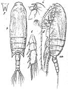 Issued from: G.O. Sars in Résult. Camp. Scient. Prince Albert I, 69, pls.1-127 (1924). [Pl.XIV, figs. 4-8]. Female: 4, habitus (dorsal); 5, idem (lateral left side); 6, forehead (lateral); 7, rostrum (frontal view); 8, exopodal segments of P3.
|
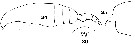 issued from : C.O. Esterly in Univ. Calif. Publs Zool., 1911, 6 (14). [Pl.28, Figs. 24, 25, 30]. As Aetideopsis pacifica. Female (from San Diego Region): 24, habitus (lateral); 25, forehead (lateral); 30, rostrum. Nota: A1 23-segmented and do not extend back to the end of the cephalothorax. Urosome 4-segmented, 1st segment longer than any of the others, 2nd segment about as long as the 3rd and 4th together.
|
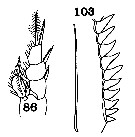 issued from : C.O. Esterly in Univ. Calif. Publs Zool., 1911, 6 (14). [Pl.31, Figs.86, 103]. As Aetideopsis pacifica. Female: 86, P1; 103, basal part of the serrate margin of the terminal bristle of exopodite segment of P4.
|
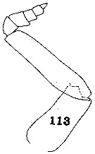 issued from : C.O. Esterly in Univ. Calif. Publs Zool., 1911, 6 (14). [Pl.32, Fig.113]. As Aetideopsis pacifica. Female: 113, Mxp (posterior).
|
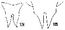 issued from : H.B. Owre & M. Foyo in Fauna Caribaea, 1967, 1, Crustacea, 1: Copepoda. Copepods of the Florida Current. [p.26, Figs.124, 125]. Female: 124, rostrum. Male: 125, rostrum.
|
 issued from : H.B. Owre & M. Foyo in Fauna Caribaea, 1967, 1, Crustacea, 1: Copepoda. [p.44, Figs.241-242]. Female (from Florida Current): 241, P2; 242, P3.
|
 Aerideopsis multiserrata Aerideopsis multiserrata female: 1 - Endopod of P1 with external lobe. 2 - Posterior corners of last thoracic segment not wing-like, not divergent (dorsal view). Genital segment (dorsal view) barrel-like without lateral swellings. 3 - Crest poorely developed, situated at the leel of A2. 1st and 2nd external spines of exopodal segments 1 and 2 of P1 reaching the base of next spine. 4 - Endopod of P2 2-segmented (though separation between segments may be incomplete). Points of last thoracic segment reaching or exceeding the midlength of genital segment. 5 - Anterior part of cephalon (lateral view) evidently prominent over rostrum (in front of it).
|
 Aerideopsis multiserrata Aerideopsis multiserrata male: 1 - Points of last thoracic segment reaching or exceeding posterior border of urosomal segment 1. 2 - Endopod of P1 with external lobe. 3 - Rostral rami spaced closely. 4 - Rostral rami not divergent.
| | | | | Ref. compl.: | | | Grice, 1963 a (p.495); Grice & Hulsemann, 1965 (p.223, 231: Rem.); Harding, 1966 (p.17); Dunbar & Harding, 1968 (p.318); Vinogradov, 1968 (1970) (p.266); Soenen, 1969 (p.72); Roe, 1972 (p.277, tabl.1, tabl.2); Björnberg, 1973 (p.322, 389); Vives & al., 1975 (p.41, tab.II, III); 1972 a (p.333); Deevey & Brooks, 1977 (p.256, tab.2, Station "S"); Pipe & Coombs, 1980 (p.223, Table II, vertical occurrence); Buchanan & Sekerak, 1982 (p.41, vertical distribution); Vives, 1982 (p.290); Kovalev & Schmeleva, 1982 (p.83); Cummings, 1984 (p.163, Table 2); Lozano Soldevilla & al., 1988 (p.58 as Aetidiopsis); Kosobokova, 1989 (p.26); Hattori, 1991 (tab.1, Appendix); Richter, 1994 (tab.4.1a); Errhif & al., 1997 (p.422); Hanssen, 1997 (tab.3.1); Suarez-Morales & Gasca, 1998 a (p107); Razouls & al., 2000 (p.343, Appendix); Holmes, 2001 (p.45); Gaard & al., 2008 (p.59, Table 1, N Mid-Atlantic Ridge); Galbraith, 2009 (pers. comm.); Park & Ferrari, 2009 (p.143, Table 4, Appendix 1, biogeography); Matsuno & al., 2012 (Table 2); El Arraj & al., 2017 (p.272, table 2); Belmonte, 2018 (p.273, Table I: Italian zones); Chaouadi & Hafferssas, 2018 (p.913, Table II: occurrence). | | | | NZ: | 15 + 1 douteuse | | |
|
Carte de distribution de Aetideopsis multiserrata par zones géographiques
|
| | | | | | | | | | Loc: | | | Antarct. (Indian, S & SE Pacif., Drake Passage), sub-Antarct. (SE Pacif.), Atlant. (tropical), Congo, G. of Guinea, Canary Is., Maroccan coast, off Madeira, Bay of Biscay, W Azores, G. of Mexico, off Bermuda: Station "S" (32°10'N, 64°30'W), Sargasso Sea, Davis Strait, Baffin Bay, Greenland Sea, Arct. (Fletcher's Ice Is.), Canadian abyssal plain, Iceland, Faroe Channel, Laptev Sea, off Ireland (S & W), Baie Ibéro-marocaine, Medit. (Alboran Sea, Habibas Is., Sidi Fredj coast, Balearic Sea, Marseille, SW Sardinia), Tyrrhenian Sea, ? Straits of Moluca, ? Halmahera Sea, Japan (Izu region, off Sanriku), Izu-Bonin Trench, Kuril-Kamtchatka, E Beaufort Sea, Wyville Thomson Ridge, Arct., Vancouver Is., NE Pacif. (off Cape Flattery), S Pacif, Chile, California, G. of California in Suarez-Morales & Gasca, 1998 a).
Type locality: North Atlantic | | | | N: | 57 | | | | Lg.: | | | (1) F: 2,8; (7) F: 2,88; (14) F: 2,8; (20) F: 3,04; 3; (37) F: 3,54-2,52; M: 3,25-2,7; (38) F: 2,64; (39) F: 3,13; M: 3,04; (59) F: 3,2-2,8; (113) F: 3,4-2,75; (143) F: 3,18; (197) F: 2,92-2,52; M: 2,86-2,7; (199) F: 2,81-2,51; (866) F: 2,2-3,54; M: 3,04; {F: 2,20-3,54; M: 2,70-3,25} | | | | Rem.: | méso-bathypélagique.
Sampling depth (Antarct., sub-Antact.): 0-1000-3000 m.
Voir aussi les remarques en anglais | | | Dernière mise à jour : 25/10/2022 | |
|
|
 Toute utilisation de ce site pour une publication sera mentionnée avec la référence suivante : Toute utilisation de ce site pour une publication sera mentionnée avec la référence suivante :
Razouls C., Desreumaux N., Kouwenberg J. et de Bovée F., 2005-2026. - Biodiversité des Copépodes planctoniques marins (morphologie, répartition géographique et données biologiques). Sorbonne Université, CNRS. Disponible sur http://copepodes.obs-banyuls.fr [Accédé le 17 juin 2026] © copyright 2005-2026 Sorbonne Université, CNRS
|
|
|
|


;)
;)
;)
;)
;)
;)
;)
;)
;)
;)

