|
|
 |
Fiche d'espèce de Copépode |
|
|
Calanoida ( Ordre ) |
|
|
|
Arietelloidea ( Superfamille ) |
|
|
|
Heterorhabdidae ( Famille ) |
|
|
|
Hemirhabdus ( Genre ) |
|
|
| |
Hemirhabdus grimaldii (Richard, 1893) (F,M) | |
| | | | | | | Syn.: | Heterochaeta grimaldii Richard, 1893 a (p.151, Descr.F);
Heterorhabdus grimaldii : Giesbrecht & Schmeil, 1898 (p.117); Wolfenden, 1905 a (p.10, Rem.F,M); Farran, 1908 b (p.66); Sewell, 1913 (p.354); Farran, 1926 (p.284); Wilson, 1936 c (p.92); Lysholm & al., 1945 (p.36); Björnberg, 1973 (p.346); Vives, 1982 (p.293);
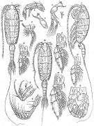
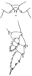
Macrorhabdus grimaldii : Sars, 1920 c (p.11) | | | | Ref.: | | | Wolfenden, 1911 (p.309, figs.F); Sars, 1925 (p.230, figs.F,M); Farran, 1926 (p.284); Sewell, 1932 (p.304, figs.F); Rose, 1933 a (p.205, figs.F,M); Jespersen, 1934 (p.108); Lysholm & al., 1945 (p.36); Sewell, 1947 (p.181); Farran, 1948 d (n°15, p.3, figs.F,M); Tanaka, 1964 a (p.26, figs.M); Tanaka & Omori, 1967 (p.252); Owre & Foyo, 1967 (p.80, figs.F,M); Chihara & Murano, 1997 (p.820, Pl.116: F,M); Ohtsuka & al., 1997 (p.579, figs.M); Park, 2000 (p.53, figs.F,M, Rem.); Vives & Shmeleva, 2007 (p.295, figs.F,M, Rem.) | 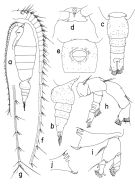 issued from : T. Park in Bull. Scripps Inst. Oceanogr. Univ. California, San Diego, 2000, 31. [p.182, Fig.30]. Female: a, habitus (left side); b, c, urosome (left, dorsal, respectively); d, forehead (ventral); e, genital somite (ventral); f, segments 1-19 of left A1 (ventral); g, segments 20-25 of left A1 (ventral); h, left A1 (posterior); i, left Md (posterior); j, masticatory edge of right Md (posterior). Nota: Genital somite (in dorsal view) with smoothly bulging lateral margins (Fig.30-c) and in ventral view with genital operculum at middle of somite (Fig.30-e).
|
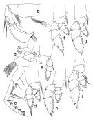 issued from : T. Park in Bull. Scripps Inst. Oceanogr. Univ. California, San Diego, 2000, 31. [p.183, Fig.31]. Female: a, Mx1 (posterior); b, left Mx2 (posterior); c, right Mxp (anterior); d, P1 (anterior); e, P1 (endopod omitted), posterior; f, P2 (anterior); g, P3 (anterior); h, P4 (anterior); i, P5 (anterior). Nota: Coxa of Mxp with 1 middle and 3 distal setae (Fig.31-c).
|
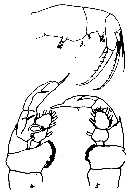
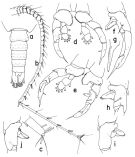 issued from : T. Park in Bull. Scripps Inst. Oceanogr. Univ. California, San Diego, 2000, 31. [p.184, Fig.32]. Male: a, urosome (dorsal); b, left A1 (ventral); c, distal segment of left A1 (ventral); d, P5 (anterior); e, P5 (right endopod omitted), posterior; f, exopod of right P5 (posterior); g, distal end of exopod of left P5 (anterior); h, i, j, second exopodal segment of right P5 (posterior, anterior, tilted clockwise, respectively). Nota: 2nd exopodal segment of right P5 with somewhat triangular medial projection (Fig.32-h)
|
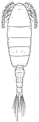
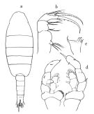 issued from : O. Tanaka in Publs Seto Mar. Biol. Lab., 1964, XII (1). [p.27, Fig.187]. Male: a, habitus (dorsal); b, Mx2; c, biting edge of Md; d, P5. Nota: The urosome segments and furca are in the proportional lengths as 17:17:17:15:17:17 = 100. Furcal rami symmetrical, 1.6 times as long as wide. Right A1 extends at least to the end of the furca.
|
 Issued from : G.O. Sars in Résult. Camp. Scient. Prince Albert I, 69, pls.1-127 (1924). [Pl.LXIII, figs.1-15]. As Macrorhabdus grimaldii. Female (from S Canary Islands & N Bay of Biscay): 1, habitus (dorsal); 2, idem (lateral left side); 3, rostrum; 4, A2; 5, left Md; 6, right Md (biting blade, without palp); 7, Mx1; 8, Mx2; 9, Mxp; 10, P1; 11, P2; 12, P3; 13, P4. Male: 14, habitus (dorsal); 15, P5.
|
 Issued from : R.B.S. Sewell in Mem. Indian Mus., 1932, X (continued). [p.305, Fig.100]. Female (from off SE Sri Lanka): a, proximal segments of A1; b, Mx1; c, Mx2; d, P1; e, P4; f, P5.
|
 Issued from : R.B.S. Sewell in Mem. Indian Mus., 1932, X (continued). [p.303, Fig.99]. Female: habitus dorsal) (distribution of the cutaneous pores).
|
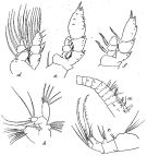
 issued from : S. Ohtsuka, H.Y. Soh & S. Nishida in J. Crustacean Biol., 1997, 17 (4). [p.581, Fig.2, D, E]. Male (from S. Japan): D, right mandibular cutting edge; E, terminal part of ventralmost tooth. v = ventral teeth; c = central teeth; d = dorsal tooth. Scale bars : 0.100 mm (D); 0.010 mm (E).
|
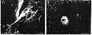 issued from : S. Ohtsuka, H.Y. Soh & S. Nishida in J. Crustacean Biol., 1997, 17 (4). [p.584, Fig.5, A-B]. Male (S Japan): A, left side of labrum (posterior surface) with gland openings (arrowed = type-2 gland opening, large arrow = gland type-1, asterisk = small gland opening); B, idem, type-3 gland opening (magnified) Scale bars: 0.100 mm (A); 0.010 mm (B).
|
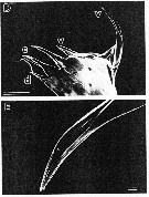
 issued from : R.N. Wolfenden in Die Marinen Copepoden der Deutschen Südpolar-Expedition 1901-1903, 1911. [p.309, Fig.56]. Female: a, forehead (ventral); b, P2.
|
 issued from : R.N. Wolfenden in Plankton Studies Part I. Copepoda. 1905 [Pl. IV, 3-5: 3]. As Heterorhabdus grimaldii. Female: 3, Mx2; Male: 4, right P5; 5, left P5.
|
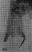 issued from : H.B. Owre & M. Foyo in Fauna Caribaea, 1, Crustacea, 1: Copepoda. Copepods of the Florida Current. 1967. [p.79, Fig.519]. Female (from 25°33'N, 79°43'W): 519, Md (biting edge of gnathobase).
|
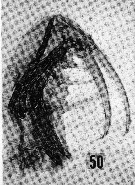 issued from : H.B. Owre & M. Foyo in Fauna Caribaea, 1, Crustacea, 1: Copepoda. Copepods of the Florida Current. 1967. [p.17, Fig.50]. Female: 50, Mx2.
|
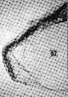 issued from : H.B. Owre & M. Foyo in Fauna Caribaea, 1, Crustacea, 1: Copepoda. Copepods of the Florida Current. 1967. [p.17, Fig.52]. Female: 52, Mxp.
|
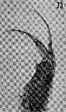 issued from : H.B. Owre & M. Foyo in Fauna Caribaea, 1, Crustacea, 1: Copepoda. Copepods of the Florida Current. 1967. [p.20, Fig.73]. Male: 73, Mx2.
| | | | | Ref. compl.: | | | Sewell, 1948 (p.503, 522, 530, 532, 547); C.B. Wilson, 1950 (p.238); Grice, 1963 a (p.496); Mazza, 1966 (p.72); Grice & Hulsemann, 1967 (p.18); Vinogradov, 1968 (1970) (p.268); Itoh, 1970 (tab.1); Roe, 1972 (p.277, tabl.1, tabl.2); Björnberg, 1973 (p.386); Deevey & Brooks, 1977 (p.256, tab.2, Station "S"); Vives, 1982 (p.293); Lozano Soldevilla & al., 1988 (p.59); Holmes, 2001 (p.15) | | | | NZ: | 12 | | |
|
Carte de distribution de Hemirhabdus grimaldii par zones géographiques
|
| | | | | | | | | | Loc: | | | Atlant. (S tropical), off Cape Verde Is., Canary Is., off N Azores, Bay of Biscay, off Ireland (S & W), Florida, Sargasso Sea, off Bermuda: Station S (32°10N, 64°30W), S Strait of Davis, off E Scotland, W Medit. (G. of Lion), Arabian Sea, S & SW Indian, Bay of Bengal, Philippines, Japan (Sagami Bay), E Tuamotu Is., Galapagos, off Peru, Pacif. (central & E, tropical & subtropical N-S), off California, Tasman Sea
Type locality : 48° N, 21° W. | | | | N: | 19 | | | | Lg.: | | | (1) F: 10,3; (10) F: 9; (38) F: 7,3; (44) F: 9,21; (47) F: 10,2; (70) F: 11,3; M: 12,2; (121) M: 10,05; (199) F: 9,6-8,96; M: 9,28-8,32; (238) F: 9; M: 9; (824) F: 11-8,41; M: 10,33-8,33; {F: 7,30-11,30; M: 8,32-12,20}.
Park (2000, p.55) emphasizes that the body size showed a wide range of variation, 8.41-11.00 in the female and 8.33-10.33 in the male. However, no morphological variation associated with the body sizes was detected. The largest specimens were found off California and the smallest off northwestern Africa. | | | | Rem.: | bathypélagique.
Cette espèce ressemble à H. amplus.
Voir aussi les remarques en anglais | | | Dernière mise à jour : 17/10/2017 | |
|
|
 Toute utilisation de ce site pour une publication sera mentionnée avec la référence suivante : Toute utilisation de ce site pour une publication sera mentionnée avec la référence suivante :
Razouls C., Desreumaux N., Kouwenberg J. et de Bovée F., 2005-2026. - Biodiversité des Copépodes planctoniques marins (morphologie, répartition géographique et données biologiques). Sorbonne Université, CNRS. Disponible sur http://copepodes.obs-banyuls.fr [Accédé le 10 juin 2026] © copyright 2005-2026 Sorbonne Université, CNRS
|
|
|
|


;)
;)
;)
;)
;)
;)
;)
;)
;)
;)
;)
;)
;)
;)

