|
|
 |
Fiche d'espèce de Copépode |
|
|
Calanoida ( Ordre ) |
|
|
|
Clausocalanoidea ( Superfamille ) |
|
|
|
Aetideidae ( Famille ) |
|
|
|
Aetideopsis ( Genre ) |
|
|
| |
Aetideopsis rostrata Sars, 1903 (F,M) | |
| | | | | | | Syn.: | Aetidiopsis rostrata Sars, 1903 (p.160, Descr.F, figs.F); With, 1915 (p.86, figs.F);
Aetideopsis divergens Tanaka, 1957 a (p.40, figs.F,M);
Aetideopsis divaricata Esterly, 1911 (p.316, figs.F); Brodsky, 1950 (1967) (p.150, figs.F); De Decker & Mombeck, 1964 (p.11); in CalCOFI regional list (MDO, Nov. 2013; M. Ohman, pers. comm.);
? Aetideopsis trichechus Vervoort, 1949 (p.7, figs.F);
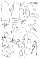
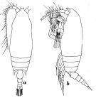
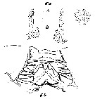
Aetideopsis inflata Park, 1978 (p.118, figs.F) (syn. n. after Markhaseva, 1996, p.42) | | | | Ref.: | | | ? A. Scott, 1909 (p.40, figs.F, Rem.); Farran, 1926 (p.248, Rem.); Sewell, 1929 (p.99); Wilson, 1932 a (p.46, figs.F); Rose, 1933 a (p.90, figs.F); Jespersen, 1934 (p.52); 1940 (p.17); Lysholm & al., 1945 (p.11, Rem.); Brodsky, 1950 (1967) (p.147, figs.F); Vervoort, 1951 (p.80, Rem.); 1952 a (n°42, p.3, figs.F); Tanaka & Omori, 1970 (p.112, figs.F, Rem.); Vidal, 1971 a (p.15, 112, figs.F); Tupitsky, 1982 a (p.110, figs.F); Shih & Stallard, 1982 (p.56, Descr.M, figs.M; Rem.); Markhaseva, 1996 (p.42, figs.F,M); Chihara & Murano, 1997 (p.681, Pl.30,31: F,M); Bradford-Grieve & al., 1999 (p.879, 920, figs.F,M); Vives & Shmeleva, 2007 (p.539, figs.F,M, Rem.); | 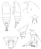 issued from : T. Park in Antarctic Res. Ser. Washington, 1978, 27. [p.120, Fig.11]. As Aetideopsis inflataFemale: A, habitus (dorsal view); B, idem (lateral view); C, posterior part of metasome and genital segment (dorsal view); E, forehead (left lateral view); F, rostrum (anterior view); G, A2.
|
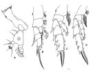 issued from : T. Park in Antarctic Res. Ser. Washington, 1978, 27. [p.121, Fig.12]. As Aetideopsis inflataFemale: A, Md; B, P1; C, P2; D, P4. Legs 1,2,4: anterior view.
|
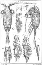
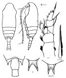
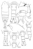 issued from : E.L. Markhaseva in Trudy Zool. Inst. RAN, St. Petersburg, 1996, 268. [p.43, Fig.26]. Female; From different specimens. R: rostrum (anterior). (a) from 71°10'N, 00°; other figures from 84°33'N, 142°17'E.
|
 issued from : E.L. Markhaseva in Trudy Zool. Inst. RAN, St. Petersburg, 1996, 268. [p.44, Fig.27]. Female (from 84°33'N, 142°17'E). P.md: mandibular palp; Gntb (part): gnathobase (incomplete
|
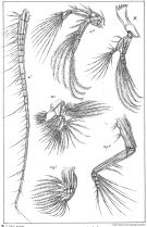
 issued from : E.L. Markhaseva in Trudy Zool. Inst. RAN, St. Petersburg, 1996, 268. [p.45, Fig.28]. Male; From different specimens. (a) from 44°10'N, 150°29'E; other figures from 83°33'N, 142°39'E.
|
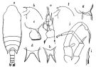
 issued from : G.O. Sars in An Account of the Crustacea of Norway. Vol. IV. Copepoda Calanoida. Published by the Bergen Museum, 1903. [Suppl. Pl.IV]. Female.
|
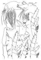
 issued from : G.O. Sars in An Account of the Crustacea of Norway. Vol. IV. Copepoda Calanoida. Published by the Bergen Museum, 1903. [Suppl. Pl.V] Female. Nota: M = Md; m = Mx1; mp1 = Mx2; mp2 = Mxp.
|
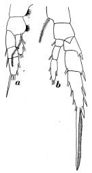
 issued from : O. Tanaka in Publ. Seto Mar. Biol. Lab., 1957, VI (1). [p.41, Fig.27]. As Aetideopsis divergens. Female: a, habitus (dorsal); b, forehead (lateral); c, last thoracic segment and genital somite (lateral left side); d, rostrum; e, P1. Nota: Articular membrane between head and 1st thoracic segment is observed both in dorsal and lateral aspects. Integuments finely granulated. Rostrum stout, bifurcate and strongly divergent. Proportional lengths of urosomites and furca 38:19:16:11:16 = 100. A1 (24-segmented) extends to distal margin of 3rd urosomal somite. Male: f, forehead (lateral); g, last thoracic segment (lateral left side); h, rostrum; i, terminal spine of exopod of P2; j, P5. Nota: Head and 1st thoracic segment fused, 4th and 5th partially fused (the line of fusion faintly visible in dorsal view). Thoracic segments finely granulated. Proportional lengths of urosomites and furca 12:31:25:18:2:12 = 100. Anal segment concealed beneath the foregoing. A1 reaches back to distal margin of 2nd thoracic segment. P5 as in Chiridius armatus (Boeck)
|
 Issued from : W. Vervoort in Zool. Verh., Leiden, 1949, 5. [p.8, Fig.2]. As Aetideopsis trichechus. With doubt. Female (Flores Sea): a, habitus (dorsal); b, idem (lateral left side). Nota: The abdominal segments and furca in the proportional lengths 38:20:14:10:18 = 100.
The present species shows some resemblance with Aetideopsis divaricata Esterly, 1911, but the proeminent frontal part of the head seems to be absent in the latter species.
|
 Issued from : W. Vervoort in Zool. Verh., Leiden, 1949, 5. [p.9, Fig.3]. As Aetideopsis trichechus. With doubt. Female (Flores Sea): a, P1; b, P4. Nota: P4 is different compared to P4 from Park (1978, p.121, Fig.12 D) and Markhaseva (1996, p.44, Fig.27)
|
 Issued from : K.A. Brodskii in Calanoida of the Far Eastern Seas and Polar Basin of the USSR. Opred. Fauna SSSR, 1950, 35 (Israel Program for Scientific Translations, Jerusalem, 1967) [p.147, Fig.61]. Female (from Arctic): habitus (dorsal and lateral left side); R, rostrum; last thoracic segment and 1st and 2nd abdominal segments (dorsal); S1, P1; S3As, apical spine of P3; R (NP), rostrum (example from NW Pacific). Nota: Head widened in middle; not fused with thorax. Boundary between 4th and 5th thorcic segments sometimes visible only at edges of carapace. Abdomen 2/7 length of cephalothorax. Genital segment slightly shorter than both following segments combined.Exopodite of A2 longer than endopodite by 1/6 its length. Outer spine of 1st exopodal segment of P1 not reaching middle of following spine.
|
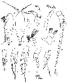
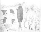 issued frm : A. Scott in Siboga-Expedition, 1909, XIX a. [Plate V, Figs.13-24]. Doutful (perhaps Aetideopsis multiserrata). Female (from Indonesia-Malaysia): 13, habitus (dorsal); 14, forehead (lateral); 15, last thoracic and genital segments (left side); 16, rostrum; 17, A1; 18, A2; 19, Md (biting blade); 20, Mx2; 21, Mxp; 22, P1; 23, P2; 24, P4. Nota: The minor points of difference with the description and figures given by Sars are: the external margins of the rostrum are more concave, and the excavation between the rami is much narrower. A comparison of the North Atlantic specimens from the Faroë Channel with the specimens from the 'Siboga' does not reveaf difference.
|
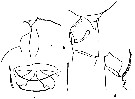
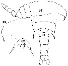 issued from : C.O. Esterly in Univ. Calif. Publs Zool., 1911, 6 (14). [Pl.28, Figs.27, 29, 31, 32]. As Aetideopsis divaricata. Female (from San Diego Region): 27, habitus (lateral); 29, forehead (lateral); 31, 4th and 5th thoracic segments and urosome (dorsal); 32, rostrum. Nota: A1 24-segmented, extend back to about the middle of the urosome.
|
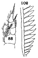 issued from : C.O. Esterly in Univ. Calif. Publs Zool., 1911, 6 (14). [Pl.31, Figs.88, 102]. As Aetideopsis divaricata. Female: 88, P1; 102, basal part of the serrate margin of the terminal bristle of exopodite of P4.
|
 issued from : C.-t. Shih & N. Stallard in Arctic, 1982, 35 (1). [p.57). Male (from Lancaster Sound, N Baffin Island): a-b, lateral and dorsal, respectively); c, rostrum (frontal view); d, left A1; e, A2; f, Md; g, Mx1; h, Mx2; i, Mxp; j-n, P1 to P5. Upper scale for a-b; lower scale for appendages. Nota: A1 23-segmented (segments 8, 9 and 10 fused; incomplete segmentation between segments 9 and 10 barely visible), reaching nearly to posterir margin of 3rd metasomal segment. A2 similar to that of female. Md with degenerate gnathobase; exopod 5-segmented, slightly longer than endopod. Mx1 degenerate; 1st and 3rd inner lobes present, with 4 and 3 setae, respectively; 2nd inner lobe represented by an indistinct outgrowth; basis with 3, endopod with 4 and 5, and exopod with 9 setae; outer lobe with 2 setae. Mx2 very much reduced in size and structure. Mxp similar, in general, to that of the female. P1 to P4 similar those of the female. P5 biramous and asymmetrical; right leg longer than left, exopod with first two segments fused, 3rd segment with a long a slightly curved terminal spine, endopod 1-segmented and reduced in size; left leg exopod 3-segmented.
|
 issued from : C. With in The Danish Ingolf-Expedition, 1915, III (4). [p.87, Text-fig.22. As Aetidiopsis rostrata. Female (65°34'N, 7°31'W): a, forehead (lateral); b, last thoracic segment and genital segment (lateral, left side); c, genital area (ventral); d, right P4 (posterior).
|
 issued from : C. With in The Danish Ingolf-Expedition, 1915, III (4). [Pl. II, Figs.6a, 6b]. As Aetidiopsis rostrata. Female: 6a, anterior portion of oral surface of labrum; 6b, lamina labialis (partly in anterior view).
|
 Aetideopsis rostrata Aetideopsis rostrata female: 1 - Endopod of P1 with external lobe. 2 - Posterior corners of last thoracic segment not wing-like, not divergent (dorsal view). Genital segment (dorsal view) barrel-like without lateral swellings. 3 - Crest present (faintly developed). 1st and 2nd external spines of exopod segments 1 and 2 of P1 exceeding base of next spine. 4 - Endopod of P2 2-segmented (though separartion between segments may be incomplete). Points of last thioracic segment reaching the midlength of geniral segment (or exceeding it). 5 - Anterior part of cephalon (lateral view) fairly smoothly transforming into rostrum, not prominent over base of rostrum. 6 -Rostrum well developed, rami not close-spaced slightly divergent. 7 -Proximal part of Mxp protopodite (externally) with well visible projection. 8 - Endopodal segment 2 of Md with 10 (8 terminal and 2 posterior) setae. A1 reaching the middle length, or end of urosomal segment 3. Body length > 3.0 mm.
|
 Aetideopsis rostrata Aetideopsis rostrata male: 1 - Points of last thoracic segment reaching or exceeding posterior border of urosomal segment 1. 2 - Endopod of P1 with external lobe. 2 - Rostral rami spaced closely. 3 - Rostral rami more or less divergent. 4 - Genital segment over half shorter than urosomal segment 2. Body length > 3.0 mm.
| | | | | Ref. compl.: | | | Damas & Koefoed, 1907 (p.400, tab.II); Sewell, 1948 (p.499, 507); Østvedt, 1955 (p.14: Table 3, p.60); Grice & Hulsemann, 1965 (p.223); Mazza, 1966 (p.70); Harding, 1966 (p.17, 65, 66); Dunbar & Harding, 1968 (p.318); Vinogradov, 1968 (1970) (p.266, 268); Shih & al., 1971 (p.201); Harding, 1974 (p.141, tab. 3, gut contents); Buchanan & Sekerak, 1982 (p.41, vertical distribution); Vives, 1982 (p.291); Kovalev & Schmeleva, 1982 (p.83); Tremblay & Anderson, 1984 (p.3); Kosobokova, 1989 (p.27); Mumm, 1993 (tab.1, fig.2); Richter, 1994 (tab.4.1a); Kouwenberg, 1994 (tab.1); Sirenko & al., 1996 (p.345); Hanssen, 1997 (tab.3.1); Kosobokova & al., 1998 (tab.2); Auel, 1999 (tab.2); Razouls & al., 2000 (p.343, tab. 5, Appendix); Holmes, 2001 (p.45); Yamaguchi & al., 2002 (p.1007, tab.1); Auel & Hagen, 2002 (p.1013, tab.2); Bradford-Grieve, 2004 (p.283); Ikeda & al., 2006 (p.1791,Table 2); Kosobokova & al., 2007 (p.923: Tab.2, 6, 7, fig.2); Blachowiak-Samolyk & al., 2007 (p.2716, Table 2); Blachowiak-Samolyk & al., 2008 (p.2210, Table 3, biomass); Morales-Ramirez & Suarez-Morales, 2008 (p.517); Darnis & al., 2008 (p.994, Table 1); Galbraith, 2009 (pers. comm.); Laakmann & al., 2009 (p.741, fig.2, vertical distribution, lipids analysis); Park & Ferrari, 2009 (p.143, Table 4, 6: bipolar distribution, , Appendix 1, biogeography); Homma & Yamaguchi, 2010 (p.965, Table 2); Mazzocchi & Di Capua, 2010 (p.423); Bucklin & al., 2010 (p.40, Table 1, Biol mol.); Dvoretsky & Dvoretsky, 2010 (p.991, Table 2); Kosobokova & al, 2011 (p.29, Table 2, figs.4, 8, Rem.: Arctic Basins); Homma & al., 2011 (p.29, Table 2, 3, abundance, feeding pattern: suspension feeders); Matsuno & al., 2012 (Table 2); Laakmann & al., 2012 (p.535, Table 1, fig.2, Rem.: mol. Biol.); Ohashi & al., 2013 (p.44, Table 1, Rem.); Zaafa & al., 2014 (p.67, Table I, occurrence); Smoot & Hopcroft, 2016 (p.1, fig.7, vertical distribution); Belmonte, 2018 (p.273, Table I: Italian zones) | | | | NZ: | 15 + 1 douteuse | | |
|
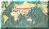
Carte de distribution de Aetideopsis rostrata par zones géographiques
|
| | | | | | | | | | | | | | |  Carte de 1996 Carte de 1996 | |
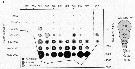 Issued from : S. Laakmann, M. Kochzius & H. Auel in Deep-Sea Res. I, 2009, 56. [p.745, Fig.2 c]. Issued from : S. Laakmann, M. Kochzius & H. Auel in Deep-Sea Res. I, 2009, 56. [p.745, Fig.2 c].
Vertical distribution abundance of copepodite stages C3 to C6 of Aetideopsis spp. and Chiridius obtusifrons. +: No occurrence; solid line depicts bottom profile (right axis); station 853 over the Yermak Plateau (81°22'N, 6°52'E) is separated from the other stations on the transect; other stations between East Greenland Current, the Fram Strait and the West Spitsbergen Current, into south return Atlantic Current (77°46'N-79°36'N, 6°20'E-7°29'W).
Deep-sea copepods collected from August 20 to September 16, 2006. |
| | | | Loc: | | | Antarct. (Atlant. SW, Pacif. S), South Africa (E), Namibia (in Carola, 1993), off Mauritania, off Woods Hole, off E Cape Cod, Ungava Bay, Baffin Bay, S Davis Strait, Iceland, Jan Mayen Is., Fram Strait, Spitsbergen, Greenland Sea, Norway Sea, Barents Sea, Nansen Basin, Laptev Sea, North Sea, off Ireland (S & W), Bay of Biscay, W Medit (M'Diq, Alboran Sea, W Basin, Ligurian Sea Tyrrhenian Sea), ? Indonesia-Malaysia, Japan (Izu), Station Knot, Kuril-Kamchatka, S Aleutian Is., Arct. (central, Fletcher's Ice Is.), Nansen Basin, Chukchi Sea, E Beaufort Sea, Canada Basin, Canadian abyssal plain, Bering Sea, S Aleutian Basin, Vancouver Is., E Pacif. (San Diego, W Costa Rica) | | | | N: | 69 | | | | Lg.: | | | (5) F: 3,5; (7) F: 3,9-3,4; (22) F: 4,4-4; (37) F: 4,6-3,8; M: 4,2-3,13; (38) F: 3,26; (39) F: 3,01; M: 3,13; (41) F: 3,38; (45) F: 4,4; (65) F: 4,4; (113) F: 3,14-3,1; (143) F: 3,36; (866) F: 3,01-4,4; M: 3,13; (1001) F: 3,9 ± 0,18; {F: 3,01-4,60; M: 3,13-4,20}
Type locality: between Jean Mayen and Finmark. | | | | Rem.: | méso à abyssopélagique.
Sampling depth (Antact.): 0-300 m.
Voir aussi les remarques en anglais | | | Dernière mise à jour : 03/12/2020 | |
|
|
 Toute utilisation de ce site pour une publication sera mentionnée avec la référence suivante : Toute utilisation de ce site pour une publication sera mentionnée avec la référence suivante :
Razouls C., Desreumaux N., Kouwenberg J. et de Bovée F., 2005-2026. - Biodiversité des Copépodes planctoniques marins (morphologie, répartition géographique et données biologiques). Sorbonne Université, CNRS. Disponible sur http://copepodes.obs-banyuls.fr [Accédé le 15 juin 2026] © copyright 2005-2026 Sorbonne Université, CNRS
|
|
|
|


;)
;)
;)
;)
;)
;)
;)
;)
;)
;)
;)
;)
;)
;)
;)
;)
;)

;)
;)
