|
|
 |
Fiche d'espèce de Copépode |
|
|
Calanoida ( Ordre ) |
|
|
|
Lucicutiidae ( Famille ) |
|
|
|
Lucicutia ( Genre ) |
|
|
| |
Lucicutia paraclausi Park, 1970 (F,M) | |
| | | | | | | Syn.: | Lucicutia clausi : Tanaka, 1963 (p.38, figs.F,M); Razouls, 1994 (p.149, figs.F,M); Chihara & Murano, 1997 (p.829, tab.5, Pl.124 (part.): F: a,M: f) | | | | Ref.: | | | Park, 1970 (p.477, 517, figs.F,M); Boxshall & Halsey, 2004 (p.133: F; p.135: M) | 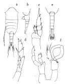 issued from : O. Tanaka in Publs Seto Mar. Biol. Lab., 1963, XI (1). [p.39, Fig.166]. As Lucicutia clausi. Female: a, habitus (dorsal); b, urosome (left lateral side); c, P1; d, P5. Nota: The urosome segments and furca are in the proportional lengths as 26:12:12:18:32 = 100. Male: e, urosome (dorsal); f, P5.
|
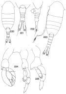 Issued from : T. S. Park in Bull. Mar. Sc., 1970, 20 (2). [p.518, Figs.220-225]. Female (from Caribbean Sea & G. of Mexico): 220, habitus (dorsal); 221, posterior part of metasome and urosome (dorsal); 222, idem (lateral left side). Nota: Head with small lateral spiniform processes pointed straight laterally (in some individuals these processes minute). Genital segment in dorsal view about as wide as long. Caudal ramus about 3.5 times as long as wide. Mx1: 2nd and 3rd inner lobes each with 3 setae; basis with 3 setae; 1st and 2nd endopodal segments with 4 and 5 setae, respectively. Male: 223, habitus (dorsal); 224, P5; 225, left P5 (anterior). Nota: Lateral spiniform processes of head as in female. Caudal ramus about 3.5 to 4.0 times as long as wide.P5: mediodistal margin of left basis with a large protrusion divided distally into 2 points, the median point with 3 small teeth.
|
 Lucicutia paraclausi Lucicutia paraclausi female: 1 - Characters following not combined : Prosome about 3 times longer than urosome. Cephalosome with slightly projecting and rounded anterior corners and well developed lateral spinous projections; anal somite about as long as wide; caudal rami 11.7 times longer than wide and bowed outwards at base, leaving elliptical space between rami proximally. 2 - P1 with 2-segmented endopod. 3 - Cephalosome not with 2 pairs of lateral spinous projections, but with other forms. 4 - A1 extending to end of caudal ramus or 1 to 2 segments beyond. 5 - Caudal rami 3 to 4 times longer than wide. 6 - Terminal setal element of P5 very short, less than 1/3 lenght of 3rd exopodal segment. 7 - A1 extending 2 segments beyond tip of caudal ramus; anal somite longer than preceding somite; cephalosome with strong spinous projection laterally.
|
 Lucicutia paraclausi Lucicutia paraclausi male: 1 - P1 with 2-segmented endopod; 2 - Caudal rami less than 10 times longer than wide. 3 - Inner distal corner of basis of P5 without strong spine. 4 - Basis of left P5 ornamented differently from L. oblonga. 5 - Caudal rami about 3 to 5 times longer than wide. 6 - A1 at most reaching to end of caudal rami; basis of left P5 different from L. longiserrata. 7 - Cephalosome with strong lateral spinous projections (in dorsal view); right A1 reaching only to tip of caudal rami; inner margin of basis of right P5 with small tooth-like projection; inner distal corner of basis of left leg not ending in single point. 8 - Spinous processes on cephalosome directed laterally; caudal rami about 3.5 to 4 times longer than wide.
| | | | | Ref. compl.: | | | Uysal & al., 2002 (p.17, tab.1); Williamson & McGowan, 2010 (p.273, Table 3, Pacific central gyres: N and S); Medellin-Mora & Navas S., 2010 (p.265, Tab. 2) | | | | NZ: | 5 | | |
|
Carte de distribution de Lucicutia paraclausi par zones géographiques
|
| | | | | | | Loc: | | | Caribbean Sea, Caribbean Colombia, G. of Mexico, E Medit. E (Lebanon Basin) (in Uysal & al., 2002), Red Sea (in Weikert, 1982), Japan (Suruga Bay); Pacific (central gyres: N and S) | | | | N: | 6 | | | | Lg.: | | | (26) F: 1,75; M: 1,86; (88) F: 1,84-1,72; M: 1,52; {F: 1,72-1,84; M: 1,52-1,86}
| | | | Rem.: | Mésopélagique (100-500 m)
D'après les illustrations de L. clausi fournies par Tanaka (1963), Park (1970, p.519) estime qu'il s'agit de cette nouvelle espèce.
| | | Dernière mise à jour : 16/01/2015 | |
|
|
 Toute utilisation de ce site pour une publication sera mentionnée avec la référence suivante : Toute utilisation de ce site pour une publication sera mentionnée avec la référence suivante :
Razouls C., Desreumaux N., Kouwenberg J. et de Bovée F., 2005-2025. - Biodiversité des Copépodes planctoniques marins (morphologie, répartition géographique et données biologiques). Sorbonne Université, CNRS. Disponible sur http://copepodes.obs-banyuls.fr [Accédé le 18 octobre 2025] © copyright 2005-2025 Sorbonne Université, CNRS
|
|
|
|


;)
;)

